The Complete Guide to Glucose Infusion Rate (GIR) Measurement in Clamp Studies: From Theory to Practice
This comprehensive guide provides researchers, scientists, and drug development professionals with a detailed framework for accurately measuring the Glucose Infusion Rate (GIR) in hyperinsulinemic-euglycemic and hyperglycemic clamp studies.
The Complete Guide to Glucose Infusion Rate (GIR) Measurement in Clamp Studies: From Theory to Practice
Abstract
This comprehensive guide provides researchers, scientists, and drug development professionals with a detailed framework for accurately measuring the Glucose Infusion Rate (GIR) in hyperinsulinemic-euglycemic and hyperglycemic clamp studies. It covers the foundational principles of the glucose clamp technique, the step-by-step methodology for GIR calculation and data acquisition, common troubleshooting scenarios and optimization strategies for data quality, and methods for validating and benchmarking GIR measurements. The article synthesizes current best practices to ensure reliable assessment of whole-body insulin sensitivity and beta-cell function in metabolic research.
Understanding GIR: The Core Metric of Glucose Clamp Studies
What is the Glucose Infusion Rate (GIR)? Defining the Key Output
Definition and Core Quantitative Data
The Glucose Infusion Rate (GIR) is the primary quantitative output of a glucose clamp study, most commonly the hyperinsulinemic-euglycemic clamp. It represents the rate (in mg/kg/min or µmol/kg/min) at which exogenous glucose must be infused to maintain target plasma glucose levels (euglycemia) under conditions of standardized, constant insulin infusion. The GIR is a direct measure of whole-body insulin sensitivity; a higher GIR indicates greater sensitivity to insulin.
Table 1: GIR Ranges and Interpretation in Humans
| Metabolic State | Typical GIR Range (mg/kg/min) | Interpretation |
|---|---|---|
| Severe Insulin Resistance | < 4.0 | Indicative of conditions like T2DM, obesity, PCOS. |
| Normal Insulin Sensitivity | 4.0 - 9.0 | Healthy, non-diabetic individuals. |
| High Insulin Sensitivity | > 9.0 | Seen in lean, aerobically trained individuals. |
Table 2: Key Clamp Parameters and Their Impact on GIR
| Parameter | Standard Value (Euglycemic Clamp) | Purpose & Impact on GIR |
|---|---|---|
| Target Plasma Glucose | ~5.0 mM (90 mg/dL) | Maintains euglycemia; provides standardized baseline. |
| Insulin Infusion Rate | 40-120 mU/m²/min (commonly 80 mU/m²/min) | Creates a steady-state hyperinsulinemic plateau. |
| Clamp Duration | 90-120 minutes (post-equilibration) | Allows time to reach steady-state for accurate GIR. |
| Sampling Interval | Every 5-10 minutes | Enables real-time adjustment of the glucose infusion. |
Experimental Protocol: Hyperinsulinemic-Euglycemic Clamp
This is the gold-standard protocol for measuring insulin sensitivity and deriving the GIR.
A. Pre-Clamp Preparation:
- Subject Preparation: Overnight fast (10-12 hours). Subject rests supine in a quiet, temperature-controlled room.
- Catheter Placement: Insert two intravenous catheters:
- Infusion catheter: For administration of insulin and glucose (e.g., in the antecubital vein).
- Sampling catheter: Placed retrograde in a contralateral hand/wrist vein for arterialized venous blood sampling, kept in a heated box (~55°C) to arterialize the blood.
B. Priming-Continuous Insulin Infusion:
- Initiate a primed, continuous intravenous infusion of regular human insulin to rapidly raise plasma insulin to a pre-determined, fixed level (e.g., 80 mU/m²/min).
- The priming dose is calculated to quickly achieve the desired plateau.
C. Variable Glucose Infusion & the "Clamp":
- Simultaneously, a variable 20% dextrose solution infusion is started.
- Measure plasma glucose every 5 minutes from the arterialized line.
- Feedback Loop: Adjust the glucose infusion rate based on a validated algorithm (e.g., the DeFronzo, Bergman, or minimal model-based algorithm) to "clamp" the plasma glucose level at the target baseline (e.g., 90 mg/dL).
- Potassium Consideration: To prevent hypokalemia, a potassium chloride (KCl) infusion is often co-administered (e.g., 0.15 mEq/min).
D. Steady-State & GIR Calculation:
- Equilibration Period: The first ~90-120 minutes are required for plasma glucose and insulin to stabilize.
- Steady-State Period: Once glucose levels are stable at the target (typically CV <5%), the final 20-30 minutes are considered the steady-state period.
- GIR Calculation: The mean glucose infusion rate (GIR) over this steady-state period is calculated. It is normalized to body weight.
Formula: GIR (mg/kg/min) = [Mean Dextrose Infusion Rate (mg/min)] / [Body Weight (kg)]
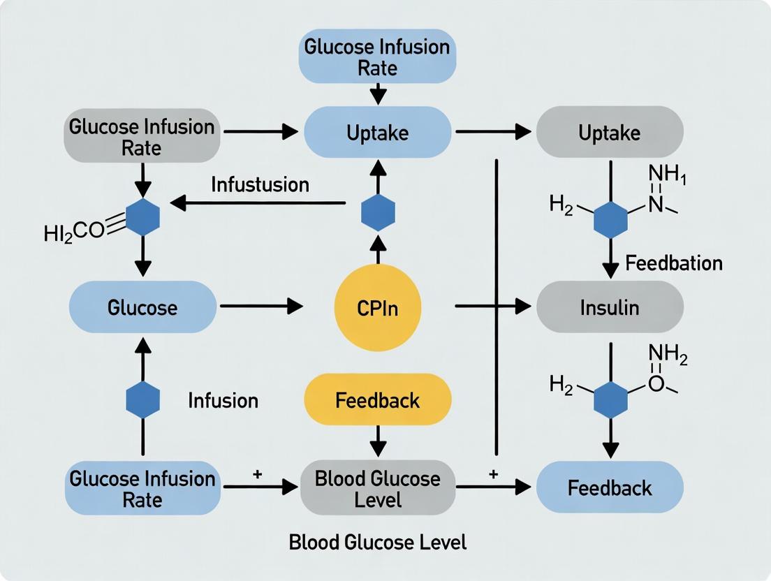
Workflow and Data Relationship Diagram
Diagram Title: Hyperinsulinemic-Euglycemic Clamp GIR Measurement Workflow
Key Physiological Pathways During the Clamp
Diagram Title: Insulin-Mediated Pathways Measured by GIR
The Scientist's Toolkit: Essential Research Reagents & Materials
Table 3: Key Reagents and Materials for Glucose Clamp Studies
| Item | Function / Purpose | Critical Specifications |
|---|---|---|
| Regular Human Insulin | Creates the hyperinsulinemic plateau. Must be IV-grade. | High purity, preservative-free recommended for research. |
| 20% Dextrose Solution | Variable infusion to maintain euglycemia. | Sterile, pyrogen-free. Concentration allows for lower infusion volumes. |
| Potassium Chloride (KCl) | Prevents insulin-induced hypokalemia. | Added to the dextrose solution or infused separately. |
| Bedside Glucose Analyzer | Provides rapid, accurate plasma glucose measurements (q5min). | Must have high precision and low sample volume requirement (e.g., YSI, Beckman). |
| Insulin Infusion Pump | Delivers a constant, precise rate of insulin. | Syringe pump with high accuracy (e.g., ±1%). |
| Variable-Rate Infusion Pump | Adjusts the dextrose infusion rate based on the algorithm. | Programmable, dual-channel pumps are ideal (one for dextrose, one for KCl). |
| Heated Venous Sampling Box | Arterializes venous blood from the hand for accurate glucose measurement. | Maintains stable temperature (~55°C) around the sampling site. |
| Blood Collection Tubes (Heparinized) | For collecting frequent plasma glucose samples. | Lithium heparin, rapid separation. |
In hyperinsulinemic-euglycemic clamp studies, the Glucose Infusion Rate (GIR) is the definitive quantitative measure of whole-body insulin sensitivity. Under conditions of standardized hyperinsulinemia and clamped euglycemia, endogenous glucose production is suppressed. The exogenous GIR required to maintain the target blood glucose concentration is therefore a direct measure of insulin-stimulated glucose disposal. A higher GIR indicates greater insulin sensitivity, while a lower GIR indicates insulin resistance. This application note details the physiological rationale and provides protocols for its accurate measurement.
Table 1: Key Physiological Parameters in a Steady-State Hyperinsulinemic-Euglycemic Clamp
| Parameter | Target / Typical Value | Physiological Rationale |
|---|---|---|
| Plasma Insulin | 40-120 mU/L (High-dose) | Creates a maximally stimulating insulin concentration to saturate insulin signaling. |
| Blood Glucose | 5.0-5.6 mmol/L (90-100 mg/dL) | Clamped at fasting euglycemia to eliminate glucose as a variable. |
| Steady-State Duration | 60-120 minutes | Allows for full suppression of hepatic glucose production and stabilization of peripheral glucose uptake. |
| Endogenous Glucose Production (EGP) | Suppressed by 80-100% | Essential precondition; remaining glucose disposal is attributed to infused glucose. |
| GIR (M-value) | 3-12 mg/kg/min (normal range) | Direct measure of insulin-mediated glucose disposal. |
Table 2: Interpreting GIR Values in Metabolic States
| Metabolic State | Typical GIR (mg/kg/min) | Pathophysiological Implication |
|---|---|---|
| Severe Insulin Resistance | < 4.0 | Impaired PI3K-Akt signaling, reduced GLUT4 translocation. |
| Mild Insulin Resistance | 4.0 - 7.5 | Suboptimal insulin action in muscle/adipose tissue. |
| Normal Insulin Sensitivity | 7.5 - 12.0 | Healthy post-receptor insulin signaling. |
| High Insulin Sensitivity (e.g., athlete) | > 12.0 | Enhanced metabolic flexibility and glucose uptake capacity. |
Detailed Experimental Protocol: Hyperinsulinemic-Euglycemic Clamp
Objective: To quantify whole-body insulin sensitivity by determining the steady-state GIR.
Materials & Pre-Clamp Preparation:
- Subjects/Animals: Fasted (10-12 hours).
- IV Lines: Two contralateral catheters (one for infusion, one for frequent sampling).
- Infusion Pumps: High-precision syringe pumps for insulin and glucose.
- Glucose Analyzer: Bedside, real-time (e.g., YSI, Beckman).
- Primed Solutions:
- Insulin Infusate: Human insulin in 0.9% NaCl with added albumin (0.1-1%) to prevent adsorption.
- 20% Dextrose Infusate: For glucose replacement.
Procedure:
- Basal Period (-30 to 0 min): Measure fasting plasma glucose and insulin levels.
- Insulin Infusion Initiation (t=0 min): Start a primed, continuous intravenous insulin infusion. A common high-dose protocol is a prime (80-120 mU/m²/min for 1 min) followed by continuous infusion (40-80 mU/m²/min).
- Glucose Clamping (t=0+ min): Begin frequent blood sampling (every 5-10 min). Measure glucose concentration immediately.
- Variable Glucose Infusion: Start and continuously adjust the 20% dextrose infusion rate based on the feedback algorithm (e.g., "DeFronzo algorithm") to maintain blood glucose at the target basal level (±5%).
- Steady-State Attainment (Typically t=60-120 min): Steady-state is defined as a period of ≥30 min where the glucose infusion rate is stable (coefficient of variation <5%) and blood glucose is constant at the target.
- GIR Calculation: The mean glucose infusion rate (mg/kg/min or µmol/kg/min) over the final 30-40 minutes of steady-state is reported as the M-value, the index of insulin sensitivity.
The Scientist's Toolkit: Essential Reagents & Materials
Table 3: Key Research Reagent Solutions for Clamp Studies
| Item | Function & Specification |
|---|---|
| Human Insulin (Recombinant) | Creates standardized hyperinsulinemia. Must be GMP-grade for clinical studies. |
| 20% Dextrose Solution | Concentrated glucose for infusion to minimize fluid load during clamping. |
| Human Serum Albumin (HSA) | Added to insulin infusate (0.1-1%) to prevent insulin adsorption to tubing and syringes. |
| Saline (0.9% NaCl) | Vehicle/diluent for insulin priming and infusion solutions. |
| Potassium Chloride (KCl) | Often added to glucose infusate (20-40 mmol/L) to prevent insulin-induced hypokalemia. |
| Bedside Glucose Analyzer & Consumables | For immediate, accurate glucose measurement to inform the feedback algorithm (e.g., YSI electrodes, test strips). |
Signaling Pathways & Experimental Workflow
1. Introduction Within the broader thesis on "How to measure glucose infusion rate in clamp studies research," understanding the distinct methodologies and applications of the hyperinsulinemic-euglycemic clamp (HEC) and the hyperglycemic clamp (HGC) is fundamental. These techniques represent the gold standard for in vivo assessment of insulin sensitivity and pancreatic beta-cell function, respectively. Both rely on the precise measurement and dynamic adjustment of the glucose infusion rate (GIR) to maintain a predefined "clamped" plasma glucose level, providing quantitative metabolic indices.
2. Comparative Overview: HEC vs. HGC The table below summarizes the core objectives, experimental conditions, and key outputs of the two clamp methodologies.
Table 1: Comparison of Hyperinsulinemic-Euglycemic and Hyperglycemic Clamp Protocols
| Parameter | Hyperinsulinemic-Euglycemic Clamp (HEC) | Hyperglycemic Clamp (HGC) |
|---|---|---|
| Primary Objective | Quantify insulin sensitivity (tissue response to insulin). | Assess pancreatic beta-cell function (insulin secretory capacity). |
| Clamp Target | Maintain euglycemia (typically ~5.0 mM or 90 mg/dL). | Maintain a steady-state hyperglycemia (typically 10-15 mM or 180-270 mg/dL). |
| Key Infusions | 1. Primed-constant insulin infusion (e.g., 40-120 mU/m²/min).2. Variable 20% glucose infusion (GIR adjusted to maintain target). | 1. Variable 20% glucose infusion (GIR adjusted to establish & maintain target hyperglycemia).2. No exogenous insulin infusion. |
| Steady-State Period | Usually 60-120 minutes after target glucose stabilization. | Usually 100-180 minutes after target glucose established (divided into first & second phase). |
| Primary Calculated Index | M-value: Mean GIR during steady-state (mg/kg/min or µmol/kg/min). Normalized to body weight and insulin level. | Acute Insulin Response (AIR): Mean plasma insulin increment during first 10 min.Steady-State Insulin Secretion: Plasma insulin concentration during final hour. |
| Interpretation | Higher M-value = greater insulin sensitivity. Lower M-value = insulin resistance. | Robust AIR and steady-state insulin = preserved beta-cell function. Blunted responses indicate dysfunction. |
3. Detailed Experimental Protocols
Protocol 3.1: Hyperinsulinemic-Euglycemic Clamp Objective: To measure whole-body insulin sensitivity by quantifying the GIR required to maintain euglycemia during a constant insulin infusion.
- Pre-Study: Overnight fast (10-12 hrs). Insert IV catheters in antecubital vein (for infusions) and contralateral dorsal hand/vein (for arterialized blood sampling, using a heated-hand box at ~55°C).
- Basal Period (-30 to 0 min): Collect baseline blood samples for plasma glucose and insulin.
- Insulin Infusion Start (t=0 min): Initiate a primed-constant intravenous infusion of human regular insulin. A common protocol uses a priming dose over 10 min followed by a constant rate of 40, 80, or 120 mU/m²/min to achieve low, medium, or high physiological insulinemia.
- Glucose Infusion & Clamping (t=0 to 120-180 min):
- Simultaneously begin a variable infusion of 20% dextrose.
- Measure plasma glucose every 5 minutes at the bedside.
- Adjust the GIR every 5-10 min using a validated algorithm (e.g., the DeFronzo, Bergman, or MINMOD models) to clamp glucose at the target basal level (e.g., 5.0 mM).
- Steady-State & Measurement: The steady-state is typically defined as the final 60 minutes of the clamp. The mean GIR over this period, normalized to body weight (M-value), is the primary measure of insulin sensitivity. Frequent sampling for insulin confirms a stable plateau.
Protocol 3.2: Hyperglycemic Clamp Objective: To assess insulin secretion by measuring the endogenous insulin response to a standardized, sustained hyperglycemic stimulus.
- Pre-Study & Basal Period: As per HEC (fasting, catheter placement, baseline sampling).
- Glucose Bolus & Clamp Establishment (t=0 min): Administer an intravenous bolus of 20% dextrose (e.g., 200 mg/kg) over 1-2 min to rapidly raise plasma glucose.
- Variable Glucose Infusion (t=0 to 180 min): Immediately initiate a variable 20% glucose infusion. Adjust the GIR every 5 minutes based on frequent plasma glucose measurements to clamp glucose at the target hyperglycemic level (e.g., 10 mM or 15 mM).
- Phases of Insulin Response:
- First Phase (0-10 min): Sample insulin at 2, 4, 6, 8, and 10 min. The Acute Insulin Response (AIR) is the mean incremental rise above basal.
- Second Phase (10-180 min): Sample insulin every 10-20 min. The mean plasma insulin concentration from 100-180 min represents the steady-state insulin secretory response.
- Interpretation: The beta-cell function is gauged by the magnitude of both the first-phase AIR and the second-phase sustained insulin secretion relative to the achieved glucose level.
4. The Scientist's Toolkit: Essential Research Reagent Solutions
Table 2: Key Materials and Reagents for Clamp Studies
| Item | Function / Explanation |
|---|---|
| Human Regular Insulin | The hormone of interest. Used at supraphysiological but standardized rates in HEC to create an insulin challenge. Not infused in HGC. |
| 20% Dextrose Solution | High-concentration glucose solution for intravenous infusion. The variable rate of this infusion (GIR) is the primary dependent variable measured. |
| Bedside Glucose Analyzer (e.g., YSI, Beckman) | Enables rapid (<2 min) and accurate plasma glucose measurement, which is critical for real-time adjustment of the GIR. |
| Heated-Hand Box (~55°C) | Arterializes venous blood from the hand by increasing blood flow and capillary permeability, providing samples that approximate arterial glucose concentration. |
| Hormone & Metabolite Assay Kits (ELISA, RIA, LC-MS) | For precise quantification of insulin, C-peptide, glucagon, and other metabolites from frequent blood samples. |
| Variable-Rate Infusion Pumps (two channels) | Precision pumps capable of adjustable infusion rates for simultaneous administration of insulin and glucose solutions. |
| IV Catheters & Tubing | For safe and continuous venous access for both infusion and sampling, often placed in contralateral arms. |
5. Visualizing Clamp Methodologies and Glucose-Insulin Dynamics
Diagram 1: Hyperinsulinemic-Euglycemic Clamp Workflow (79 chars)
Diagram 2: Hyperglycemic Clamp Phases and Outputs (66 chars)
Diagram 3: GIR Role in Clamp Study Types (55 chars)
Within the context of measuring the glucose infusion rate (GIR) in hyperinsulinemic-euglycemic clamp studies, the precision and integration of core instrumentation are paramount. The GIR is the primary quantitative output of the clamp, representing the amount of glucose required to maintain euglycemia under steady-state hyperinsulinemia, and thus a direct measure of whole-body insulin sensitivity. This application note details the essential components—infusion pumps, glucose analyzers, and data logging systems—and provides protocols for their integrated use to ensure accurate, reproducible GIR determination in both clinical and preclinical research.
Infusion Pumps: Precision Delivery
Infusion pumps are responsible for the controlled administration of insulin (to induce hyperinsulinemia) and variable-rate glucose (to maintain euglycemia). Their accuracy directly defines the GIR measurement.
Key Specifications & Selection Criteria:
| Parameter | Critical Requirement | Impact on GIR Measurement |
|---|---|---|
| Flow Rate Range | Insulin: 0.1 - 10 mL/hr; Glucose: 0.1 - 500 mL/hr | Must accommodate both low basal and high peak GIR scenarios. |
| Flow Rate Accuracy | ≤ ±2% of set rate across full range. | Inaccuracy causes systematic error in calculated GIR. |
| Resolution | ≤ 0.1 mL/hr for glucose infusion. | Fine adjustment is needed for precise clamp control. |
| Communication Interface | RS-232, Ethernet, or USB for external control. | Enables automated, dynamic adjustment via clamp algorithm. |
| Syringe/Reservoir Capacity | Glucose: 50-100 mL (preclinical) to 500+ mL (clinical). | Must sustain infusion for full study duration without interruption. |
Protocol: Pump Calibration and Setup
Objective: To verify infusion pump accuracy prior to clamp initiation.
- Gravimetric Calibration:
- Place a precision balance (0.001g resolution) on a vibration-free surface.
- Prime the infusion line with the test solution (e.g., 0.9% saline).
- Attach a pre-weighed, empty collection vessel to the line outlet.
- Program the pump to infuse at three critical rates (e.g., 1, 10, 100 mL/hr) for a set duration (e.g., 10 minutes each).
- Weigh the vessel after each infusion period. Calculate actual flow rate: (Weight gain in g) / (Duration in hr) / (Solution density ~1.0 g/mL).
- Compare to set rate. Deviation >2% necessitates pump service or calibration factor application.
Glucose Analyzers: Real-Time Feedback
Continuous or frequent intermittent blood glucose measurement is the feedback signal for the clamp controller.
Key Specifications & Selection Criteria:
| Parameter | Critical Requirement | Rationale |
|---|---|---|
| Measurement Technique | Glucose oxidase or hexokinase preferred (YSI/LinkedIn). | High specificity and accuracy vs. home glucometers. |
| Sample Volume | ≤ 10 µL per measurement (preclinical); 100-500 µL (clinical). | Minimizes blood loss, especially in rodent studies. |
| Measurement Interval | ≤ 5 minutes for continuous analyzers. | Shorter interval allows tighter glycemic control. |
| Accuracy & Precision | ≤ ±2% vs. reference standard. | Bias or noise in reading causes oscillation or drift in GIR. |
| Calibration Stability | Stable for ≥ 8 hours. | Critical for long-duration clamp studies. |
Protocol: Glucose Analyzer Calibration and Use
Objective: To ensure accurate plasma glucose readings throughout the clamp.
- Pre-study Calibration:
- Use manufacturer-provided standard solutions (e.g., 0, 50, 100, 200, 300 mg/dL).
- Follow instrument-specific priming and calibration sequence.
- Record calibration coefficients/factors.
- In-study Quality Control:
- Run a known-concentration control sample at least every 60-90 minutes.
- If deviation exceeds ±3%, pause the clamp, recalibrate the analyzer, and repeat the last 1-2 glucose measurements before resuming.
Data Logging & Control Systems: Integration
The control system integrates the glucose analyzer reading and computes the required glucose infusion rate, commanding the pump in real-time.
Core Functional Requirements:
| Module | Function | Key Feature |
|---|---|---|
| Data Acquisition | Logs glucose readings (time, value) and pump rates (time, set rate). | Timestamp synchronization within ±1 second. |
| Control Algorithm | Computes new GIR based on glucose error (target - measured). | Implement a validated PID or model-predictive algorithm. |
| Command Interface | Sends new infusion rate commands to glucose pump. | Robust error-handling for communication failures. |
| Real-time Visualization | Displays glucose trace and GIR over time. | Allows manual intervention if needed. |
Protocol: Integrated Clamp Control Workflow
Objective: To execute a standardized hyperinsulinemic-euglycemic clamp.
- System Initialization:
- Prime insulin and glucose infusion lines.
- Start insulin pump at constant rate (e.g., 1 mU/kg/min).
- Start glucose pump at a low basal rate (e.g., 2 mg/kg/min).
- Calibrate glucose analyzer and establish sampling line (arterial or venous).
- Clamp Phase (Steady-State Attainment & Maintenance):
- Measure glucose every 5 minutes.
- The control algorithm calculates adjustment:
GIR_new = GIR_old + Kp * (G_measured - G_target)(simplified). - Send updated GIR command to the glucose pump every 5-10 minutes.
- Steady-state is achieved when glucose is within ±5% of target for ≥ 30 minutes and GIR coefficient of variation is < 5%.
- Data Recording:
- The system logs: Timestamp, Measured Glucose, GIR, Insulin Rate.
- The mean GIR over the final 30-60 minutes of steady-state is reported as the study outcome.
Visualizations
Title: Glucose Clamp Control Loop Workflow
Title: Research Reagent & Equipment Solutions Table
The Scientist's Toolkit: Key Research Reagent Solutions
| Component | Example Product/Solution | Primary Function in Clamp Studies |
|---|---|---|
| Human Insulin Infusate | Humulin R (Eli Lilly) diluted in saline with <1% albumin. | Induces standardized hyperinsulinemia. |
| Dextrose Infusate (20%) | Sterile 20% Dextrose Injection, USP. | Concentrated solution for variable glucose infusion. |
| Glucose Assay Standards | YSI Multipoint Calibration Standards. | Calibrates analyzer for accurate absolute values. |
| Tracer for HGP Assessment | [6,6-²H₂]-Glucose (Cambridge Isotopes). | Quantifies endogenous Ra (hepatic glucose production). |
| Blood Sampling Kit | Heparinized syringes, microcentrifuge tubes. | For manual sampling and plasma separation. |
| Vascular Access Supplies | In-dwelling catheters, surgical tape. | Maintains patency for infusion and sampling lines. |
The reliable measurement of GIR in clamp studies is entirely dependent on the performance and synergistic operation of infusion pumps, glucose analyzers, and data logging systems. By adhering to the detailed calibration protocols and selection criteria outlined herein, researchers can minimize instrumental error, thereby ensuring that the calculated GIR robustly reflects the underlying metabolic physiology of insulin action. This rigorous approach to core instrumentation is foundational for generating high-quality data in metabolic research and drug development.
Step-by-Step Protocol: Calculating and Measuring GIR Accurately
Within the thesis framework of How to measure glucose infusion rate (GIR) in clamp studies, achieving a stable target clamp is the foundational prerequisite. The Glucose-Insulin Clamp, specifically the hyperinsulinemic-euglycemic clamp, is the gold standard for quantifying whole-body insulin sensitivity. Phase 1 focuses on establishing and maintaining the target steady-state condition where plasma glucose is "clamped" at a predefined level (typically euglycemia) through a variable glucose infusion, while insulin is held at a constant, elevated concentration. The subsequent measurement of the GIR required to maintain this steady state directly reflects insulin-mediated glucose disposal.
Key Quantitative Parameters & Data
Table 1: Standardized Hyperinsulinemic-Euglycemic Clamp Protocol Parameters
| Parameter | Typical Range / Value | Purpose & Rationale |
|---|---|---|
| Target Plasma Glucose | 90 mg/dL (5.0 mmol/L) | Represents physiological fasting euglycemia; minimizes counter-regulatory hormone response. |
| Insulin Infusion Rate (Priming) | 10-20 mU/m²/min | Rapidly achieves and sustains a steady-state hyperinsulinemic plateau (~80-120 µU/mL). |
| Duration of Clamp | 90-120 minutes (steady-state) | Allows sufficient time for insulin levels to plateau and glucose turnover to reach equilibrium. |
| Sampling Interval (Glucose) | Every 5-10 minutes | Enables frequent feedback for the glucose infusion rate (GIR) adjustment algorithm. |
| Coefficient of Variation (CV) for Steady-State | <5% (Plasma Glucose) | Defines an acceptable stable clamp; lower CV indicates tighter control. |
| Steady-State GIR | Variable (e.g., 4-12 mg/kg/min in healthy subjects) | Primary Outcome Measure: The mean glucose infusion rate during the final 30-40 minutes of the clamp quantifies insulin sensitivity. |
Detailed Experimental Protocol: Achieving the Target Clamp
A. Pre-Clamp Preparations
- Subject Preparation: Overnight fast (10-12 hours). Insert two intravenous catheters: one in an antecubital vein for infusions, another in a contralateral heated-hand vein (~55°C) for arterialized venous blood sampling.
- Solution Preparation:
- Insulin Solution: Dilute human regular insulin in 0.9% NaCl with added subject's own blood (1 mL per 49 mL) to prevent adsorption.
- 20% Dextrose Solution: For glucose infusion. Concentration can vary (e.g., 10-20%) based on expected GIR.
- Baseline Sampling: Collect at least two baseline blood samples (-30 and -10 min) for plasma glucose and insulin.
B. Initiation of the Clamp (Time 0 min)
- Start Insulin Infusion: Begin a primed, continuous intravenous infusion of insulin. A common protocol: a priming dose administered over 10 min in a decreasing manner, followed immediately by a continuous fixed-rate infusion.
- Initiate Variable Glucose Infusion: Simultaneously, begin a variable 20% dextrose infusion. The initial rate is often estimated based on subject weight (e.g., 2 mg/kg/min). Adjustments are driven by frequent glucose measurements.
C. The Clamp Algorithm & Maintenance of Target Glucose
- Frequent Plasma Glucose Measurement: Obtain blood samples every 5 minutes. Use a rapid, accurate bedside glucose analyzer.
- GIR Adjustment Formula: Apply a feedback algorithm to adjust the dextrose infusion pump. A common proportional-derivative formula is:
GIR_new = GIR_previous + ΔGwhereΔG = [ (G_target - G_measured) * Kp ] + [ (G_previous - G_measured) * Kd ]G_target: Desired glucose level (e.g., 90 mg/dL).G_measured: Current measured glucose.G_previous: Glucose from the prior measurement.Kp(Proportional gain): e.g., 0.1-0.2 (mg/kg/min per mg/dL error).Kd(Derivative gain): e.g., 0.02-0.05.
- Achieving Steady-State: The clamp is considered at steady-state when plasma glucose is stable at the target (±5%) with a CV <5% and the GIR shows minimal fluctuation for at least 30 minutes. This typically occurs from 90-120 minutes onwards.
D. Sample Collection & Calculations
- Steady-State Sampling: During the final 30 minutes, collect blood samples at 10-minute intervals for later confirmation of insulin and glucose levels.
- GIR Calculation: The primary metric is the mean GIR (mg/kg/min) during the steady-state period, often normalized to fat-free mass.
Visualizations
Feedback Loop for Glucose Clamp Control
Clamp Steady-State Output Variables
The Scientist's Toolkit: Essential Reagents & Materials
Table 2: Key Research Reagent Solutions for the Clamp Protocol
| Item | Function & Specification |
|---|---|
| Human Regular Insulin | Pharmacological agent to create a standardized hyperinsulinemic state. Must be of high purity and diluted appropriately in saline with a blood carrier protein. |
| 20% Dextrose Solution | The variable infusion solution used to counteract insulin-induced glucose disposal and maintain target glycemia. Must be sterile and pyrogen-free. |
| Heparinized Saline | Used to maintain patency of sampling catheters (low concentration, e.g., 1-2 U/mL). |
| Subject Blood (Autologous) | Added to the insulin infusion bag (typically 1-2% v/v) to prevent insulin adsorption to tubing and bag surfaces. |
| Bedside Glucose Analyzer & Strips | Critical for rapid (<60 sec), accurate feedback of plasma glucose levels. Must be calibrated and validated against lab reference methods. |
| Heated Hand Box (~55°C) | Device to arterialize venous blood from the sampling site, providing plasma glucose values equivalent to arterial levels. |
| Precision Infusion Pumps (x2) | One for the fixed-rate insulin infusion, another for the variable-rate glucose infusion. Requires high accuracy at low flow rates. |
The accurate measurement of the Glucose Infusion Rate (GIR) during hyperinsulinemic-euglycemic clamp studies is the definitive method for assessing in vivo insulin sensitivity. The validity of this measurement is entirely contingent upon achieving and maintaining a physiological and metabolic steady state. This document outlines the explicit criteria for verifying steady state and details protocols for ensuring reliable GIR data, a core component of research on metabolic diseases and therapeutic development.
Fundamental Criteria for Steady State
A true steady state is declared only when the following quantitative and qualitative conditions are met simultaneously for a predefined period (typically ≥30 minutes).
Table 1: Quantitative Criteria for Steady-State Declaration
| Parameter | Acceptance Criterion | Physiological Rationale | Typical Monitoring Interval |
|---|---|---|---|
| Plasma Glucose Concentration | Coefficient of Variation (CV) < 5% around target (e.g., 90-100 mg/dL or 5.0-5.5 mmol/L) | Essential for eliminating confounding effects of hypo- or hyperglycemia on glucose metabolism. | Every 5 minutes (Bedside Analyzer). |
| Glucose Infusion Rate (GIR) | CV < 5-10% during the evaluation period. | Indicates stable peripheral and hepatic glucose disposal matching the exogenous insulin stimulus. | Calculated per 5-10 min interval. |
| Insulin Infusion Rate | Constant, as per predefined protocol (e.g., 40 mU/m²/min or 1 mU/kg/min). | Provides the constant stimulus necessary for stable insulin receptor signaling and action. | Verified continuously by pump. |
| Plasma Insulin Concentration | Stable plateau, CV < 10-15% after distribution phase. | Confirms adequate and stable hormonal milieu for interpreting tissue response (GIR). | Measured every 10-20 minutes. |
Table 2: Qualitative/Ancillary Steady-State Criteria
| Criterion | Description & Importance |
|---|---|
| Counterregulatory Hormone Absence | Plasma glucagon, cortisol, epinephrine, and growth hormone should not be elevated. Stress hormones antagonize insulin action, invalidating GIR. |
| Suppression of Endogenous Glucose Production (EGP) | Hepatic glucose output must be maximally suppressed (typically >80-90% in normal subjects). Verified using tracer methods (e.g., [6,6-²H₂]-glucose). |
| Stable Cardiovascular Parameters | Heart rate and blood pressure should be stable. Significant changes may indicate physiological stress. |
| Subject Comfort | Subject reports no symptoms of hypoglycemia (sweating, anxiety, hunger), which would trigger counterregulation. |
Detailed Experimental Protocol for Valid GIR Measurement
Protocol: Standard Hyperinsulinemic-Euglycemic Clamp with Steady-State Verification
Objective: To measure whole-body insulin sensitivity as the GIR required to maintain euglycemia during a constant insulin infusion.
Pre-Clamp Preparation:
- Subject Preparation: 10-12 hour overnight fast. Cannulation of two intravenous lines: one for infusion (antecubital vein) and one for frequent blood sampling (heated contralateral hand vein or distal forearm vein).
- Priming-Continuous Insulin Infusion: Initiate a fixed-rate infusion of human insulin (e.g., 40 mU/m²/min or 1 mU/kg/min) via a precision infusion pump. This continues for the duration of the clamp (commonly 120-180 min).
- Variable Glucose Infusion: Simultaneously, initiate a variable 20% dextrose infusion. The infusion rate is adjusted based on frequent plasma glucose measurements.
Steady-State Attainment & GIR Measurement Phase (Critical Period: Minutes 80-120):
- Frequent Glucose Monitoring: Measure plasma glucose at 5-minute intervals using a validated bedside glucose analyzer.
- Feedback Algorithm: Adjust the 20% dextrose infusion rate using a standardized algorithm (e.g., the DeFronzo, Andres, or Tobin formula) to reach the target euglycemic level within 20-40 minutes.
- Steady-State Evaluation Window: Once glucose is stabilized at target, begin the formal 30-minute steady-state evaluation period (e.g., minutes 90-120).
- Data Collection for Steady-State Validation:
- Record the dextrose infusion rate (GIR) every 5-10 minutes.
- Collect plasma samples for insulin measurement at times 0, 60, 90, 100, 110, and 120 minutes.
- If using a glucose tracer, ensure the tracer infusion has reached isotopic equilibrium (constant glucose-specific activity or enrichment).
- Steady-State Declaration & GIR Calculation: If all criteria in Tables 1 & 2 are met during the evaluation window, steady state is validated. The mean GIR (in mg/kg/min or μmol/kg/min) over this final 30-minute period is reported as the measure of insulin sensitivity (M-value).
Visualizing the Steady-State Principle
Diagram 1: Decision Logic for Valid GIR Measurement (Steady-State Validation)
Diagram 2: Experimental Workflow for Hyperinsulinemic-Euglycemic Clamp
The Scientist's Toolkit: Essential Reagents & Materials
Table 3: Key Research Reagent Solutions for Clamp Studies
| Item | Function & Specification | Critical Notes |
|---|---|---|
| Human Insulin (Regular) | Provides the constant hyperinsulinemic stimulus. Must be of high purity and diluted in saline with added albumin (e.g., 0.1-1%) to prevent adsorption to tubing. | Infusion rate is protocol-dependent (e.g., low-dose: 10 mU/m²/min; high-dose: 40-120 mU/m²/min). |
| 20% Dextrose Solution | The variable infusion to maintain euglycemia. The high concentration minimizes fluid volume load. | Must be USP sterile. The infusion rate (GIR) is the primary outcome measure. |
| Glucose Tracer ([6,6-²H₂]-glucose or [3-³H]-glucose) | Enables calculation of endogenous glucose production (EGP) rates and glucose disposal (Rd) via dilution methodology. | Essential for confirming hepatic insulin sensitivity and complete EGP suppression during steady state. |
| Sterile Saline with Albumin (0.1-1% HSA) | Diluent for insulin and tracer infusions. Albumin prevents adhesion of peptides to plastic surfaces. | Ensures accurate delivery of the intended insulin dose. |
| Quality-Controlled Bedside Glucose Analyzer | Provides rapid (<60 sec), accurate plasma glucose measurements for real-time adjustment of the dextrose infusion. | Requires frequent calibration. YSI 2300 STAT Plus or similar analyzers are the historical gold standard. |
| Heparinized Saline | Used to keep the sampling catheter patent. | Concentration must be optimized to avoid interference with subsequent hormone assays. |
| Standardized Assay Kits | For precise measurement of plasma insulin, C-peptide, and counterregulatory hormones (glucagon, cortisol) from clamp samples. | Multiplex or ELISA kits with high sensitivity and specificity are required for reliable concentration data. |
| Precision Infusion Pumps (x2) | One for the constant insulin infusion, one for the variable glucose infusion. Must have high accuracy at low flow rates. | Syringe pumps are commonly used for insulin; peristaltic or syringe pumps for glucose. |
In hyperinsulinemic-euglycemic clamp studies, the Glucose Infusion Rate (GIR) is the primary measure of whole-body insulin sensitivity. The direct calculation of GIR and correct interpretation of its units (mg/kg/min vs. µmol/kg/min) are fundamental for accurate data reporting and cross-study comparison. This protocol details the methodology and unit conversions essential for clamp research.
The GIR Calculation Formula
The steady-state GIR is calculated as the mean glucose infusion rate required to maintain euglycemia during the final 30-60 minutes of the clamp. The formula accounts for the glucose concentration of the infused solution and the subject's body weight.
Core Formula:
GIR = (G_inf * IR) / BW
Where:
G_inf= Concentration of glucose in the infusate (mg/mL or mmol/mL)IR= Infusion rate of the glucose solution (mL/min)BW= Subject's body weight (kg)
Unit Considerations and Conversion
GIR is most commonly reported in mg/kg/min. For molecular studies, it may be converted to µmol/kg/min using the molecular weight of glucose (180.156 g/mol).
Conversion Formula:
GIR (µmol/kg/min) = [GIR (mg/kg/min) / Molecular Weight of Glucose (mg/µmol)]
1 µmol/kg/min = 0.180156 mg/kg/min
1 mg/kg/min = 5.551 µmol/kg/min
Table 1: GIR Unit Conversion Reference
| Value in mg/kg/min | Equivalent in µmol/kg/min | Common Interpretation |
|---|---|---|
| 2.0 | 11.1 | Low insulin sensitivity |
| 5.0 | 27.8 | Moderate insulin sensitivity |
| 10.0 | 55.5 | High insulin sensitivity |
| 15.0 | 83.3 | Very high insulin sensitivity |
Detailed Experimental Protocol: Hyperinsulinemic-Euglycemic Clamp
Materials and Reagents
Table 2: Research Reagent Solutions Toolkit
| Item | Function in Clamp Study |
|---|---|
| 20% or 25% Glucose Infusion Solution | Dextrose solution for intravenous administration to maintain euglycemia. |
| Regular Human Insulin | Used to create and maintain a hyperinsulinemic plateau (e.g., 40-120 mU/m²/min). |
| 0.9% Sodium Chloride (Saline) | Diluent for insulin priming and for flushing intravenous lines. |
| Potassium Chloride (KCl) | Often added to the glucose infusate (e.g., 20 mmol/L KCl) to prevent insulin-induced hypokalemia. |
| Bedside Glucose Analyzer | Must be calibrated and provide rapid, accurate plasma glucose readings every 5-10 minutes. |
| Variable-Rate Infusion Pump | Precisely controls the administration rate of the glucose solution. |
| Double-Lumen Catheter or Separate IV Lines | For simultaneous insulin/glucose infusion and blood sampling to avoid interference. |
Protocol Steps
- Pre-Clamp Preparation: After an overnight fast, insert intravenous catheters. One catheter is for the infusion of insulin and glucose, and a second, placed in a contralateral heated hand vein, is for arterialized venous blood sampling.
- Priming Insulin Infusion: Initiate a primed, continuous intravenous insulin infusion at a constant rate (e.g., 40-120 mU/m²/min) to rapidly raise plasma insulin to a desired physiological or supraphysiological plateau.
- Variable Glucose Infusion: Simultaneously, begin a variable-rate 20% glucose infusion. The initial rate is often estimated based on subject weight.
- Euglycemic Maintenance: Measure plasma glucose at 5-minute intervals. Adjust the glucose infusion rate (GIR) empirically using a standardized algorithm (e.g., the DeFronzo algorithm) based on the current glucose level and its rate of change to maintain the target euglycemia (typically 90-100 mg/dL or 5.0-5.5 mmol/L).
- Steady-State Period: The clamp lasts 100-120 minutes. The final 30-60 minutes, when glucose infusion rates are stable (typically ±10% coefficient of variation), constitute the steady-state period.
- GIR Calculation: The mean glucose infusion rate (in mL/min) during the steady-state period is recorded. This value is used in the GIR formula with the known glucose concentration of the infusate and the subject's body weight to calculate the final M-value (GIR in mg/kg/min).
Title: Euglycemic Clamp Feedback Loop for GIR Determination
Title: GIR Calculation and Unit Conversion Workflow
1. Introduction and Thesis Context Within the broader thesis on "How to measure glucose infusion rate in clamp studies research," the analysis of the Glucose Infusion Rate (GIR) over time is paramount. The dynamic GIR profile, rather than a single averaged value, provides critical insights into the time course of insulin action, tissue responsiveness, and potential counter-regulatory responses. The culmination of this analysis is the derivation of the M-value, a standardized measure of whole-body insulin sensitivity calculated during steady-state periods of a hyperinsulinemic-euglycemic clamp. This application note details the protocols for data collection, processing, and interpretation necessary for robust dynamic GIR analysis and M-value calculation.
2. Key Quantitative Parameters in GIR Analysis Table 1: Core Quantitative Metrics for Dynamic GIR Analysis
| Metric | Definition | Typical Units | Interpretation |
|---|---|---|---|
| Time to Steady-State (Tss) | Time from clamp initiation until GIR stabilizes (e.g., CV < 5% over 30 min). | minutes (min) | Indicates rapidity of insulin action onset. |
| Mean Steady-State GIR | Average GIR during the pre-defined steady-state period (e.g., last 60-120 min of clamp). | mg/kg/min or µmol/kg/min | Primary measure of insulin-mediated glucose disposal. |
| Coefficient of Variation at S.S. | (Standard Deviation / Mean GIR) x 100 during steady-state. | percent (%) | Quality control metric; indicates clamp stability (<5-10% ideal). |
| M-Value | Mean Steady-State GIR normalized to body mass (often expressed per min). | mg/kg/min | Gold-standard index of whole-body insulin sensitivity. |
| GIR AUC | Area Under the GIR-time curve from 0 to Tss. | mg/kg or related | Quantifies total glucose disposed prior to steady-state. |
| Half-Maximal Effective Time (ET50) | Time to achieve 50% of the mean steady-state GIR. | minutes (min) | Pharmacodynamic parameter for insulin speed of action. |
3. Experimental Protocols
Protocol 3.1: Performing the Hyperinsulinemic-Euglycemic Clamp Objective: To create a controlled physiological state of steady-state hyperinsulinemia and maintained euglycemia, allowing for the direct measurement of the GIR required to offset insulin-induced glucose disposal. Materials: See "Scientist's Toolkit" (Section 6). Procedure:
- Pre-Clamp Preparation: After an overnight fast, insert intravenous cannulae in an antecubital vein (for infusions) and a contralateral dorsal hand or wrist vein (for blood sampling, placed in a heated box ~55°C for arterialized venous blood).
- Basal Period (-30 to 0 min): Collect baseline plasma glucose and insulin samples.
- Insulin Infusion Priming: Initiate a primed-continuous intravenous infusion of human insulin. A common protocol is a prime of 80 mU/m²/min for 5 min, 60 mU/m²/min for 5 min, followed by a continuous infusion at 40 or 80 mU/m²/min (depending on desired insulin level) for the remainder of the clamp (typically 120-240 min).
- Variable Glucose Infusion: Simultaneously, initiate a variable 20% dextrose infusion. The infusion rate is adjusted based on plasma glucose measurements performed at 5-minute intervals using a bedside glucose analyzer.
- Glucose Monitoring & Feedback: Measure plasma glucose every 5 min. Adjust the glucose infusion rate using a validated algorithm (e.g., the DeFronzo, Bergman, or microcomputer-based algorithm) to rapidly achieve and maintain the target euglycemia (e.g., 5.0 mM or 90 mg/dL).
- Steady-State Definition: The clamp is considered at steady-state when the glucose infusion rate varies by <10% (ideally <5%) for at least 30 minutes and plasma glucose is within ±10% of the target. This period is typically the final 60-120 minutes of the clamp.
- Sample Collection: Collect plasma samples for insulin, C-peptide, and counter-regulatory hormones (e.g., glucagon, cortisol) at baseline and during the steady-state period.
Protocol 3.2: Dynamic GIR Calculation and M-Value Derivation Objective: To process raw clamp data to generate the time-course GIR profile and calculate the M-value. Procedure:
- Data Compilation: Compile time-stamped data for: a) Plasma glucose concentration (measured every 5 min), b) Variable glucose infusion rate (recorded every 5-10 min), c) Plasma insulin concentration (from periodic samples).
- GIR Time Course Plotting: Plot the glucose infusion rate (y-axis) against clamp time (x-axis). Smooth the curve if needed (e.g., moving average) to reduce noise from infusion pump adjustments.
- Identify Steady-State Period: Visually and statistically identify the period where GIR is stable (see Table 1, CV<5-10%).
- Calculate Mean Steady-State GIR: Average all GIR values within the identified steady-state period.
- Derive the M-Value: Compute the M-value as the mean steady-state GIR (in mg/min) divided by the subject's body weight (in kg). M-value (mg/kg/min) = Mean Steady-State GIR (mg/min) / Body Weight (kg).
- Correct for Glycemia (if applicable): For high-precision studies, the M-value may be corrected to the exact target glucose level (Mcorr) using a standard formula, though the uncorrected M is widely accepted.
4. Visualizing Key Concepts and Workflows
Diagram 1: Hyperinsulinemic-Euglycemic Clamp Feedback Loop (93 chars)
Diagram 2: From Insulin Action to M-Value Derivation (84 chars)
5. Data Presentation: Example GIR Dataset Table 2: Example Dynamic GIR Data from a 120-Minute Clamp
| Clamp Time (min) | Plasma Glucose (mg/dL) | GIR (mg/kg/min) | Notes |
|---|---|---|---|
| 0 | 95 | 0.0 | Basal period end. Insulin infusion starts. |
| 30 | 92 | 3.5 | Early insulin action. |
| 60 | 90 | 5.8 | Approaching steady-state. |
| 75 | 89 | 6.1 | Steady-State Period Begins |
| 90 | 90 | 6.0 | |
| 105 | 91 | 5.9 | |
| 120 | 90 | 6.2 | Steady-State Period Ends |
| Analysis | Mean (75-120 min) | 6.0 | M-Value = 6.0 mg/kg/min |
| CV (75-120 min) | 2.1% | Indicates excellent clamp stability. |
6. The Scientist's Toolkit: Essential Research Reagents & Materials Table 3: Key Reagent Solutions for Hyperinsulinemic-Euglycemic Clamp Studies
| Item / Reagent | Function / Purpose |
|---|---|
| Human Insulin (Regular) | The primary pharmacological agent to create a steady-state hyperinsulinemic plateau. Must be for intravenous use. |
| 20% Dextrose Solution | The exogenous glucose source for the variable infusion. Concentration is high to minimize fluid volume administered. |
| Potassium Chloride (KCl) | Often added to the dextrose infusion (e.g., 20-40 mmol/L) to prevent insulin-induced hypokalemia. |
| Bedside Glucose Analyzer | Critical for rapid (≤5 min), accurate plasma glucose measurement to inform the feedback algorithm. |
| Arterialized Venous Blood Sampling Setup | Heated hand box (~55°C) to "arterialize" venous blood from a dorsal hand vein, providing samples that approximate arterial glucose. |
| Hormone Assay Kits (ELISA/RIA) | For precise measurement of plasma insulin, C-peptide, and counter-regulatory hormones (glucagon, cortisol, epinephrine) at key time points. |
| Clamp Data Acquisition Software | Specialized software (e.g, ClampA, iHEC) to log infusion rates, glucose readings, and calculate adjustment algorithms in real-time. |
The hyperinsulinemic-euglycemic clamp is the gold standard for assessing insulin sensitivity by quantifying the glucose infusion rate (GIR) required to maintain euglycemia during a constant insulin infusion. Historically, GIR calculation and clamp management were manual, relying on spreadsheets and clinician intuition. This has evolved toward sophisticated, automated software solutions that integrate real-time data acquisition, algorithmic glucose dosing, and comprehensive data analysis, enhancing accuracy, reproducibility, and researcher throughput.
Evolution of GIR Measurement Tools
Table 1: Comparison of GIR Measurement Methodologies
| Feature | Manual Spreadsheet Method | Automated Clamp Software |
|---|---|---|
| Primary Interface | Microsoft Excel, Google Sheets | Dedicated GUI (e.g., ClampArt, AICS) |
| Data Input | Manual entry of glucose meter readings | Direct interface with glucose analyzer |
| GIR Calculation | Researcher-calculated, periodic (e.g., every 5-10 min) | Real-time, continuous algorithm |
| Glucose Infusion Control | Manual pump adjustment | Closed-loop control of infusion pump |
| Error Handling | Prone to transcription/calculation errors | Automated error detection & alerts |
| Data Output | Static tables & basic graphs | Dynamic visualization & exportable reports |
| Protocol Standardization | Low (user-dependent) | High (embedded clamp protocols) |
| Throughput | Low (1-2 clamps/tech/day) | High (potential for multiple simultaneous clamps) |
Detailed Protocol: Automated Hyperinsulinemic-Euglycemic Clamp
Materials and Reagent Solutions
Table 2: Essential Research Reagent Solutions for Clamp Studies
| Item | Function |
|---|---|
| Human Insulin (Regular) | To create a steady hyperinsulinemic plateau (typically 40-120 mU/m²/min). |
| Dextrose (20% solution) | Variable infusion to maintain target blood glucose (e.g., 90 mg/dL). |
| Potassium Chloride (KCl) | Co-infused to prevent insulin-induced hypokalemia. |
| Glucose Analyzer | Device for frequent (e.g., every 5 min) plasma glucose measurement. |
| Variable Infusion Pump | For precise, software-controlled dextrose infusion. |
| Primed Insulin Infusion Pump | For constant, fixed-rate insulin delivery. |
| Automated Clamp Software | Platform for data integration, GIR calculation, and pump control. |
Procedure
- Pre-Clamp Setup: Prime lines with respective solutions. Calibrate glucose analyzer. In software, enter subject parameters (weight, target glucose, insulin dose) and select clamp algorithm.
- Basal Period: Measure fasting plasma glucose (FPG) and insulin. Start insulin infusion at time zero.
- Clamp Initiation: Begin variable dextrose infusion 4 minutes after insulin start. Initiate real-time glucose sampling (e.g., every 5 minutes).
- Glucose Monitoring & Feedback: Software receives glucose value, compares to target, and calculates required GIR using a proportional-integral-derivative (PID) or similar control algorithm.
- Infusion Adjustment: Software sends command to variable pump to adjust dextrose infusion rate (DIR) every 1-5 minutes to minimize glucose deviation.
- Steady-State: Clamp is typically maintained for 90-120 minutes. Steady-state is defined as a stable GIR for ≥30 minutes with glucose within ±10% of target.
- Data Collection: Software logs all glucose values, DIR/GIR, timestamps, and pump commands. Mean GIR during steady-state is the primary outcome (M-value, mg/kg/min).
- Termination: Stop all infusions. Process and store samples for subsequent analysis (e.g., insulin, counter-regulatory hormones).
Key Calculations
- GIR (mg/min): Equals the dextrose infusion rate (DIR) corrected for glucose concentration.
- M-Value (mg/kg/min):
Mean Steady-State GIR (mg/min) / Subject Body Weight (kg)– normalized index of insulin sensitivity.
Visualization of Systems and Workflows
Automated Clamp System Architecture
Automated Clamp Experimental Workflow
Common Pitfalls and Pro Tips for Reliable GIR Data
Within glucose clamp studies, the accurate measurement of the Glucose Infusion Rate (GIR) is the critical endpoint for assessing insulin sensitivity or beta-cell function. A foundational assumption is that the system has reached a metabolic steady-state, where the GIR plateaus to match the exogenous insulin's effect. However, unrecognized non-plateaus—periods where GIR continues to trend upward or downward—lead to steady-state errors, misrepresenting the true metabolic state and corrupting research data. This application note provides protocols for recognizing and correcting these errors, framed within the essential methodology of clamp research.
Recognizing Non-Plateaus: Data Analysis Protocols
The first step is rigorous, real-time assessment of whether a true plateau has been achieved. The following protocol should be implemented during the clamp procedure.
Protocol 1.1: Real-Time Plateau Verification
- Data Window: During the predefined steady-state period (e.g., the final 30 minutes of a clamp step), calculate the moving average GIR over a 5-10 minute sliding window.
- Trend Analysis: Perform linear regression on the GIR values within the final 20-30 minutes.
- Acceptance Criteria: A plateau is confirmed if both of the following criteria are met:
- The slope of the regression line is not statistically significantly different from zero (p ≥ 0.05).
- The coefficient of variation (CV) of GIR over the period is ≤ 5%.
- Action: If criteria are not met, extend the clamp duration for an additional 15-20 minutes and re-evaluate. Persistent non-conformation indicates a systemic error (see Section 2).
Table 1: Quantitative Criteria for Plateau Identification
| Metric | Calculation | Acceptance Threshold for Steady-State |
|---|---|---|
| Slope Significance | Linear regression of GIR vs. time (final 30 min) | p-value ≥ 0.05 |
| Coefficient of Variation (CV) | (Standard Deviation / Mean GIR) * 100 | ≤ 5% |
| Mean Absolute Deviation | Average of absolute differences from the mean | < 0.1 mg/kg/min |
Correcting for Common Causes of Non-Plateaus
Non-plateaus arise from physiological or technical sources. The corrective protocols below must be followed.
Protocol 2.1: Addressing Inadequate Insulin Priming or Equilibrium (Rising GIR)
- Symptom: A continuously rising GIR during the putative steady-state period.
- Root Cause: Insulin action has not reached equilibrium due to insufficient priming or an underestimation of the required insulin infusion rate for the target hyperinsulinemia.
- Corrective Methodology:
- Pre-clamp Calculation: Use validated pharmacokinetic models to determine the insulin priming dose and constant infusion rate required to achieve target plasma insulin levels rapidly.
- Verification: Measure plasma insulin at 20-minute intervals during the early clamp phase. Levels should be stable within ±15% of the target.
- Correction: If GIR is rising and insulin levels are below target, increase the insulin infusion rate by 10-15% and re-evaluate plateau after 30 minutes.
Protocol 2.2: Correcting for Counter-Regulatory Hormone Response (Falling GIR)
- Symptom: A declining GIR after an initial period of stability.
- Root Cause: Activation of counter-regulatory hormones (e.g., glucagon, catecholamines, cortisol) often due to hypoglycemia or stress.
- Corrective Methodology:
- Monitoring: Continuously monitor blood glucose with high temporal resolution. Watch for rapid declines below the target glucose level.
- Prevention: Implement a "glucose cushion" by setting the target glucose no lower than 4.4 mmol/L (80 mg/dL) for hyperinsulinemic-euglycemic clamps, unless specifically required.
- Intervention: If a downward trend in GIR coincides with glucose ≤4.0 mmol/L, temporarily increase the glucose infusion to raise blood glucose to 5.0 mmol/L, then resume the clamp. Data from the period of counter-regulation should be excluded from analysis.
Protocol 2.3: Technical & Infusion System Calibration
- Symptom: Erratic GIR without clear trend, or consistent drift.
- Root Cause: Inaccurate syringe pumps, glucose assay drift, or delays in the feedback loop.
- Corrective Methodology:
- Pre-study Validation: Calibrate all infusion pumps with the intended solutions (20% glucose, insulin diluent) at multiple flow rates using gravimetric methods.
- System Lag Time Measurement: Prior to human/animal studies, measure the total system lag (from infusion pump start to glucose sensor detection) using a in vitro setup. Incorporate this lag time into the clamp algorithm.
- Internal QC: Use control solutions at known concentrations for glucose analyzers every 30 samples.
The Scientist's Toolkit: Research Reagent Solutions
Table 2: Essential Materials for Accurate GIR Measurement
| Item | Function & Importance |
|---|---|
| High-Precision Dual-Syringe Pumps | Infuse insulin and dextrose simultaneously. Must have ≤1% flow rate accuracy and RS-232/analog control for computer integration. |
| Rapid-Response Glucose Analyzer (e.g., YSI 2900) | Provides near-real-time plasma glucose measurements (<30 sec delay). Essential for tight feedback control. |
| Stable Isotope Tracers (e.g., [6,6-²H₂]Glucose) | Allows calculation of endogenous glucose production (Ra) and glucose disposal (Rd). Critical for distinguishing changes in GIR due to hepatic vs. peripheral effects. |
| Human Insulin for Infusion (Recombinant) | Minimizes antibody formation in long-term studies. Use a dedicated, calibrated preparation. |
| Standardized Dextrose Solution (20% w/v) | High concentration minimizes fluid volume load. Must be prepared under aseptic, pyrogen-free conditions. |
| Physiological Variable Monitoring (ECG, BP, Temp) | To detect physiological stress (which alters GIR) and ensure subject safety during prolonged studies. |
Visualizing the Clamp Feedback Loop and Error Points
Title: Glucose Clamp Feedback Loop and Error Sources
Title: Protocol for Plateau Verification and Correction
Glucose Analyzer Calibration and Sampling Frequency Best Practices
The accurate measurement of plasma glucose concentration is a foundational requirement for hyperinsulinemic-euglycemic and hyperglycemic clamp studies, the gold-standard methodologies for assessing insulin sensitivity and beta-cell function, respectively. The precision of the derived glucose infusion rate (GIR) is directly contingent upon the reliability of the glucose analyzer. This document outlines critical best practices for glucose analyzer calibration and sampling frequency to ensure data integrity in clamp research and drug development.
Glucose Analyzer Calibration: Protocols and Validation
Multi-Point Calibration Protocol
A robust calibration curve is essential. A two-point calibration is minimum; a multi-point calibration is recommended for high-precision work.
Detailed Protocol:
- Preparation of Calibration Standards: Prepare a series of certified glucose standards spanning the expected experimental range (e.g., 40 mg/dL, 100 mg/dL, 200 mg/dL, 400 mg/dL) from a primary stock solution using a gravimetric method or certified commercial ampoules.
- Analyzer Preparation: Follow manufacturer startup procedures, including priming with calibration solution and system checks.
- Calibration Sequence: Analyze each standard in triplicate, in ascending concentration order.
- Curve Fitting & Acceptance Criteria: The analyzer software typically performs linear regression. The correlation coefficient (R²) must be ≥ 0.995. The slope should be within the manufacturer's specified range.
- Validation with Quality Controls (QCs): Post-calibration, analyze independent low, medium, and high QC solutions. Measured values must fall within ±5% of the target value for the calibration to be accepted.
Frequency of Calibration
- Start of Day: Full multi-point calibration.
- During Long Experiments (>8 hours): A two-point (low/high) recalibration is recommended every 6-8 hours.
- After Maintenance or Reagent Change: Full recalibration is mandatory.
- Following Any Anomalous Result: Recalibrate after troubleshooting.
Table 1: Calibration Schedule and QC Criteria
| Event | Calibration Type | Frequency | Acceptance Criteria (QC) |
|---|---|---|---|
| Daily Start-up | Multi-point (≥3 points) | Each experimental day | R² ≥ 0.995; QCs within ±5% |
| Extended Clamp | Two-point (low/high) | Every 6-8 hours | QCs within ±5% |
| Post-Maintenance | Multi-point (≥3 points) | After any system intervention | R² ≥ 0.995; QCs within ±5% |
Sampling Frequency During Clamp Studies
The sampling frequency dictates the temporal resolution of the GIR calculation. Insufficient frequency can miss critical dynamics, while excessive frequency is wasteful and can deplete subject blood volume.
Recommended Sampling Protocol
Phase-Based Sampling Strategy:
- Baseline Period (-30 to 0 min): Samples at -30, -15, and 0 min to establish a reliable fasting baseline.
- Clamp Establishment (0 to 120 min): Frequent sampling is critical due to rapid dynamics. Sample every 5-10 minutes.
- Steady-State Period (120 min onward): Once glucose levels are stabilized at the target (e.g., 90-100 mg/dL for euglycemic clamp), sampling can be reduced to every 5-10 minutes. The GIR is averaged over this period (typically 120-180 min).
- Hyperglycemic Clamp (First Phase): Sample every 2-5 minutes for the first 10 minutes to capture acute insulin response, then every 5-10 minutes.
Table 2: Recommended Sampling Frequency for Euglycemic Clamp
| Clamp Phase | Time Period (Minutes) | Sampling Interval | Primary Purpose |
|---|---|---|---|
| Baseline | -30 to 0 | 15 minutes | Establish baseline glucose |
| Ramp-up & Stabilization | 0 to 120 | 5-10 minutes | Achieve and confirm target glycemia |
| Steady-State | 120 to 180 | 5-10 minutes | Calculate mean GIR (primary outcome) |
Integrated Workflow for GIR Measurement
Title: Glucose Analyzer & GIR Feedback Loop in Clamp Studies
The Scientist's Toolkit: Essential Research Reagent Solutions
Table 3: Key Reagents and Materials for Glucose Clamp Analysis
| Item | Function & Importance | Specification Notes |
|---|---|---|
| Certified Glucose Standards | Primary reference for analyzer calibration. Provides traceability and accuracy. | NIST-traceable, ampouled solutions recommended. Multiple concentrations (e.g., 40, 100, 400 mg/dL). |
| Quality Control (QC) Sera | Validates calibration stability and daily performance. Monitors precision. | Commercial assayed human serum-based controls at low, normal, and high glucose levels. |
| Enzymatic Glucose Reagent Kit | Core chemistry for glucose measurement (e.g., glucose oxidase or hexokinase). | Must be compatible with analyzer. Check lot-to-lot consistency and stability. |
| Anticoagulant Tubes | For blood collection. Prevents clotting and preserves glucose stability. | Lithium Heparin or Fluoride/oxalate (gray top) tubes. Fluoride inhibits glycolysis. |
| Pipettes & Calibrated Dispensers | For precise handling of reagents, standards, and plasma samples. | Regular calibration is essential for volumetric accuracy. |
| Hemolysis Removal Filter | Removes RBCs from small-volume capillary samples pre-analysis. | Critical for bedside analyzers to prevent interference from hemolysis. |
| Stable Isotope Glucose Tracer ([6,6-²H₂]-Glucose) | For sophisticated clamp studies measuring endogenous glucose production. | Requires specialized analytical equipment (GC-MS or LC-MS/MS) for detection. |
Managing Lag Time and Adapting Infusion Algorithms (e.g., PID Controllers)
Within the critical research context of the hyperinsulinemic-euglycemic clamp—the gold standard for quantifying in vivo insulin sensitivity—the precise measurement and control of the Glucose Infusion Rate (GIR) is paramount. The core challenge is the inherent physiological lag time between insulin infusion, its action on glucose disposal, and the measurable change in plasma glucose. Uncompensated lag times lead to oscillation, overshoot, and inaccurate steady-state GIR measurement. This note details the application of adaptive infusion algorithms, specifically Proportional-Integral-Derivative (PID) controllers, to manage this lag and ensure robust clamp performance.
Quantifying the Lag Time Challenge
Lag time (t_lag) is a composite of pharmacokinetic (PK) and pharmacodynamic (PD) delays. PK lag includes mixing time in circulation and interstitial fluid equilibration. PD lag involves signal transduction time within insulin-sensitive tissues. The following table summarizes typical ranges and sources:
Table 1: Components of Lag Time in Clamp Studies
| Component | Typical Duration (Minutes) | Description & Impact on GIR |
|---|---|---|
| Circulatory Mixing | 2 - 5 | Time for infused insulin/glucose to equilibrate in bloodstream. Causes initial delay in sensor response. |
| Interstitial Equilibrium | 5 - 15 | Time for insulin to reach interstitial fluid and bind receptors. Major source of primary lag. |
| Cellular Signaling | 10 - 20 | Intracellular signal transduction (e.g., PI3K/Akt pathway activation). Determines onset of glucose disposal. |
| Glucose Distribution | 2 - 10 | Time for infused glucose to distribute into its volume of distribution. Affects early GIR calculations. |
| Total Apparent Lag | 20 - 50 | Net effect observed in the GIR response to an insulin rate change. Critical for controller tuning. |
PID Controller Fundamentals for Clamp Studies
A PID controller calculates the required GIR at time t based on the error e(t) between the setpoint (target glucose, e.g., 5.0 mM) and the measured glucose G(t).
Where:
- Proportional (P): Responds to current error. High
K_pcan cause oscillation. - Integral (I): Eliminates steady-state error by accounting for past error. Essential for achieving euglycemia.
- Derivative (D): Predicts future error based on rate of change. Can counteract lag but amplifies noise.
Diagram: PID Controller Feedback Loop in a Clamp
Title: PID Control Loop in Euglycemic Clamp
Adaptive Protocols for Lag Management
Protocol 4.1: Empirical Determination of System Lag Time
Objective: To experimentally measure the total apparent lag time for a specific clamp setup and subject population.
Methodology:
- Stabilization: Establish a fixed, moderate insulin infusion rate (e.g., 40 mU/m²/min) and allow plasma glucose to stabilize at the target.
- Step Change: Implement a significant upward step change in insulin infusion (e.g., to 80 mU/m²/min). Maintain constant glucose monitoring (2-5 min intervals).
- Data Analysis: Plot GIR over time. Identify
t_step(time of insulin change) andt_response(time when GIR definitively increases from baseline, defined as >10% change). - Calculation:
t_lag = t_response - t_step. Perform inn≥6subjects to establish population mean.
Protocol 4.2: Tuning a PID Controller with Lag Compensation
Objective: To determine optimal PID gains (K_p, K_i, K_d) using a model that incorporates the empirically derived t_lag.
Methodology (Ziegler-Nichols Tuning Adaptation):
- Initialize: Set
K_i=0,K_d=0. Begin clamp with a lowK_p. - Increase
K_p: Gradually increaseK_pduring the clamp until the glucose trace exhibits sustained, constant oscillations (neither dampening nor amplifying). Record this as the ultimate gain (K_u) and measure the oscillation period (P_u). - Apply Tuning Rules (with Lag Modifier):
- Use standard Ziegler-Nichols rules as a starting point:
K_p = 0.6 * K_u,K_i = 2 * K_p / P_u,K_d = K_p * P_u / 8. - Adapt for Lag: If
t_lag > 0.25 * P_u, reduceK_pby 20% and increaseK_dby 30% to improve stability.
- Use standard Ziegler-Nichols rules as a starting point:
- Validate & Fine-Tune: Run a validation clamp using tuned parameters. Performance is optimal when the Mean Absolute Error (MAE) of glucose is <5% of target and time to reach ±5% of target (settling time) is minimized.
Table 2: PID Tuning Parameters & Performance Metrics
| Parameter / Metric | Symbol | Typical Range (Clamp Studies) | Target Performance |
|---|---|---|---|
| Proportional Gain | K_p |
0.05 - 0.2 (mg/kg/min per mM) | Prevents large overshoot/undershoot. |
| Integral Gain | K_i |
0.005 - 0.02 (mg/kg/min per mM•min) | Drives steady-state error to zero. |
| Derivative Gain | K_d |
0.1 - 0.5 (mg/kg/min per mM/min) | Damps oscillation from lag. |
| Sampling Interval | Δt |
1 - 5 min | Must be < t_lag/2 for effective control. |
| Glucose MAE | MAE | < 0.25 mM (4.5 mg/dL) | Measure of overall control accuracy. |
| Settling Time | T_s |
30 - 60 min | Time to enter & maintain ±5% target zone. |
Advanced Adaptive Algorithm: Model Predictive Control (MPC)
MPC uses an internal model of the subject's glucose-insulin dynamics (including lag) to predict future glucose levels and optimize a sequence of GIR adjustments.
Diagram: Model Predictive Control (MPC) Workflow
Title: Model Predictive Control for Clamp Studies
The Scientist's Toolkit: Research Reagent Solutions
Table 3: Essential Materials for Advanced Clamp Algorithms
| Item | Function in Lag/Algorithm Research | Example/Note |
|---|---|---|
| High-Fidelity Glucose Analyzer | Provides rapid, accurate glucose measurements at short intervals (<2 min) critical for feedback control. | YSI 2900 Series, Beckman Glucose Analyzer 2. |
| Programmable Infusion Pumps | Allow precise, computer-controlled delivery of insulin and dextrose based on algorithmic output. | Harvard Apparatus PHD Ultra, Alaris IVAC. |
| Clamp Control Software | Implements PID/MPC algorithms, logs data, and provides a user interface for tuning and monitoring. | ClampGen, ePID, Biostator GCRS (legacy). |
| Insulin Formulation | Stable, rapid-acting analog (e.g., Lispro, Aspart) reduces PK lag component versus human regular insulin. | Humalog, Novolog. |
| Physiological Kinetic Model | Mathematical model (e.g., Minimal Model, Bergman Model) used for simulation, MPC, and lag analysis. | Frequently implemented in MATLAB/Simulink. |
| Stable Isotope Tracers | ([³H]- or [¹⁴C]-glucose) Used to measure endogenous Ra and total Rd, validating GIR accuracy during non-steady-state. | [6,6-²H₂]-glucose for GC-MS. |
| Signal Filtering Software | Applies real-time smoothing (e.g., Kalman filter) to noisy glucose data before derivative calculation. | Prevents K_d term from causing instability. |
Application Notes
In hyperinsulinemic-euglycemic clamp studies, the glucose infusion rate (GIR) is the primary metric of whole-body insulin sensitivity. However, inter-individual variability in GIR is significantly influenced by subject-specific factors unrelated to the primary metabolic intervention. Proper accounting for these factors is critical for accurate data interpretation and cohort stratification.
1. Body Composition: The primary site of insulin-mediated glucose disposal is skeletal muscle. Therefore, GIR must be normalized to metrics of lean body mass (LBM) or fat-free mass (FFM) rather than total body weight to avoid misclassification. Individuals with higher adiposity, particularly visceral fat, exhibit inherent insulin resistance, which confounds baseline GIR.
2. Acute Stress: Elevations in stress hormones (catecholamines, cortisol) directly induce insulin resistance by impairing insulin signaling pathways and promoting hepatic glucose production. Pre-procedural anxiety or physical discomfort can thus acutely lower measured GIR.
3. Prior Diet: Short-term dietary intake (e.g., high-carbohydrate vs. fasting) directly influences glycogen stores and substrate metabolism. Longer-term patterns, including high-fat diets, can induce muscle and hepatic insulin resistance. Standardization of prior diet is essential for reproducible results.
Table 1: Impact of Subject-Specific Factors on Glucose Infusion Rate (GIR)
| Factor | Primary Metabolic Effect | Impact on GIR | Typical Adjustment/Method |
|---|---|---|---|
| High Adiposity | Reduced insulin-stimulated glucose disposal in muscle; Increased lipolysis. | Decrease | Normalize GIR to Fat-Free Mass (FFM). |
| Low Lean Mass | Reduced total skeletal muscle glucose sink. | Decrease | Express GIR per kg of FFM. |
| Acute Stress | Increased catecholamines/cortisol; Promotes gluconeogenesis. | Decrease | Standardized calming protocol; acclimatization period. |
| High-Fat Prior Diet | Induces muscle & hepatic insulin resistance; Increases intramyocellular lipids. | Decrease | ≥3-day isocaloric diet control (55-65% carbs) prior to clamp. |
| Carbohydrate Loading | Replenishes glycogen stores; increases non-oxidative glucose disposal. | Increase | Standardized diet (as above) to control glycogen levels. |
| Fasting / Very Low-Calorie | Depletes glycogen; alters substrate preference. | Variable (can decrease) | Overnight fast (10-12h) standardized for all subjects. |
Experimental Protocols
Protocol 1: Pre-Clamp Subject Preparation & Standardization Objective: To minimize variability from prior diet, activity, and acute stress.
- Dietary Control: Provide subjects with a weight-maintaining, standardized isocaloric diet (55-65% carbohydrate, 15-20% protein, 20-25% fat) for a minimum of 3 days prior to the clamp procedure.
- Activity Control: Instruct subjects to refrain from strenuous exercise for 72 hours prior to the study. Record habitual activity levels for potential covariance analysis.
- Pre-Study Fast: Subjects fast for 10-12 hours overnight, consuming only water.
- Stress Mitigation: On clamp day, allow the subject to rest in a quiet, temperature-controlled room for 30 minutes upon arrival. Insert venous cannulae for infusion and sampling and allow a further 20-30 minute acclimatization period before initiating baseline measurements.
Protocol 2: Body Composition Assessment for GIR Normalization Objective: To acquire accurate body composition metrics for normalizing the steady-state GIR (mg/kg/min).
- Method: Perform Dual-Energy X-ray Absorptiometry (DXA) scan within 1 week of the clamp study.
- Analysis: Derive total Fat-Free Mass (FFM) and Fat Mass from the whole-body DXA scan.
- Calculation: Compute the normalized GIR: GIR_normalized (mg/kg FFM/min) = (Steady-state GIR in mg/min) / (FFM in kg) Report both absolute (mg/min) and normalized GIR.
Protocol 3: Assessment of Stress Hormones (Optional Add-on) Objective: To quantify acute stress as a potential covariate.
- Sampling: Collect a venous blood sample during the pre-clamp acclimatization period (after cannulation).
- Analysis: Measure plasma epinephrine, norepinephrine (HPLC or LC-MS/MS), and/or serum cortisol (ELISA).
- Statistical Adjustment: Use hormone levels as continuous covariates in the analysis of variance (ANCOVA) when comparing GIR between study groups.
Visualizations
The Scientist's Toolkit
Table 2: Key Research Reagent Solutions & Materials
| Item | Function/Application |
|---|---|
| Dual-Energy X-ray Absorptiometry (DXA) | Gold-standard for in-vivo measurement of fat mass, lean mass, and bone mineral density to normalize GIR. |
| High-Precision Variable-Rate Infusion Pump | For accurate, adjustable infusion of 20% glucose solution to maintain euglycemia during the clamp. |
| Standardized Liquid Meal Formulas | For precise dietary control in the 3-day lead-up to the clamp study (e.g., Ensure Plus, Glucerna). |
| HPLC or LC-MS/MS Kits | For precise quantification of plasma catecholamines (epinephrine, norepinephrine) as stress biomarkers. |
| Cortisol ELISA Kit | For measurement of serum/plasma cortisol levels, another key stress hormone influencing insulin resistance. |
| Bedside Glucose Analyzer (e.g., YSI) | For rapid, frequent (every 5 min) plasma glucose measurement to guide the GIR adjustment in real-time. |
| Insulin (Human Recombinant) | For the creation of the hyperinsulinemic plateau (often at 40 or 80 mU/m²/min). |
| 20% Dextrose Solution | The concentrated glucose solution infused to maintain euglycemia; the rate of infusion = GIR. |
Within the broader thesis on "How to measure glucose infusion rate (GIR) in clamp studies research," the optimization of insulin dosing and clamp duration is paramount. The hyperinsulinemic-euglycemic clamp is the gold standard for assessing insulin sensitivity in vivo, quantifying the glucose infusion rate required to maintain euglycemia under steady-state hyperinsulinemia. Precise protocol design directly impacts the accuracy, reproducibility, and physiological relevance of the measured GIR. This document provides application notes and detailed protocols for these critical parameters.
Table 1: Standard Insulin Infusion Dose Protocols and Their Applications
| Insulin Dose (mU/m²/min) | Steady-State Plasma [Insulin] (pM) | Target Physiology | Typical Clamp Duration (min) | Primary Application |
|---|---|---|---|---|
| 10 - 20 | 100 - 200 | Low Physiological | 120 - 180 | Assessing hepatic insulin sensitivity |
| 40 (or 1 mU/kg/min) | 400 - 500 | High Physiological | 120 - 180 | Standard whole-body insulin sensitivity (Muscle) |
| 80 - 120 | 800 - 1200 | Supra-physiological | 80 - 120 | Maximizing tissue response; β-cell function tests |
| 240 (or 5 mU/kg/min) | >2500 | Maximal Stimulation | 60 - 90 | Assessing maximal glucose disposal (M value) |
Table 2: Impact of Clamp Duration on GIR Stability & Metrics
| Duration (min) | Time to Steady-State (GIR) | Advantage | Disadvantage | Recommended For |
|---|---|---|---|---|
| 60 - 90 | ~30-40 min | Shorter subject burden; lower glucose pool turnover | Higher risk of non-steady-state; more noise | Supra/maximal dose clamps |
| 120 | ~60-80 min | Reliable steady-state for most protocols | Longer procedure | Standard 40 mU/m²/min clamp |
| 180 - 240 | 80 - 120 min | Excellent steady-state; allows tracer equilibration | Significant subject burden; risk of hypoglycemia | Low-dose clamps; tracer kinetic studies |
Detailed Experimental Protocols
Protocol 3.1: Standard Whole-Body Insulin Sensitivity Clamp
Objective: To measure insulin-stimulated whole-body glucose disposal (M-value) in healthy or insulin-resistant individuals.
Materials:
- Intravenous catheters (one for infusion, one for sampling).
- Variable-rate infusion pumps (for insulin and 20% dextrose).
- Bedside glucose analyzer (YSI or equivalent).
- Human insulin (regular) solution.
- Sterile 20% dextrose solution.
- Potassium chloride (KCl) for supplementation.
Procedure:
- Baseline Period (t = -30 to 0 min): Insert IV catheters. Obtain baseline plasma glucose and insulin levels.
- Primed-Continuous Insulin Infusion (t = 0 min): Initiate insulin infusion at 40 mU/m²/min (or 1.0 mU/kg/min). This is a primed infusion: a priming dose is often given in the first 10 minutes (e.g., double the rate) to rapidly raise insulin levels.
- Variable Glucose Infusion (t = 0 min onward): Simultaneously, begin a variable 20% dextrose infusion. The initial rate can be estimated from subject weight (e.g., 2 mg/kg/min).
- Glucose Monitoring & Adjustment: Measure arterialized venous glucose every 5 minutes. Adjust the dextrose infusion rate (GIR) using a validated algorithm (e.g., the DeFronzo method) to clamp glucose at the target euglycemia (typically 5.0 mM or 90 mg/dL).
- Steady-State Period: The clamp duration is 120 minutes. Steady-state is typically defined as the final 60 minutes (t=60-120 min) where glucose levels are stable (±5%) and dextrose infusion rate shows minimal fluctuation.
- Calculation: The M-value (mg/kg/min) is the mean GIR during the steady-state period, normalized to body weight. Plasma insulin is measured at 10-20 minute intervals to confirm steady-state hyperinsulinemia.
Protocol 3.2: Low-Dose Insulin Clamp for Hepatic Sensitivity
Objective: To specifically assess insulin's suppression of endogenous (primarily hepatic) glucose production (EGP).
Modifications from Protocol 3.1:
- Insulin Dose: 10 mU/m²/min. No priming dose is used to avoid overshoot.
- Clamp Duration: 180-240 minutes. EGP suppression takes longer to reach steady-state.
- Tracer Requirement: MUST be combined with a stable isotope glucose tracer (e.g., [6,6-²H₂]-glucose) infused to measure EGP kinetics.
- Analysis: Steady-state GIR is lower. Insulin sensitivity is derived from the percent suppression of EGP from baseline, calculated using tracer-determined rates.
Mandatory Visualizations
Diagram Title: Clamp Protocol Decision Logic (100 chars)
Diagram Title: Hyperinsulinemic-Euglycemic Clamp Workflow (100 chars)
The Scientist's Toolkit: Research Reagent Solutions
Table 3: Essential Materials for Glucose Clamp Studies
| Item | Function & Rationale | Example/Specification |
|---|---|---|
| Human Regular Insulin | The stimulus. Must be pharmaceutical grade for consistent pharmacokinetics. | 100 U/mL solution, diluted in normal saline with added albumin (e.g., 0.1-0.2%) to prevent surface adsorption. |
| 20% Dextrose Solution | The variable infusion to maintain euglycemia. Higher concentration minimizes fluid load. | Sterile, pyrogen-free. Often supplemented with KCl (20 mmol/L) to prevent insulin-induced hypokalemia. |
| Glucose Tracer (Stable Isotope) | Essential for distinguishing endogenous glucose production from exogenous infusion. | [6,6-²H₂]-glucose or [U-¹³C]-glucose. Requires specialized infusion protocol and mass spectrometry analysis. |
| Arterialized Venous Blood Sampling | Provides accurate plasma glucose concentration. "Arterialization" via heated hand vein mimics arterial blood. | Heated hand box or pad (maintained at ~55°C) applied 10-15 min before and during sampling. |
| Bedside Glucose Analyzer | Enables rapid (≤5 min) glucose measurement for real-time adjustment of dextrose infusion. | YSI 2300 STAT Plus or similar. Must be precisely calibrated. |
| Variable-Rate Infusion Pumps | Precisely control the infusion rates of insulin and dextrose. Syringe pumps are often used for insulin. | Two pumps required. Must have fine rate resolution (e.g., 0.1 mL/h increments). |
| PID Control Algorithm | Software or manual calculation sheet to determine dextrose rate adjustments based on glucose error. | Based on the formula: GIRnew = GIRold + (ΔG * CF) where ΔG is glucose deviation and CF is an empirical correction factor. |
Benchmarking GIR: Validation, Interpretation, and Comparative Methods
Within the thesis on "How to measure glucose infusion rate in clamp studies," validating the precision of GIR measurements is paramount. The Glucose Infusion Rate (GIR) is the primary quantitative output of a hyperinsulinemic-euglycemic clamp, the gold standard for assessing insulin sensitivity. The reliability of conclusions drawn from clamp research hinges on the assay's reproducibility. This Application Note details protocols and standards for determining the Intra-Assay Coefficient of Variation (CV) as a core metric for validating GIR measurement precision, ensuring data integrity for research and drug development.
Core Concepts: Intra-Assay CV for GIR
The Intra-Assay CV measures the precision of replicate GIR measurements within a single clamp experiment under identical conditions. A low CV indicates high repeatability, confirming that observed changes in GIR are due to experimental intervention rather than methodological noise.
- Acceptance Criteria: While standards vary by study rigor, a target Intra-Assay CV of < 5-10% for the steady-state GIR is often cited as indicative of a well-controlled clamp procedure in human metabolic research. Higher variability can obscure the detection of treatment effects.
Experimental Protocol: Determining Intra-Assay CV for GIR
Title: Protocol for Intra-Assay GIR Precision Assessment.
Principle: Multiple, consecutive GIR calculations are performed during the steady-state period of a single clamp. The mean, standard deviation (SD), and CV of these values are computed.
Materials & Pre-Clamp Setup:
- Validated clamp system (automated or manual) with calibrated infusion pumps.
- High-frequency blood glucose analyzer (e.g., YSI STAT, Abbott Biosen, or equivalent) with QC passed.
- Standardized insulin and glucose (dextrose) infusion solutions.
- Study subject/animal model under standardized basal conditions.
Procedure:
- Achieve Euglycemic Steady-State: Conduct the hyperinsulinemic-euglycemic clamp per established protocol. The steady-state is defined as a minimum 30-minute period where blood glucose is maintained at the target level (e.g., 5.0 mmol/L ± 5%) with glucose infusion rate adjustments not exceeding ±5% of the mean.
- Data Sampling: Once steady-state is confirmed, record the GIR at frequent, regular intervals. The interval should be the shortest sustainable by the clamp protocol (e.g., every 5-10 minutes for a human clamp).
- Replicate Collection: Collect a minimum of n=6 consecutive GIR data points from the steady-state period. More points (n≥8) improve statistical reliability.
- Calculation:
- Calculate the Mean GIR (x̄) from the n data points.
- Calculate the Standard Deviation (SD).
- Calculate the Intra-Assay CV (%): (SD / x̄) * 100.
Data Analysis & Interpretation:
- The calculated CV is compared against pre-defined acceptance criteria (e.g., <7%).
- A CV exceeding the threshold necessitates investigation into sources of error: glucose analyzer drift, unstable insulin infusion, procedural inconsistencies, or subject non-compliance.
Data Presentation: Example CV Calculation
Table 1: Example Intra-Assay GIR Data from a Single Clamp Steady-State Period
| Time Point (min) | Blood Glucose (mmol/L) | GIR (mg/kg/min) | Notes |
|---|---|---|---|
| 90 | 5.1 | 8.2 | Start of steady-state sampling |
| 95 | 5.0 | 8.0 | |
| 100 | 4.9 | 7.9 | |
| 105 | 5.0 | 8.3 | |
| 110 | 5.1 | 8.1 | |
| 115 | 5.0 | 8.2 | End of sampling period |
| Mean (x̄) | 5.02 | 8.12 | |
| SD | 0.07 | 0.15 | |
| CV (%) | 1.4% | 1.85% | PASS (<5%) |
Signaling Pathway & Workflow Visualization
Title: GIR Intra-Assay CV Validation Workflow
Title: GIR as a Measure of Insulin Signaling Output
The Scientist's Toolkit: Key Research Reagent Solutions
Table 2: Essential Materials for GIR Measurement & Validation
| Item | Function in GIR Validation |
|---|---|
| High-Precision Glucose Analyzer (e.g., YSI 2900, Biosen C-Line) | Provides the frequent, accurate blood glucose measurements essential for real-time GIR calculation and defining steady-state. Regular calibration is critical. |
| Calibrated Infusion Pumps (Syringe or Peristaltic) | Deliver insulin (fixed rate) and variable-rate glucose with high accuracy. Pump calibration ensures the GIR is a true reflection of the metabolic need. |
| Certified Reference Standards for Glucose Analyzer | Used for daily calibration and quality control of the glucose analyzer, ensuring measurement traceability and accuracy. |
| Standardized Insulin Infusate | A consistently prepared solution (e.g., human insulin in saline with added albumin) to ensure stable and predictable insulin action across clamps. |
| Variable Dextrose Infusate (e.g., 20% solution) | The solution whose infusion rate is adjusted to maintain euglycemia. Concentration must be precisely known for correct GIR calculation. |
| Data Acquisition/Clamp Software | Facilitates real-time glucose recording, GIR calculation, and pump control, enabling precise determination of steady-state for CV analysis. |
The Glucose Infusion Rate (GIR) is the primary quantitative endpoint of the hyperinsulinemic-euglycemic clamp, the gold standard method for assessing in vivo insulin sensitivity. Within a broader thesis on measuring GIR in clamp studies, interpreting the resultant values requires a rigorous understanding of normative physiological ranges and their alteration in disease states or following therapeutic intervention. This document provides application notes and protocols for the correct interpretation of GIR data.
Normative GIR Ranges: Quantitative Reference Data
GIR values are typically normalized to body weight (mg/kg/min) or fat-free mass. Normative ranges vary based on population characteristics and clamp protocol specifics (e.g., insulin infusion rate). The following table summarizes reference data from key studies.
Table 1: Normative GIR Ranges in Adult Populations
| Population Cohort | Clamp Insulin Dose (mU/m²/min) | Mean GIR ± SD (mg/kg/min) | Classification Range | Key Study Reference |
|---|---|---|---|---|
| Lean, Healthy | 40 | 7.3 ± 1.8 | Normal Insulin Sensitivity | DeFronzo (1979) |
| Obese, Non-Diabetic | 40 | 4.1 ± 1.4 | Insulin Resistant | DeFronzo (1979) |
| Type 2 Diabetes | 40 | 2.3 ± 0.7 | Severe Insulin Resistance | DeFronza (1979) |
| Healthy Young Adults | 80 | 12.0 ± 2.5 | High-Dose Reference | Rizza (1981) |
| Elderly, Healthy | 40 | 5.8 ± 1.5 | Age-Related Decline | Fink (1983) |
Table 2: GIR-Based Classification of Insulin Sensitivity States
| GIR Range (mg/kg/min, 40 mU/m²/min clamp) | Insulin Sensitivity Category | Typical Clinical/Research Associations |
|---|---|---|
| > 7.5 | High | Athletes, lean individuals |
| 4.5 - 7.5 | Normal | Healthy, metabolically normal |
| 2.5 - 4.5 | Mild-Moderate Resistance | Obesity, PCOS, metabolic syndrome |
| < 2.5 | Severe Resistance | Type 2 Diabetes, NAFLD/NASH |
Experimental Protocols for GIR Determination
Standard Hyperinsulinemic-Euglycemic Clamp Protocol
Objective: To measure the steady-state GIR required to maintain euglycemia (typically 90 mg/dL or 5.0 mmol/L) during a fixed, supra-physiological insulin infusion, thereby quantifying whole-body insulin sensitivity.
Materials & Reagents: See The Scientist's Toolkit (Section 5.0).
Detailed Methodology:
- Pre-Study Preparation: Subject fasts for 8-12 hours. Insert two intravenous catheters: one in an antecubital vein for insulin/glucose infusion and one retrograde in a contralateral hand vein for arterialized blood sampling (using a heated box ~55°C).
- Basal Period (-30 to 0 min): Collect baseline plasma glucose and insulin samples.
- Insulin Infusion Initiation (0 min): Begin a primed-continuous intravenous infusion of regular human insulin at a fixed rate (e.g., 40 or 80 mU/m²/min). The prime is calculated as the desired rate multiplied by body surface area (m²) multiplied by 6 min.
- Variable Glucose Infusion (0-120+ min): Simultaneously, begin a variable 20% dextrose infusion. Plasma glucose is measured every 5 minutes using a bedside glucose analyzer.
- Feedback Algorithm: The dextrose infusion rate is adjusted every 5-10 minutes using a standardized algorithm (e.g., the "DeFronzo algorithm") to clamp plasma glucose at the target basal level (e.g., 90 mg/dL). The algorithm is based on the current glucose level, the rate of change of glucose, and the current GIR.
- Steady-State Period (Last 30 minutes): Once the glucose infusion rate has stabilized (coefficient of variation <5% for plasma glucose and <10% for GIR) for at least 30 minutes, the study is considered at steady state.
- GIR Calculation: The mean glucose infusion rate (in mg/min) over the final 30-minute steady-state period is calculated. This value is then normalized to body weight (mg/kg/min) or fat-free mass.
Multi-Step Clamp Protocol for Dose-Response
Objective: To assess the dynamic relationship between insulin concentration and tissue sensitivity by performing sequential clamps at increasing insulin doses.
Methodology:
- Perform the standard clamp (Section 3.1) at a low insulin dose (e.g., 10 mU/m²/min) for 120 minutes until steady-state GIR₁ is achieved.
- Without stopping the insulin infusion, increase the rate to a medium dose (e.g., 40 mU/m²/min). Re-adjust the glucose infusion to maintain euglycemia. After a new 60-90 min steady state, record GIR₂.
- Finally, increase the insulin infusion to a high dose (e.g., 120 mU/m²/min). After achieving a final steady state, record GIR₃.
- Plot insulin dose vs. GIR to generate an insulin dose-response curve. The slope indicates tissue insulin responsiveness.
Visualizations
Figure 1: Hyperinsulinemic-Euglycemic Clamp Workflow
Figure 2: Insulin Signaling & GIR Determinants
The Scientist's Toolkit
Table 3: Essential Research Reagents and Materials for Clamp Studies
| Item | Function & Specification | Critical Notes |
|---|---|---|
| Regular Human Insulin | Provides the constant hyperinsulinemic stimulus. Pharmaceutical grade, 100 U/mL. | Must be diluted in saline with a small amount (0.1-1%) of subject's own serum or albumin to prevent adsorption to tubing. |
| 20% Dextrose Solution | The variable glucose infusion to maintain euglycemia. Sterile, pyrogen-free. | High concentration minimizes infusion volume. Must be warmed to room/body temperature to prevent venous discomfort. |
| Bedside Glucose Analyzer | For rapid (≤5 min turnaround) plasma glucose measurement. (e.g., YSI, Beckman). | Accuracy and precision are critical. Requires frequent calibration. |
| Heated Hand Box | Arterializes venous blood from the sampling site by warming to ~55°C. | Essential for obtaining accurate arterial-like glucose values from venous blood. |
| Insulin Infusion Pump | Syringe or volumetric pump for precise, continuous insulin delivery. | Must have high precision at low flow rates. |
| Variable Glucose Infusion Pump | Volumetric pump capable of rapid rate changes controlled by the operator/algorithm. | Often a dual-channel pump is used for flexibility. |
| Standardized Algorithm Sheet/Software | Provides the formula for adjusting the glucose infusion based on current glucose and GIR. | The DeFronzo algorithm is the classic reference. Custom software can automate calculations. |
| Tubing with 3-Way Stopcocks | For safe, simultaneous infusion and sampling. | Prevents backflow and allows for flushing of lines. |
Within the broader thesis on measuring the glucose infusion rate (GIR) in clamp studies, a critical research objective is to contextualize this gold-standard measure against simpler, surrogate indices of insulin sensitivity. This document provides application notes and protocols for comparing GIR derived from the hyperinsulinemic-euglycemic clamp (HEC) to indices like HOMA-IR, Matsuda, and those from oral glucose tolerance tests (OGTT). The clamp's GIR is the definitive measure of whole-body insulin sensitivity but is labor-intensive. Surrogate indices offer practical alternatives for large-scale or clinical studies, though with limitations in accuracy and physiological scope.
Quantitative Comparison of Insulin Sensitivity Indices
Table 1: Characteristics of Primary Insulin Sensitivity Measures
| Index | Full Name | Primary Inputs | Physiological Scope | Complexity | Correlation with Clamp (Typical r-value) | Key Limitations |
|---|---|---|---|---|---|---|
| GIR (HEC) | Glucose Infusion Rate (Hyperinsulinemic-Euglycemic Clamp) | Steady-state insulin, variable glucose infusion | Whole-body (primarily muscle) | Very High | 1.00 (Reference) | Invasive, resource-intensive, non-physiological |
| HOMA-IR | Homeostatic Model Assessment of Insulin Resistance | Fasting glucose, fasting insulin | Hepatic (fasting state) | Very Low | -0.6 to -0.8 | Poor dynamic assessment, reflects hepatic more than peripheral sensitivity |
| Matsuda | Matsuda Index | OGTT glucose & insulin (0, 30, 60, 90, 120 min) | Whole-body (fasting + postprandial) | Low | 0.7 to 0.8 | Influenced by beta-cell function, GI absorption |
| OGTT-Si | OGTT-derived Insulin Sensitivity Index | OGTT glucose & insulin (frequent sampling) | Whole-body (dynamic) | Medium | 0.7 to 0.85 | Model-dependent, requires precise sampling |
Table 2: Typical Experimental Parameters and Outputs
| Parameter | Hyperinsulinemic-Euglycemic Clamp | 2-hr OGTT (for Matsuda/Si) | Fasting Sample (for HOMA-IR) |
|---|---|---|---|
| Duration | 120-180 min | 120 min | 5 min |
| Key Samples | Glucose (q5-10 min), insulin (q10-30 min) | Glucose & Insulin: 0, 30, 60, 90, 120 min | Glucose & Insulin: single fasted |
| Calculated Metric | GIR (mg/kg/min) at steady-state (last 30 min) | Matsuda = 10,000 / √[(G0*I0) * (mean OGTT G * mean OGTT I)] | HOMA-IR = (G0 * I0) / 405 (G in mg/dL) or / 22.5 (G in mmol/L) |
| Insulin Level | Pharmacologically raised (~80-120 mU/L) | Physiological response | Basal fasting |
| Cost & Labor | Very High | Moderate | Very Low |
Detailed Experimental Protocols
Protocol 1: Hyperinsulinemic-Euglycemic Clamp for GIR Measurement
Objective: To directly measure whole-body insulin sensitivity as the steady-state Glucose Infusion Rate (GIR) required to maintain euglycemia during hyperinsulinemia.
Pre-test Preparation:
- Subjects fast for 10-12 hours overnight.
- Insert two intravenous catheters: one in an antecubital vein for insulin/glucose infusion, and one retrograde in a contralateral hand vein for arterialized blood sampling (hand kept in a heated box at ~55°C).
- Basal blood samples are drawn for fasting glucose and insulin.
Clamp Procedure:
- A primed, continuous infusion of human insulin (e.g., 40 mU/m²/min or 1 mU/kg/min) is started (t=0 min).
- A variable 20% dextrose infusion is simultaneously started and adjusted every 5-10 minutes based on plasma glucose measurements from the heated hand.
- Plasma glucose is measured at 5-minute intervals using a bedside glucometer or rapid analyzer.
- The goal is to clamp plasma glucose at the fasting level (typically 90-100 mg/dL or 5.0-5.5 mmol/L).
Steady-State & GIR Calculation:
- Steady-state is typically achieved after 90-120 minutes.
- The steady-state period is defined as a minimum 30-minute window where glucose infusion rate is stable and plasma glucose is within ±10% of target.
- Blood for precise insulin assay is drawn at 10-30 minute intervals.
- GIR is calculated as the mean glucose infusion rate (mg/kg/min) during the steady-state period, often normalized to steady-state insulin level (M-value/I).
Protocol 2: Oral Glucose Tolerance Test (OGTT) for Surrogate Indices
Objective: To obtain dynamic glucose and insulin data for calculating Matsuda, OGTT-Si, and other surrogate indices.
Pre-test Preparation:
- Subjects fast for 10-12 hours overnight.
- Insert a venous catheter for serial blood sampling.
OGTT Procedure:
- Draw baseline (0 min) blood samples for plasma glucose and insulin.
- Ingest a standardized 75g glucose solution within 5 minutes.
- Draw blood samples at 30, 60, 90, and 120 minutes post-ingestion. For more precise models (e.g., OGTT-Si), samples at 10, 20, 30, 60, 90, 120 min may be needed.
- Process plasma immediately for glucose and insulin analysis.
Index Calculation:
- Matsuda Index: = 10,000 / √[ (FPG x FPI) x (mean OGTT glucose x mean OGTT insulin) ]. (FPG: fasting plasma glucose in mg/dL; FPI: fasting plasma insulin in µU/mL).
- OGTT-Si (Cederholm/Wibell Model): Requires specialized software/model fitting to integrated glucose and insulin areas.
Protocol 3: Fasting Sample Collection for HOMA-IR
Objective: To obtain a single time-point measure of basal (hepatic) insulin resistance.
- After an overnight fast (10-12 hours), draw a venous blood sample.
- Centrifuge and collect plasma for analysis of fasting plasma glucose (FPG) and fasting plasma insulin (FPI).
- Calculation: HOMA-IR = (FPG [mg/dL] x FPI [µU/mL]) / 405. (Use 22.5 as the denominator if FPG is in mmol/L).
Visualizations
Title: Hyperinsulinemic-Euglycemic Clamp Protocol Workflow
Title: Decision Logic for Selecting an Insulin Sensitivity Measure
The Scientist's Toolkit: Key Research Reagent Solutions
Table 3: Essential Materials for Insulin Sensitivity Assessment Studies
| Item | Function/Description | Example/Supplier Note |
|---|---|---|
| Human Insulin (for Clamp) | Provides the standardized hyperinsulinemic stimulus during HEC. Must be pharmaceutical grade for IV infusion. | Humulin R (Eli Lilly) or Actrapid (Novo Nordisk). |
| 20% Dextrose Solution | The variable glucose source titrated to maintain euglycemia during the clamp. | Prepared under sterile, pyrogen-free conditions, often in pharmacy. |
| IV Catheters & Pumps | For precise, dual-channel infusion of insulin/dextrose and for frequent blood sampling. | Precision syringe pumps for infusions; heated-hand box for arterialized sampling. |
| GLP-compliant Glucometer | For rapid (≤5 min) plasma glucose measurement to guide dextrose infusion adjustments during clamp. | YSI 2900 Analyzer (historical gold standard) or newer point-of-care devices (e.g., Nova StatStrip). |
| Insulin Immunoassay Kit | For accurate quantification of plasma insulin levels from clamp steady-state and OGTT samples. | Meso Scale Discovery (MSD) ELISA, Millipore RIA, or automated chemiluminescence assays. |
| Standardized 75g Glucose Drink | Provides the consistent carbohydrate challenge for OGTTs used in surrogate index calculation. | Trutol, Polycose, or equivalent WHO-approved formulation. |
| Software for Index Calculation | For computing HOMA, Matsuda, and model-based indices (e.g., OGTT-Si, HEC M-value). | HOMA2 Calculator (University of Oxford), custom scripts (R/Python), or MINMOD. |
| Reference Standard Serum | For calibration and validation of glucose and insulin assays across batches. | NIST-traceable materials for glucose; WHO International Reference Reagents for insulin. |
Glucose Infusion Rate (GIR) in hyperinsulinemic-euglycemic clamps is the gold standard for quantifying whole-body insulin sensitivity. Integrating tissue-specific metabolic tracer studies with clamp-GIR data enables precise dissection of organ-level glucose metabolism, a critical advancement for metabolic disease research and drug development. These application notes detail protocols for combining stable isotope tracers with clamp studies to partition GIR into its tissue-specific components, framed within the thesis of refining in vivo metabolic phenotyping.
While GIR measures systemic glucose disposal (Rd), it cannot delineate contributions from skeletal muscle, liver, adipose tissue, or brain. The infusion of stable isotope tracers (e.g., [6,6-²H₂]glucose, [U-¹³C]glucose) during a clamp allows for the calculation of tissue-specific substrate utilization via arterial-venous difference sampling or isotopic enrichment analysis in conjunction with GIR.
Core Principles & Quantitative Data
Key parameters derived from combining GIR and tracers are summarized below.
Table 1: Key Metabolic Parameters from Integrated GIR-Tracer Studies
| Parameter | Symbol | Typical Unit | Derivation Method | Primary Tissue Inferred |
|---|---|---|---|---|
| Whole-Body Glucose Disposal | Rd (≈GIR at steady state) | mg/kg/min | GIR during clamp | Whole-body |
| Endogenous Glucose Production | EGP | mg/kg/min | Isotope dilution ([6,6-²H₂]glucose) | Liver (primarily) |
| Tissue Glucose Uptake | - | mg/kg/min or µmol/100g/min | Arterial-Venous difference × Plasma Flow | Sampled tissue (e.g., leg muscle, brain) |
| Tissue-Specific Glucose Oxidation | - | mg/kg/min | ¹³CO₂ excretion from [U-¹³C]glucose | Skeletal muscle, heart, brain |
| Non-Oxidative Glucose Disposal | - | mg/kg/min | Rd - Oxidative Disposal | Primarily skeletal muscle glycogen synthesis |
Table 2: Example Data from a Combined Clamp-Tracer Study in Humans
| Subject Group (n=10) | GIR (mg/kg/min) | EGP Suppression (%) | Leg Muscle Glucose Uptake (µmol/100g/min) | Whole-Body Glucose Oxidation (% of Rd) |
|---|---|---|---|---|
| Healthy Controls | 8.5 ± 1.2 | 85 ± 5 | 45 ± 12 | 42 ± 6 |
| Type 2 Diabetes | 4.1 ± 0.9* | 62 ± 8* | 18 ± 7* | 58 ± 7* |
| p < 0.01 vs. Controls |
Detailed Experimental Protocols
Protocol 1: Whole-Body & Hepatic Metabolism ([6,6-²H₂]Glucose + Clamp)
Objective: To measure whole-body insulin sensitivity (GIR) and hepatic insulin sensitivity (EGP suppression) simultaneously.
Materials: See "Scientist's Toolkit" below.
Procedure:
- Primed-Constant Tracer Infusion: After an overnight fast, initiate a primed (bolus: 4.4 mg/kg), constant (0.044 mg/kg/min) infusion of [6,6-²H₂]glucose via a peripheral IV line. Allow 120 min for isotopic equilibration (basal period).
- Basal Sampling: At t = -30, -20, -10, and 0 min pre-clamp, collect arterialized venous blood for plasma glucose, insulin, and tracer enrichment.
- Hyperinsulinemic-Euglycemic Clamp Initiation:
- Start a constant IV infusion of insulin (e.g., 40 mU/m²/min or 1 mU/kg/min).
- Simultaneously, begin a variable 20% dextrose infusion, spiked with [6,6-²H₂]glucose (typically 2.5% of the dextrose infusion rate) to maintain tracer enrichment constant ("hot GINF" method).
- Adjust the dextrose infusion rate based on plasma glucose measurements every 5 min to maintain euglycemia (~5.0-5.5 mM).
- Clamp Steady-State Sampling: From t=90 to 120 min of the clamp, sample blood every 10 min for glucose, insulin, and tracer enrichment. The GIR required to maintain euglycemia (averaged over the final 30 min) is the primary measure of insulin sensitivity.
- Calculations:
- Steady-State Rd = GIR + (EGP corrected for tracee).
- EGP = (Tracer Infusion Rate / Enrichment in Plasma) - GIR (during clamp).
Protocol 2: Skeletal Muscle-Specific Metabolism (Forearm/Leg Balance + Clamp)
Objective: To partition GIR into skeletal muscle and non-muscle components.
Procedure:
- Set up as in Protocol 1. Additionally, catheterize the arterial inflow (e.g., brachial or femoral artery) and venous outflow (e.g., deep forearm or femoral vein) of the limb of interest.
- Measure or estimate limb blood flow using strain-gauge plethysmography or Doppler ultrasound at basal and during clamp steady-state.
- During clamp steady-state, simultaneously collect arterial and venous blood for glucose, lactate, free fatty acids, and isotopic enrichment.
- Calculation:
- Net Tissue Glucose Uptake = (Arterial [Glucose] - Venous [Glucose]) × Plasma Flow.
- Fractional extraction can be calculated if tracer data is available.
The Scientist's Toolkit: Research Reagent Solutions
Table 3: Essential Materials for Integrated GIR-Tracer Studies
| Item | Function & Rationale |
|---|---|
| Stable Isotope Tracers ([6,6-²H₂]Glucose, [U-¹³C]Glucose) | Metabolic probes that allow quantification of glucose turnover, oxidation, and tissue-specific fluxes without radioactivity. |
| High-Purity Human Insulin (Recombinant) | For achieving and maintaining precise hyperinsulinemic plateaus during clamps. |
| Variable-Infusion Pump Systems (Dual Channel) | One channel for insulin, one for variable dextrose/tracer infusion. Critical for clamp accuracy. |
| GC-MS or LC-MS/MS System | For high-precision measurement of isotopic enrichment in plasma metabolites (glucose, lactate, etc.). |
| Arterialized Venous Blood Sampling Kit (Heated hand box, arterial line catheters) | Creates arterial-like blood samples from a warmed peripheral vein for accurate systemic concentration measurement. |
| Blood Flow Measurement Device (Plethysmograph, Doppler Ultrasound) | Quantifies plasma flow to specific tissues (forearm, leg) for arteriovenous difference calculations. |
| Automated Glucose Analyzer (YSI or equivalent) | Provides real-time, precise plasma glucose measurements (<2% CV) for immediate GIR adjustment. |
| Specialized Clamp Software | Software (e.g, Biostator emulation programs) to assist in calculating and adjusting the glucose infusion rate in real-time. |
Visualizations
Title: GIR-Tracer Study Integrated Workflow
Title: Tissue-Specific Partitioning of GIR-Derived Rd
Within the broader thesis on measuring the Glucose Infusion Rate (GIR) in clamp studies, consistent and comprehensive reporting is fundamental for validating metabolic research and drug development. This document outlines the essential GIR-related data that must be included in publications to ensure reproducibility, transparency, and scientific rigor.
Essential Quantitative Data for Reporting
The following tables summarize the core quantitative data that must be reported in any publication involving hyperinsulinemic-euglycemic or hyperglycemic clamp studies.
Table 1: Participant/Subject Characteristics and Pre-Clamp Baseline Data
| Data Category | Specific Metrics | Reporting Unit |
|---|---|---|
| Demographics | Number, Age, Sex, Ethnicity (if relevant) | n, years, M/F count, group |
| Anthropometrics | Body Weight, Height, BMI, Body Composition (if measured) | kg, m, kg/m², % fat / fat-free mass |
| Metabolic Baseline | Fasting Plasma Glucose, Fasting Insulin, HbA1c | mmol/L or mg/dL, pmol/L or µU/mL, % or mmol/mol |
| Study Design | Group allocation (e.g., Control vs. Treatment) | n per group |
Table 2: Clamp Procedure and Steady-State Conditions
| Data Parameter | Description | Reporting Unit |
|---|---|---|
| Clamp Target | Euglycemic level (e.g., 5.0 mmol/L) or hyperglycemic level | mmol/L or mg/dL |
| Insulin Infusion Rate | Fixed rate used (e.g., 40 mU/m²/min) | mU/m²/min or pmol/kg/min |
| Steady-State Definition | Duration and glucose/insulin stability criteria (e.g., ±5% for 30 min) | minutes, % CV |
| Achieved Steady-State | Mean plasma glucose and insulin during the steady-state period | mmol/L, pmol/L |
| Clamp Duration | Total duration of the insulin/glucose infusion | minutes |
Table 3: Glucose Infusion Rate (GIR) Data and Derived Indices
| Data Parameter | Calculation / Description | Reporting Unit |
|---|---|---|
| Raw GIR Data | The glucose infusion rate at each time point (e.g., every 5-10 min) | mg/kg/min or µmol/kg/min |
| Mean Steady-State GIR | Average GIR during the defined steady-state period | mg/kg/min |
| GIR Time Course | Plot of GIR vs. time (typically last 60-120 min) | Graph (Time vs. GIR) |
| Coefficient of Variation (CV) | Variability of GIR during steady-state | % |
| M-value | Whole-body glucose uptake per unit metabolic body size (often GIR normalized to fat-free mass) | mg/kg_FFM/min |
| Insulin Sensitivity Index (ISI) | M-value / mean steady-state insulin | Common unit: [mg/kg_FFM/min] / [pmol/L] |
Detailed Experimental Protocol: Hyperinsulinemic-Euglycemic Clamp
Adapted from DeFronzo et al. (1979) and contemporary best practices.
Objective: To quantify insulin sensitivity by determining the glucose infusion rate required to maintain euglycemia during a constant intravenous insulin infusion.
Principle: A primed, continuous intravenous insulin infusion creates a steady-state of hyperinsulinemia. A variable-rate 20% glucose infusion is adjusted based on frequent plasma glucose measurements to "clamp" blood glucose at a target basal (euglycemic) level. The GIR during the steady-state period reflects whole-body insulin sensitivity.
Materials & Reagents: See "The Scientist's Toolkit" section.
Pre-Procedure:
- Subject Preparation: After a 10-12 hour overnight fast, insert two intravenous catheters: one in an antecubital vein for infusions and one in a contralateral dorsal hand or wrist vein for blood sampling. The sampling hand should be placed in a heated box (~55°C) for arterialized venous blood.
- Baseline Sampling: Collect at least two baseline blood samples (-30 and -10 min) for measurement of fasting plasma glucose and insulin.
Procedure:
- Insulin Infusion Start (t=0 min): Begin a primed, continuous intravenous infusion of human regular insulin. A common protocol uses a priming dose over 10 minutes followed by a constant infusion of 40 mU/m²/min for 120 minutes.
- Glucose Infusion Start (t=4 min): Begin a variable 20% glucose infusion. The initial rate can be estimated from body weight.
- Plucose Monitoring & Clamping: Measure plasma glucose every 5 minutes from a bedside analyzer.
- GIR Adjustment: Adjust the glucose infusion rate every 5-10 minutes using a validated algorithm (e.g., the "DeFronzo algorithm" or computerized system) to reach and maintain the target glucose level (e.g., 5.0 mmol/L [90 mg/dL]).
- Steady-State Period: The final 60 minutes (t=60-120 min) are typically considered the metabolic steady-state. Plasma glucose and insulin levels should be stable (CV <5%).
- Blood Sampling: Collect blood at t=100, 110, and 120 min for precise central laboratory measurement of plasma glucose and insulin to confirm steady-state.
Post-Procedure Calculations:
- Mean Steady-State GIR: Average the glucose infusion rates (in mg/kg/min) from t=60-120 min.
- Mean Steady-State Insulin & Glucose: Average the values from the t=100, 110, and 120 min samples.
- Derive Indices: Calculate M-value (M = GIR / body weight or fat-free mass) and ISI.
Critical Reporting Notes: The specific insulin dose, glucose target, duration of steady-state, and normalization method (per kg body weight vs. fat-free mass) must be explicitly stated.
The Scientist's Toolkit: Key Research Reagent Solutions
| Item | Function in Clamp Study |
|---|---|
| Human Regular Insulin | The standardized hormone to create a steady-state hyperinsulinemic plateau. Must be of high purity and stated concentration. |
| 20% Dextrose Solution | The concentrated glucose solution for intravenous infusion. Concentration must be precisely verified. |
| Potassium Chloride (KCl) | Often added to the glucose infusion (e.g., 20-40 mmol/L) to prevent insulin-induced hypokalemia. |
| Bedside Glucose Analyzer | A precise and accurate device (e.g., YSI, Beckman) for rapid plasma glucose measurement to guide the clamp. |
| Arterialized Blood Sampling Setup | Heated hand box or warming pad to arterialize venous blood from the sampling site, providing metabolic arterial-equivalent samples. |
| Heparin or Saline Flush | To maintain patency of the sampling catheter without interfering with assays. |
| Standardized ELISA/RIA Kits | For accurate post-hoc measurement of plasma insulin, C-peptide, and counterregulatory hormones (if needed). |
Visualizations
Diagram 1: Hyperinsulinemic-Euglycemic Clamp Workflow
Diagram 2: Physiological Principle of the GIR Clamp
Conclusion
Accurate measurement of the Glucose Infusion Rate is paramount for deriving valid, reproducible conclusions from clamp studies, which remain the gold standard for assessing insulin sensitivity and beta-cell function. This guide has detailed the journey from foundational theory through precise methodological execution, troubleshooting, and final validation. Mastery of GIR calculation requires strict adherence to steady-state principles, vigilant technical oversight, and proper contextual interpretation. As metabolic research evolves, integrating GIR with advanced techniques like stable isotope tracers and digital health tools promises deeper insights into tissue-specific metabolism. Implementing these robust practices ensures that GIR data continues to reliably inform drug development, mechanistic physiology, and our understanding of diseases like diabetes and obesity.