The Caco-2/TC7 Model: A Comprehensive Guide to Assessing Intestinal Glucose Uptake for Drug and Nutraceutical Research
This article provides researchers, scientists, and drug development professionals with a detailed, current guide to the Caco-2/TC7 intestinal epithelial cell model for glucose uptake assessment.
The Caco-2/TC7 Model: A Comprehensive Guide to Assessing Intestinal Glucose Uptake for Drug and Nutraceutical Research
Abstract
This article provides researchers, scientists, and drug development professionals with a detailed, current guide to the Caco-2/TC7 intestinal epithelial cell model for glucose uptake assessment. We explore the foundational biology of these cells and their relevance in mimicking human intestinal absorption. A step-by-step methodological framework is presented for establishing and conducting robust glucose uptake assays, including radiolabeled and fluorescent techniques. Critical troubleshooting and optimization strategies are discussed to address common challenges like transepithelial electrical resistance (TEER) variability and differentiation consistency. Finally, we examine the model's validation against in vivo data and compare it to other in vitro systems, highlighting its strengths, limitations, and appropriate applications in screening bioactive compounds, drug candidates, and understanding transport mechanisms.
Caco-2 vs. TC7 Cells: Understanding the Gold Standard for Intestinal Glucose Transport Research
The Physiological Process of Intestinal Glucose Absorption
Glucose absorption in the small intestine is a critical process for maintaining systemic energy homeostasis. It occurs primarily in the duodenum and jejunum via two distinct mechanisms:
1. SGLT1-mediated Active Transport: The Sodium-Glucose Linked Transporter 1 (SGLT1) co-transports glucose with sodium ions across the apical membrane of enterocytes, against glucose's concentration gradient. This secondary active transport is driven by the sodium gradient established by the basolateral Na⁺/K⁺-ATPase. 2. GLUT2-mediated Facilitated Diffusion: At high luminal glucose concentrations, glucose also enters via the facilitative glucose transporter GLUT2 on the apical membrane. Once inside the enterocyte, glucose exits across the basolateral membrane into the bloodstream via the facilitative transporter GLUT2.
Recent research highlights the role of rapid trafficking of GLUT2 to the apical membrane in response to high luminal sugar, a process regulated by protein kinase C βII (PKCβII) and the sweet taste receptor T1R3.
Table 1: Key Transporters in Intestinal Glucose Absorption
| Transporter | Location (Enterocyte) | Mechanism | Primary Role | Inhibition Constant (Ki) / Km |
|---|---|---|---|---|
| SGLT1 | Apical Membrane | Sodium-Glucose Co-transport (2 Na⁺:1 Glucose) | Active absorption of dietary glucose & galactose | Km for glucose: ~0.5-2 mM |
| GLUT2 | Basolateral & (Apical upon induction) | Facilitated Diffusion | Basolateral efflux & high-capacity apical influx | Km for glucose: ~17-20 mM |
| GLUT5 | Apical Membrane | Facilitated Diffusion | Fructose transport | N/A |
| Na⁺/K⁺-ATPase | Basolateral Membrane | Active Pump | Maintains Na⁺ gradient for SGLT1 activity | N/A |
The Caco-2/TC7 Cell Model: Relevance for Glucose Uptake Studies
The human colon adenocarcinoma cell line Caco-2, and its clone TC7, spontaneously differentiate into enterocyte-like cells under standard culture conditions. This model is a cornerstone for studying intestinal glucose transport mechanisms and screening modulators (e.g., SGLT1 inhibitors). Differentiated cells express key brush border enzymes and nutrient transporters, including SGLT1 and GLUT2, forming polarized monolayers with tight junctions suitable for transport studies.
Application Notes & Protocols
Protocol 1: Culture and Differentiation of Caco-2/TC7 Cells for Glucose Uptake Assays
Objective: To establish a confluent, differentiated monolayer of Caco-2/TC7 cells expressing functional glucose transporters.
Materials:
- Caco-2 or TC7 cells (e.g., from ATCC or ECACC)
- Dulbecco's Modified Eagle Medium (DMEM), high glucose
- Fetal Bovine Serum (FBS), heat-inactivated
- Non-essential amino acids (NEAA), 100x
- L-Glutamine, 200 mM
- Penicillin-Streptomycin, 100x
- Trypsin-EDTA solution (0.05%)
- Transwell polycarbonate inserts (e.g., 12-mm diameter, 0.4 µm pore)
- 12-well cell culture plates
Procedure:
- Seeding: Trypsinize sub-confluent stock cultures. Seed cells onto Transwell inserts at a density of 1.0 x 10⁵ cells/cm². Add complete growth medium (DMEM with 10% FBS, 1% NEAA, 2 mM L-Glutamine, 1% Pen/Strep) to both the apical (insert) and basolateral (well) chambers.
- Differentiation: Change medium every 48 hours. Allow cells to differentiate for 18-21 days post-confluence. Monitor Transepithelial Electrical Resistance (TEER) regularly using an epithelial volt-ohm meter to confirm monolayer integrity (TEER > 300 Ω·cm² is typical for mature monolayers).
- Pre-Assay Preparation: 24 hours before the experiment, replace the medium with glucose-free DMEM supplemented with 0.5% FBS to induce expression of SGLT1.
Protocol 2: Radioisotopic (¹⁴C-D-Glucose) Uptake Assay in Caco-2/TC7 Monolayers
Objective: To quantitatively measure apical, SGLT1-mediated glucose uptake.
Materials:
- Hanks' Balanced Salt Solution (HBSS), pH 7.4
- ¹⁴C-D-Glucose (specific activity ~250-300 mCi/mmol)
- Unlabeled D-Glucose
- Phlorizin (specific SGLT1 inhibitor)
- Stop solution: Ice-cold HBSS containing 0.1 mM phlorizin
- Lysis buffer: 0.1% (v/v) Triton X-100 in PBS
- Scintillation cocktail and vials
- Liquid Scintillation Counter
Procedure:
- Inhibition Control Preparation: Prepare 0.5 mM phlorizin in uptake buffer (HBSS with 10 mM HEPES, pH 7.4).
- Uptake Buffer Preparation: Prepare uptake buffer containing a trace amount of ¹⁴C-D-glucose (e.g., 0.1 µCi/mL) and unlabeled D-glucose for a final concentration of 0.5 mM (within SGLT1's linear range).
- Assay Execution: a. Wash cell monolayers 3x with pre-warmed (37°C), glucose-free HBSS. b. For inhibition control wells, add phlorizin-containing buffer to the apical side and incubate for 15 min. c. Aspirate buffers. Add 0.2 mL of the radioactive uptake buffer to the apical side of all inserts. Add 0.6 mL of plain HBSS to the basolateral side. d. Incubate at 37°C for precisely 2 minutes (within initial linear uptake rate). e. Terminate uptake by aspirating radioactive buffer and washing the apical side 3x rapidly with ice-cold stop solution.
- Sample Processing: Excise the membrane from the insert. Place in a scintillation vial with 0.5 mL lysis buffer for 1 hour. Add 4 mL of scintillation cocktail, vortex, and count ¹⁴C activity.
- Data Analysis: Calculate glucose uptake (nmol/mg protein/min). Subtract phlorizin-insensitive uptake (non-SGLT1 mediated) from total uptake to determine specific SGLT1 activity.
Table 2: Example Radioisotopic Uptake Data (Glucose Concentration: 0.5 mM)
| Condition | Radioactivity (DPM/mg protein) | Uptake Rate (nmol/mg protein/min) | % of Total Uptake |
|---|---|---|---|
| Total Uptake | 15,450 ± 1,210 | 1.52 ± 0.12 | 100% |
| + 0.5 mM Phlorizin | 4,635 ± 405 | 0.46 ± 0.04 | 30% |
| SGLT1-specific | 10,815 | 1.06 | 70% |
Protocol 3: Non-Radioactive, Colorimetric/Fluorometric Glucose Uptake Assay
Objective: To measure glucose uptake using a fluorescence-based, non-radioactive method.
Materials:
- 2-Deoxy-D-glucose (2-DG)
- 2-Deoxy-D-glucose Assay Kit (e.g., colorimetric/fluorometric)
- Glucose-free HBSS
- Insulin (positive control for GLUT4/GLUT2 translocation studies)
- Microplate reader (for absorbance/fluorescence)
Procedure:
- Cell Preparation: Differentiate Caco-2/TC7 cells in 96-well plates.
- Uptake Phase: Wash cells 2x with glucose-free HBSS. Add 100 µL/well of uptake buffer containing 1 mM 2-DG. Incubate for 20 min at 37°C.
- Reaction: Follow kit instructions. Typically, cells are lysed, and lysate is incubated with a reaction mix that converts accumulated 2-DG-6-phosphate to NADPH, measured at Ex/Em = 535/587 nm.
- Calculation: Determine 2-DG uptake from a standard curve. Normalize to total cellular protein.
The Scientist's Toolkit: Key Research Reagent Solutions
Table 3: Essential Materials for Caco-2/TC7 Glucose Uptake Studies
| Item | Function & Rationale | Example/Specification |
|---|---|---|
| Caco-2/TC7 Cells | Differentiate into enterocyte-like monolayers; express functional SGLT1/GLUT2. | ATCC HTB-37; ECACC 86010202. |
| Transwell Inserts | Provide a polarized environment for apical/basolateral separation and TEER measurement. | Corning, 0.4 µm pore, polycarbonate. |
| Phlorizin | Specific, competitive inhibitor of SGLT1. Used to define SGLT1-specific uptake component. | ≥98% purity, prepare 100 mM stock in DMSO. |
| ¹⁴C-D-Glucose | Radioactive tracer for sensitive, quantitative measurement of glucose transport kinetics. | PerkinElmer, NEC043X. |
| 2-Deoxy-D-Glucose | Non-metabolizable glucose analog for safe, non-radioactive uptake assays. | ≥98% purity. |
| DMEM, High Glucose | Standard growth medium. Pre-assay shift to low/glucose-free medium upregulates SGLT1. | Gibco, 4.5 g/L D-Glucose. |
| TEER Measurement System | Monitors monolayer integrity and differentiation state. | EVOM2 Voltohmmeter with chopstick electrode. |
| GLUT2 Antibody | Detect and quantify GLUT2 expression and membrane localization via WB/IF. | Rabbit monoclonal, Cell Signaling Technology. |
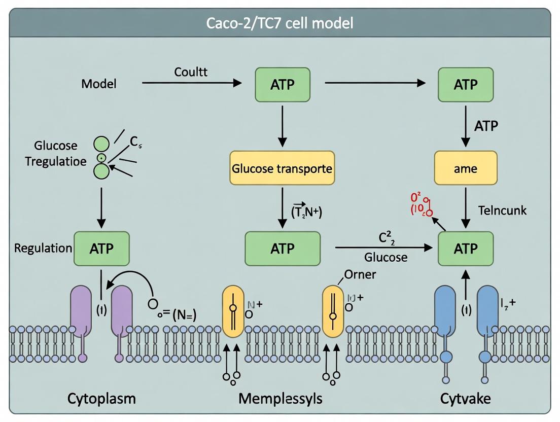
Pathway and Workflow Diagrams
Diagram 1: SGLT1/GLUT2 Mediated Glucose Absorption
Diagram 2: Caco-2/TC7 Glucose Uptake Assay Workflow
The Caco-2 Cell Line Origin and Spontaneous Enterocytic Differentiation
Application Notes
The Caco-2 cell line is a cornerstone in vitro model for intestinal absorption studies, particularly within research focused on nutrient transport and drug permeability. Derived from a human colorectal adenocarcinoma, these cells undergo spontaneous enterocytic differentiation upon reaching confluence, forming polarized monolayers with well-defined tight junctions and brush border membranes expressing digestive hydrolases and transporters. This makes them highly relevant for assessing mechanisms of glucose uptake.
Within the context of a thesis on the Caco-2/TC7 subclone for glucose uptake assessment, understanding the origin and differentiation dynamics is critical. The TC7 clone, selected for its more homogeneous and accelerated differentiation phenotype, provides a robust platform for high-throughput screening of SGLT1 and GLUT2-mediated glucose transport modulation. The following notes and protocols detail the characterization and utilization of this model system.
Key Data on Caco-2 Differentiation and Glucose Transporters
Table 1: Timeline of Key Differentiation Markers in Parental Caco-2 vs. TC7 Clone
| Parameter | Parental Caco-2 (Days Post-Confluence) | TC7 Clone (Days Post-Confluence) | Detection Method |
|---|---|---|---|
| Transepithelial Electrical Resistance (TEER) Peak | 14-21 days | 7-10 days | Voltohmmeter / EVOM |
| Sucrase-Isomaltase (SI) Activity | Detectable at ~7 days, plateaus at 14-20 days | Detectable at ~4 days, plateaus at 10-14 days | Biochemical assay (Dahlqvist) |
| SGLT1 mRNA/Protein Expression | Significant increase from day 5-20 | Rapid increase from day 2, stable by day 10 | qPCR, Western Blot |
| GLUT2 Apical Membrane Recruitment | Induced by high glucose (~25 mM) | Enhanced responsiveness to glucose stimulus | Immunofluorescence, Surface Biotinylation |
| Peak Glucose Uptake Rate | ~15-20 days | ~8-12 days | Radiolabeled (³H- or ¹⁴C-) or fluorescent 2-NBDG uptake |
Table 2: Representative Quantitative Glucose Uptake Parameters in Differentiated Caco-2/TC7 Monolayers
| Condition | Apparent Km for α-MG (SGLT1) | Maximal Uptake Velocity (Vmax) | Contributing Transporter |
|---|---|---|---|
| Basal (5mM Glucose) | ~0.7 - 1.2 mM | ~1.5 - 3.0 nmol/mg protein/min | Primarily SGLT1 |
| High Glucose (25mM) or PMA Stimulation | N/A (induces facilitative component) | Increases by 50-150% | SGLT1 + Apical GLUT2 |
| With SGLT1 Inhibitor (Phloridzin 0.1-0.5 mM) | Uptake largely abolished | >90% inhibition | Confirms SGLT1 activity |
Detailed Experimental Protocols
Protocol 1: Culture and Differentiation of Caco-2/TC7 Cells for Glucose Uptake Assays Objective: To establish fully differentiated, polarized Caco-2/TC7 monolayers on permeable filter supports.
- Culture Maintenance: Grow Caco-2/TC7 cells in high-glucose (25 mM) Dulbecco's Modified Eagle Medium (DMEM), supplemented with 10% (v/v) heat-inactivated fetal bovine serum (FBS), 1% non-essential amino acids (NEAA), 2 mM L-glutamine, and 1% penicillin/streptomycin at 37°C, 10% CO₂.
- Seeding for Assays: Detach cells at ~80% confluence. Seed on collagen-coated polycarbonate Transwell filters (12-well, 1.12 cm², 0.4 µm pore) at a density of 6-8 x 10⁴ cells/cm².
- Differentiation & Monitoring: Change media every 48 hours. Monitor Transepithelial Electrical Resistance (TEER) regularly using a chopstick electrode. Monolayers are typically ready for functional assays when TEER exceeds 500 Ω·cm² (TC7: days 10-12).
- Pre-Assay Preparation: 24 hours before the uptake experiment, replace medium with glucose-free DMEM supplemented with 5 mM D-glucose (physiological mimic) to standardize transporter expression.
Protocol 2: Radiolabeled Glucose Transporter Kinetic Assay (SGLT1 Focus) Objective: To determine the kinetic parameters (Km and Vmax) of SGLT1-mediated uptake.
- Solution Preparation: Prepare uptake buffer (Hanks' Balanced Salt Solution, HBSS, with 10 mM HEPES, pH 7.4). Prepare a dilution series of unlabeled α-Methyl-D-Glucoside (α-MG, a non-metabolizable SGLT1 substrate) from 0.1 to 20 mM, each spiked with a constant trace amount of ¹⁴C-α-MG.
- Uptake Procedure: Wash differentiated monolayers 3x with pre-warmed, glucose-free HBSS. Add donor solution (0.5 mL apical, 1.5 mL basolateral). For inhibition control, include 0.5 mM phloridzin in the apical solution. Incubate for 3-5 minutes (within linear uptake phase) at 37°C.
- Termination & Quantification: Rapidly aspirate solutions and wash filters 3x with ice-cold PBS containing 0.5 mM phloridzin. Dissolve filters in 0.5 mL of 0.1% SDS. Transfer lysate to scintillation vials, add cocktail, and count radioactivity (CPM).
- Data Analysis: Normalize CPM to protein content (BCA assay). Plot uptake rate vs. substrate concentration. Perform nonlinear regression (Michaelis-Menten) to derive Km and Vmax.
Protocol 3: Immunofluorescence Staining for Tight Junctions and Transporters Objective: To visualize epithelial polarization and transporter localization.
- Fixation & Permeabilization: Wash monolayers on filters with PBS. Fix with 4% paraformaldehyde (15 min, RT). Permeabilize with 0.1% Triton X-100 (10 min, RT). Block with 1% BSA in PBS (1 hour, RT).
- Primary Antibody Incubation: Incubate with primary antibodies diluted in blocking buffer overnight at 4°C (e.g., mouse anti-ZO-1 for tight junctions, rabbit anti-SGLT1).
- Secondary & Imaging: Wash and incubate with Alexa Fluor-conjugated secondary antibodies (1 hour, RT, dark). Stain nuclei with DAPI (5 min). Mount filters on slides using antifade mounting medium. Image using a confocal microscope.
Signaling Pathways and Workflows
Title: Signaling Pathways in Caco-2 Enterocytic Differentiation
Title: Workflow for Caco-2/TC7 Glucose Uptake Kinetics Study
The Scientist's Toolkit: Research Reagent Solutions
Table 3: Essential Materials for Caco-2/TC7 Glucose Uptake Research
| Reagent/Material | Function/Application | Example Product/Catalog |
|---|---|---|
| Caco-2/TC7 Cell Line | Differentiating intestinal epithelial model. | ECACC 86010202 or equivalent. |
| Collagen-Coated Transwell Filters | Provides surface for polarization and monolayer formation. | Corning 3460 (Collagen I, 0.4 µm pore). |
| High-Glucose DMEM with Supplements | Standard growth medium promoting differentiation. | Gibco DMEM, 25 mM Glucose, with NEAA, FBS. |
| Phloridzin | Specific, competitive inhibitor of SGLT1; essential for control experiments. | Sigma-Aldrich P3449. |
| ¹⁴C-α-Methyl-D-Glucoside (¹⁴C-α-MG) | Radiolabeled, non-metabolizable SGLT1 substrate for kinetic uptake assays. | PerkinElmer NET461A. |
| 2-NBDG (Fluorescent Glucose Analog) | Non-radioactive alternative for real-time or high-throughput glucose uptake screening. | Thermo Fisher Scientific N13195. |
| Anti-SGLT1 Antibody | For validation of transporter expression and localization via Western Blot/IF. | Santa Cruz Biotechnology sc-393350. |
| EVOM Voltohmmeter | For non-invasive, routine measurement of Transepithelial Electrical Resistance (TEER). | World Precision Instruments EVOM2. |
| TEER Measurement Electrodes (Chopstick) | Paired with EVOM for monolayer integrity assessment. | World Precision Instruments STX2. |
Within the established paradigm of using Caco-2 cells for intestinal glucose transport research, the TC7 clone emerges as a critical tool for enhancing data reproducibility and throughput. This application note, framed within a broader thesis on optimizing the Caco-2/TC7 model for glucose uptake assessment, details the specific advantages of the TC7 clone for mechanistic and inhibitor studies targeting Sodium-Glucose Linked Transporter 1 (SGLT1) and Glucose Transporter 2 (GLUT2). The clone's homogeneous genetic background and stable phenotypic expression make it ideal for standardized, high-throughput screening in drug development pipelines.
Advantages of TC7 over Parental Caco-2
The parental Caco-2 line exhibits significant heterogeneity in differentiation and transporter expression between passages and laboratories. The TC7 clone, selected for its stable and homogeneous expression of differentiated enterocyte markers, offers distinct benefits for transporter studies.
Table 1: Quantitative Comparison of Caco-2 vs. TC7 for Transporter Studies
| Parameter | Parental Caco-2 | TC7 Clone | Implication for SGLT1/GLUT2 Studies |
|---|---|---|---|
| Differentiation Time | 15-21 days | 12-15 days | Faster assay turnaround, increased throughput. |
| SGLT1 Expression (mRNA) | Variable (CV ~30-40%) | High & Consistent (CV <15%) | Reduced inter-experiment variability in phlorizin-sensitive uptake. |
| GLUT2 Apical Recruitment | Heterogeneous | Reproducible, high-glucose inducible | Reliable model for studying GLUT2 trafficking and high-capacity sugar absorption. |
| Transepithelial Electrical Resistance (TEER) | Variable plateau | Consistent, high plateau (~500 Ω·cm²) | Robust monolayer integrity for reliable transport/uptake assays. |
| Suitability for HTS | Low | High | Amenable to 96/384-well format for compound screening. |
Key Experimental Protocols
Protocol 1: High-Throughput Glucose Uptake Assay in 96-Well Format
Objective: Quantify initial rates of Na⁺-dependent (SGLT1) and Na⁺-independent (GLUT2) glucose uptake. Materials: TC7 monolayers (12-15 days post-seeding in 96-well plates), Krebs-Ringer HEPES (KRH) buffer, ²H- or ¹⁴C-labeled D-glucose, unlabeled D-glucose, phlorizin (SGLT1 inhibitor), phloretin (GLUT inhibitor), cell lysis buffer, scintillation cocktail. Procedure:
- Pre-incubation: Wash monolayers twice with warm Na⁺-containing (for SGLT1 activity) or Na⁺-free (choline substitution) KRH buffer.
- Inhibition (Optional): Add KRH buffer containing 500 µM phlorizin (for SGLT1-specific inhibition) or 200 µM phloretin (for total GLUT inhibition) for 15 min.
- Uptake Phase: Replace buffer with uptake solution (KRH with 100 µM labeled glucose ± inhibitors). Incubate for 2-5 minutes (linear uptake phase) at 37°C.
- Termination: Rapidly aspirate uptake solution and wash 3x with ice-cold PBS.
- Lysis & Quantification: Add lysis buffer, shake 30 min. Transfer lysate to scintillation vials, add cocktail, and count radioactivity.
- Calculation: Na⁺-dependent uptake = (Uptake in Na⁺ buffer) - (Uptake in Na⁺-free buffer). SGLT1-specific = Phlorizin-sensitive component.
Protocol 2: GLUT2 Apical Recruitment Induction & Assessment
Objective: Induce and measure apical membrane recruitment of GLUT2 in response to high glucose. Materials: TC7 monolayers on filters, high-glucose (25 mM) DMEM, immunofluorescence staining reagents. Procedure:
- Induction: Maintain TC7 monolayers in standard (5 mM) glucose medium until fully differentiated. Switch experimental group to high-glucose (25 mM) DMEM for 4-6 hours.
- Fixation & Staining: Fix cells with 4% PFA, permeabilize, and block. Stain with anti-GLUT2 primary antibody and appropriate fluorescent secondary antibody. Use phalloidin for actin.
- Imaging & Analysis: Acquire confocal Z-stacks. Quantify apical membrane fluorescence intensity (vs. cytoplasmic) using image analysis software (e.g., ImageJ). Compare high-glucose vs. control groups.
Signaling Pathways in TC7 Glucose Transporter Regulation
Diagram Title: Signaling Pathways Regulating GLUT2 in TC7 Cells
Experimental Workflow for Inhibitor Screening
Diagram Title: HTS Inhibitor Screening Protocol with TC7 Cells
The Scientist's Toolkit: Essential Research Reagents & Materials
Table 2: Key Research Reagent Solutions for TC7-based Glucose Uptake Studies
| Item | Function & Rationale |
|---|---|
| TC7 Cell Clone | Homogeneous, stable enterocyte model providing reproducible SGLT1/GLUT2 expression. Foundational reagent. |
| High-Glucose (25 mM) DMEM | Induction medium for stimulating apical membrane recruitment of GLUT2. Critical for studying transporter trafficking. |
| Phlorizin | High-affinity, specific competitive inhibitor of SGLT1. Used to define SGLT1-specific component of total Na⁺-dependent uptake. |
| Phloretin | Broad-spectrum inhibitor of facilitative GLUTs. Used to define total GLUT-mediated (including GLUT2) uptake component. |
| Chloride-free / Choline-based Buffers | Allows creation of Na⁺-free uptake buffers to dissect Na⁺-dependent (SGLT1) vs. Na⁺-independent (GLUT) transport mechanisms. |
| ³H- or ¹⁴C-labeled D-Glucose | Tracer for sensitive, quantitative measurement of initial uptake rates in intact monolayers. |
| Anti-GLUT2 Antibody (Validated for IF) | Essential tool for visualizing and quantifying the subcellular localization and induction of GLUT2 via microscopy. |
| 96-well Scintillation Plates / LumaPlates | Enables direct, high-throughput measurement of radioactivity in adherent cell monolayers without liquid scintillation vials. |
Application Notes
The Caco-2/TC7 cell line, a differentiated subclone of human colorectal carcinoma cells, serves as a robust in vitro model for studying intestinal glucose and fructose transport and its regulation. This model reliably expresses the key apical and basolateral transporters found in the human small intestine. Understanding their coordinated expression and hormonal/nutritional regulation is crucial for research in metabolism, nutrition, and drug development targeting diabetes and obesity.
1. Transporter Profiles and Kinetic Parameters
| Transporter | Gene | Location (Caco-2/TC7) | Substrate | Transport Mechanism | Approx. Km (mM) | Key Regulators & Notes |
|---|---|---|---|---|---|---|
| SGLT1 | SLC5A1 | Apical Membrane | Glucose, Galactose | Na+-dependent active (secondary active) | 0.5 - 1.0 (Glucose) | Upregulated by high luminal glucose, SGLT2 inhibitors, cAMP/PKA. Constitutively expressed. |
| GLUT2 | SLC2A2 | Basolateral & Apical* | Glucose, Fructose, Galactose | Facilitated diffusion (bidirectional) | 10 - 20 (Glucose) | Rapid apical insertion triggered by high luminal glucose. Regulated by insulin, T1R3, PKCβII. |
| GLUT5 | SLC2A5 | Apical Membrane | Fructose | Facilitated diffusion | 6 - 12 (Fructose) | Highly specific for fructose. Upregulated by luminal fructose, PPARγ, glucocorticoids. |
Note: Apical GLUT2 insertion is a dynamic, diet-responsive regulatory phenomenon.
2. Key Regulatory Signaling Pathways
Experimental Protocols
Protocol 1: Assessment of Glucose Uptake in Differentiated Caco-2/TC7 Monolayers
Objective: To measure Na+-dependent (SGLT1-mediated) and Na+-independent (GLUT-mediated) glucose uptake.
Materials:
- Differentiated Caco-2/TC7 monolayers (21-28 days post-seeding on Transwell or multiwell plates).
- Uptake Buffer (pH 7.4): 137 mM NaCl, 5.4 mM KCl, 2.8 mM CaCl2, 1.2 mM MgSO4, 10 mM HEPES. For Na+-free buffer, replace NaCl with equimolar choline-Cl or N-methyl-D-glucamine (NMDG)-Cl.
- Radiolabeled substrate: ¹⁴C-D-Glucose or ³H-OMG (non-metabolizable analog).
- Wash Buffer: Ice-cold PBS with 0.1 mM phloretin (GLUT inhibitor) to stop transport.
- Cell lysis buffer: 0.1% SDS in 0.1 M NaOH.
- Scintillation counter and vials.
Procedure:
- Pre-incubation: Wash monolayers 3x with pre-warmed (37°C) uptake buffer (with or without Na+). Incubate for 10 min.
- Uptake Phase: Replace buffer with uptake buffer containing radiolabeled glucose (typical final concentration: 0.1-1 mM for SGLT1 kinetics). Incubate for precisely 1-3 minutes (linear uptake phase).
- Termination: Rapidly remove radioactive buffer and wash monolayers 4x with ice-cold stop/wash buffer.
- Lysis & Quantification: Lyse cells in 0.1% SDS/NaOH. Transfer lysate to scintillation vials, add cocktail, and count radioactivity. Measure protein content of parallel wells (BCA assay) for normalization.
- Calculation: Na+-dependent uptake = (Uptake in Na+ buffer) - (Uptake in Na+-free buffer).
Protocol 2: Investigating GLUT2 Apical Recruitment via Immunofluorescence
Objective: To visualize the dynamic insertion of GLUT2 into the apical membrane in response to high luminal glucose.
Materials:
- Differentiated Caco-2/TC7 monolayers on Transwell filters.
- Stimulation medium: DMEM with 25 mM Glucose (high) vs. 5 mM Glucose (control).
- Fixative: 4% paraformaldehyde (PFA) in PBS.
- Permeabilization/Blocking buffer: PBS with 0.1% Triton X-100 and 5% normal goat serum.
- Primary Antibody: Anti-GLUT2 antibody (validated for immunofluorescence).
- Secondary Antibody: Fluorophore-conjugated (e.g., Alexa Fluor 488).
- Actin stain: Phalloidin (e.g., Alexa Fluor 594 conjugate).
- Mounting medium with DAPI.
- Confocal microscope.
Procedure:
- Stimulation: Treat the apical side of differentiated monolayers with high-glucose or control medium for 30-60 min.
- Fixation: Wash with PBS and fix with 4% PFA for 15 min at RT.
- Permeabilization & Blocking: Permeabilize and block for 1 hour.
- Staining: Incubate with anti-GLUT2 primary antibody (overnight, 4°C). Wash, then incubate with fluorophore-conjugated secondary antibody and phalloidin (for F-actin) for 1 hour at RT in the dark.
- Mounting & Imaging: Excise membrane, mount on slides. Acquire Z-stack images using a confocal microscope. Apical co-localization with actin or specific apical markers (e.g., villin) can be analyzed.
Protocol 3: qRT-PCR Analysis of Transporter Gene Expression Regulation
Objective: To quantify changes in SLC5A1 (SGLT1), SLC2A2 (GLUT2), and SLC2A5 (GLUT5) mRNA levels in response to treatments (e.g., fructose, hormones, drug candidates).
Materials:
- Treated Caco-2/TC7 cells (e.g., with 100 nM insulin, 10 mM fructose, or PPARγ agonist for 24-48h).
- RNA extraction kit (e.g., TRIzol or column-based).
- cDNA synthesis kit.
- qPCR Master Mix (SYBR Green or TaqMan).
- Validated primer/probe sets for target genes and housekeeping genes (e.g., GAPDH, HPRT1, B2M).
- Real-time PCR system.
Procedure:
- RNA Extraction: Homogenize cells in lysis reagent. Isolate total RNA following kit protocol. Assess purity and concentration.
- cDNA Synthesis: Reverse transcribe 1 µg of total RNA using a high-capacity cDNA kit.
- qPCR Setup: Prepare reactions with master mix, primers, and cDNA template. Run in triplicate.
- Data Analysis: Calculate ΔΔCt values. Normalize target gene Ct values to the geometric mean of housekeeping genes and compare to the control group.
The Scientist's Toolkit: Essential Research Reagent Solutions
| Item | Function in Caco-2/TC7 Glucose Uptake Research |
|---|---|
| Caco-2/TC7 Cell Line | Differentiated human intestinal epithelial model expressing functional SGLT1, GLUT2, and GLUT5. |
| Transwell Permeable Supports | Provides polarized cell growth with distinct apical and basolateral compartments for transport studies. |
| ¹⁴C-D-Glucose or ³H-OMG | Radiolabeled tracers for sensitive and specific quantification of glucose uptake rates. |
| Phlorizin | Specific, competitive inhibitor of SGLT1; used to define SGLT1-mediated transport component. |
| Phloretin | Broad-spectrum inhibitor of facilitative GLUTs (including GLUT2); used in stop solutions. |
| Validated Anti-GLUT2 Antibody | Critical for detecting low-abundance, dynamically trafficked GLUT2 protein via WB or IF. |
| SGLT2 Inhibitor (e.g., Dapagliflozin) | At high doses, can also inhibit SGLT1; used in pharmacological studies of transporter interplay. |
| Fructose | Primary substrate and transcriptional inducer of GLUT5 expression. |
| Insulin | Hormonal regulator that modulates GLUT2 expression and trafficking. |
| PPARγ Agonist (e.g., Rosiglitazone) | Potent inducer of SLC2A5 (GLUT5) gene transcription. |
Why This Model? Relevance to Human Drug Absorption and Nutrient Bioavailability.
Within the ongoing thesis on the application of the Caco-2/TC7 cell monolayer model for glucose uptake assessment, a fundamental question must be addressed: "Why this model?" The Caco-2 cell line, and its subclone TC7, has become the de facto gold standard in vitro model for predicting intestinal permeability for over three decades. Its relevance extends from pharmaceutical drug absorption to the study of nutrient and bioactive compound bioavailability. This application note details the model's physiological foundation, provides standardized protocols, and synthesizes current data supporting its use, thereby justifying its central role in the thesis's experimental framework on intestinal transport mechanisms.
Physiological & Functional Basis of the Caco-2/TC7 Model
The Caco-2 cell line, derived from a human colorectal adenocarcinoma, spontaneously differentiates under standard culture conditions into a monolayer of polarized enterocytes. These cells exhibit key morphological and functional characteristics of the small intestinal epithelium:
- Formation of Tight Junctions: Creating a physiologically relevant barrier with measurable Transepithelial Electrical Resistance (TEER).
- Expression of Brush Border Enzymes: Such as sucrase-isomaltase, aminopeptidases, and alkaline phosphatase.
- Polarized Expression of Transporters: Apical (AP) and basolateral (BL) localization of influx (e.g., SGLT1, PEPT1) and efflux transporters (e.g., P-glycoprotein, MRPs).
- Viability on Permeable Supports: Enabling independent access to AP and BL compartments for transport studies.
The TC7 subclone is selected for its more homogeneous and robust expression of differentiated enterocyte markers, particularly relevant for sugar transport studies due to its consistent expression of SGLT1.
Table 1: Comparison of Key Functional Markers in Caco-2 vs. Caco-2/TC7 Cells
| Parameter | Human Small Intestine (Proximal) | Standard Caco-2 | Caco-2/TC7 Subclone | Measurement Method |
|---|---|---|---|---|
| TEER (Ω·cm²) | ~30-70 in vivo | 200-600 | 250-500 | Voltohmmeter / EVOM |
| Sucrase-Isomaltase Activity | High | Variable, Moderate | High & Consistent | Biochemical assay |
| Alkaline Phosphatase Activity | High | Moderate | High & Consistent | Biochemical assay |
| SGLT1 mRNA/Protein Expression | High | Moderate, Variable | High, Stable | qPCR / Western Blot |
| P-glycoprotein (MDR1) Expression | Present | Present, Variable | Present | Functional assay / WB |
Table 2: Representative Apparent Permeability (Papp) Coefficients for Model Validation
| Compound (Class) | Papp (AP→BL) x10⁻⁶ cm/s | Human Fa% (Absorbed) | Caco-2 Prediction | Primary Transport Route |
|---|---|---|---|---|
| Metoprolol (High Perm) | 25-35 | ~95% | High Absorption | Passive Transcellular |
| Ranitidine (Low Perm) | 0.5-2.0 | ~50% | Low Absorption | Paracellular / Carrier |
| Glucose (Nutrient) | 15-25 * | ~100% | High Absorption | SGLT1-mediated (Na+-dep.) |
| Atenolol (Low/Mod Perm) | 1.5-3.5 | ~50% | Low Absorption | Paracellular |
*Papp for glucose is highly dependent on SGLT1 expression and sodium gradient.
Detailed Experimental Protocols
Protocol 4.1: Cell Culture and Monolayer Differentiation on Transwell Inserts
Purpose: To establish differentiated, confluent Caco-2/TC7 monolayers for transport studies. Materials: Caco-2/TC7 cells, DMEM (high glucose, GlutaMAX), Fetal Bovine Serum (FBS, 10%), Non-Essential Amino Acids (1%), Penicillin-Streptomycin, HEPES, 12-well or 24-well polycarbonate Transwell inserts, collagen coating (optional). Procedure:
- Culture Maintenance: Grow cells in T-flasks in complete DMEM at 37°C, 5% CO₂. Passage at 80-90% confluence using Trypsin-EDTA.
- Seeding: Seed cells onto the apical compartment of Transwell inserts at a density of 60,000-100,000 cells/cm² (e.g., ~60,000 cells for a 0.33 cm² 24-well insert).
- Media Schedule: Replace media in both AP and BL compartments every 48 hours. Use complete DMEM for the first 7 days.
- Differentiation: After 7 days, switch to differentiation medium (complete DMEM with reduced FBS to 5-7%). Culture for an additional 14-21 days.
- Quality Control: Monitor TEER weekly using an epithelial voltohmmeter. Accept monolayers for experiments when TEER > 300 Ω·cm² (for 0.33 cm² inserts).
Protocol 4.2: Glucose Uptake Assay (SGLT1-Mediated)
Purpose: To quantify active, sodium-dependent glucose transport across the apical membrane. Materials: Uptake buffer (UB: 137mM NaCl, 5.4mM KCl, 2.8mM CaCl₂, 1.2mM MgCl₂, 10mM HEPES, pH 7.4), Sodium-free UB (NaCl replaced with Choline-Cl or NMDG-Cl), ³H- or ¹⁴C-labeled D-glucose (or fluorescent analog 2-NBDG), unlabeled D-glucose for competition, phlorizin (SGLT1 inhibitor), stop/wash buffer (UB with 0.1mM phlorizin, ice-cold). Procedure:
- Pre-incubation: Wash differentiated monolayers 2x with pre-warmed (37°C) UB or Na+-free UB. Incubate for 20 min in respective buffer.
- Uptake Phase: Replace AP buffer with uptake solution containing radiolabeled/fluorescent glucose tracer (e.g., 1 μCi/mL ³H-D-glucose, 100μM cold D-glucose) ± inhibitor (e.g., 0.5mM phlorizin). Incubate for a defined time (e.g., 2-10 minutes) at 37°C.
- Termination: Rapidly aspirate uptake solution and wash AP side 3x with ice-cold stop buffer.
- Lysis & Quantification: Lysate cells (e.g., with 1% Triton X-100 in PBS). Measure radioactivity via scintillation counting or fluorescence. Normalize to total protein (BCA assay).
- Calculation: Sodium-dependent uptake = (Uptake in Na+ buffer) - (Uptake in Na+-free buffer).
Protocol 4.3: Transepithelial Transport Assay (AP→BL)
Purpose: To determine the apparent permeability (Papp) of a test compound (drug/nutrient). Materials: Hank's Balanced Salt Solution (HBSS) with 10mM HEPES, pH 7.4 (transport buffer), test compound, integrity marker (e.g., ¹⁴C-mannitol or Lucifer Yellow), receiver plate. Procedure:
- Pre-equilibration: Wash monolayers and equilibrate in transport buffer at 37°C for 20 min.
- Dosing: Add fresh transport buffer to the BL compartment. Add test compound in transport buffer to the AP compartment (donor). Include an integrity marker in a control well.
- Sampling: At defined time points (e.g., 30, 60, 90, 120 min), sample aliquots (e.g., 100 μL) from the BL receiver compartment and replace with fresh pre-warmed buffer.
- Analysis: Quantify compound concentration in samples via HPLC-MS, scintillation counting, or plate reader.
- Calculation:
- Calculate cumulative amount transported (Q).
- Plot Q vs. time. The slope (dQ/dt) is the steady-state flux rate (J).
- Papp (cm/s) = J / (A * C₀), where A is the insert membrane area (cm²) and C₀ is the initial donor concentration.
Diagrams
Diagram Title: Intestinal Glucose Transport Pathway in Caco-2/TC7 Cells
Diagram Title: Caco-2/TC7 Experimental Workflow
The Scientist's Toolkit: Essential Research Reagents & Materials
Table 3: Key Research Reagent Solutions for Caco-2/TC7 Studies
| Reagent/Material | Function/Application | Example & Notes |
|---|---|---|
| Caco-2/TC7 Cell Line | The biological model itself. TC7 clone offers more consistent differentiation. | Obtain from reputable cell bank (e.g., ECACC, ATCC). Passage number < 40. |
| Transwell Permeable Supports | Physical scaffold for polarized monolayer growth and independent compartment access. | Polycarbonate membrane, 0.4 μm pore, various sizes (12-well, 24-well). |
| Differentiation Media | Promotes enterocyte differentiation and functional protein expression. | DMEM + 5-7% FBS, NEAA, Pen/Strep. Reduced serum post-confluence. |
| TEER Measurement System | Non-destructive integrity check of tight junction formation. | Epithelial Voltohmmeter (e.g., EVOM) with STX2 chopstick electrodes. |
| SGLT1 Inhibitor (Phlorizin) | Specific pharmacological tool to confirm sodium-dependent glucose uptake component. | Use at 0.1-0.5 mM in uptake buffer. High solubility in DMSO. |
| Paracellular Integrity Marker | Validates monolayer integrity during transport studies. | ¹⁴C-Mannitol or Lucifer Yellow. Low Papp indicates intact tight junctions. |
| Radiolabeled/Fluorescent Tracers | Enables sensitive, quantitative tracking of nutrient/drug transport. | ³H- or ¹⁴C-D-Glucose; ³H-Mannitol; 2-NBDG (fluorescent glucose analog). |
| LC-MS/MS System | Gold-standard for quantitative analysis of unlabeled test compounds. | Enables precise measurement of Papp for novel drugs or nutraceuticals. |
Step-by-Step Protocol: Establishing and Running a Caco-2/TC7 Glucose Uptake Assay
Thesis Context
This application note details standardized protocols for the culture and differentiation of the human intestinal epithelial Caco-2/TC7 cell subclone, a cornerstone model for studying intestinal glucose uptake and transporter (SGLT1, GLUT2) regulation within the broader thesis research on nutraceutical and pharmaceutical modulation of intestinal absorption.
Quantitative Parameters for Caco-2/TC7 Culture & Differentiation
Table 1: Essential Quantitative Parameters for a Standard 21-Day Protocol
| Parameter | Specification | Rationale/Notes |
|---|---|---|
| Seeding Density | 5.0 x 10⁴ cells/cm² | Optimal for forming confluent, differentiated monolayers. Higher densities accelerate confluence but may compromise barrier formation. |
| Seeding Volume | 1.5 mL for 12-well plate (3.8 cm²/well) | Ensures even distribution. Adjust proportionally for other formats (e.g., 0.5 mL for 24-well). |
| Initial Media | High-glucose DMEM (4.5 g/L D-Glucose), 20% FBS, 1% Non-Essential Amino Acids (NEAA), 2 mM L-Glutamine, 1% Penicillin/Streptomycin. | Supports rapid proliferation post-seeding. High serum promotes cell attachment and initial growth. |
| Differentiation Media | High-glucose DMEM (4.5 g/L), 10% FBS, 1% NEAA, 2 mM L-Glutamine, 1% Penicillin/Streptomycin. | Reduced serum (to 10%) initiates contact inhibition and differentiation post-confluence. |
| Media Change Schedule | Every 48 hours. | Maintains nutrient and growth factor levels, removes metabolites. Critical for reproducible differentiation. |
| Time to Confluence | Days 3-4 post-seeding. | Visual confirmation required before differentiation timeline begins. |
| Differentiation Start | Day 0 (Day of Confluence). | The 21-day clock starts at confirmed confluence. |
| Full Differentiation | Day 21 post-confluence. | Mature enterocyte phenotype with established tight junctions, brush border enzymes (e.g., sucrase-isomaltase), and polarized transporter expression. |
| Passage Number Range | 25-40 | Use cells within this range to ensure stable genotype/phenotype. Avoid high-passage cells (>45) which may show reduced differentiation capacity. |
Detailed Experimental Protocols
Protocol 1: Subculture and Seeding for Differentiation
Objective: To passage and seed Caco-2/TC7 cells at the correct density to initiate a 21-day differentiation study. Materials: T75 flask of Caco-2/TC7 cells (80-90% confluent), DPBS (Ca²⁺/Mg²⁺ free), 0.25% Trypsin-EDTA, Initial Media (see Table 1), 12-well cell culture plates, hemocytometer or automated cell counter. Procedure:
- Aspirate media from the T75 flask and wash cell monolayer gently with 10 mL pre-warmed DPBS.
- Add 3 mL of pre-warmed 0.25% Trypsin-EDTA. Incubate at 37°C for 3-5 minutes until cells detach.
- Neutralize trypsin by adding 7 mL of Initial Media. Pipette gently to create a single-cell suspension.
- Centrifuge the cell suspension at 200 x g for 5 minutes. Aspirate supernatant.
- Resuspend the cell pellet in 5 mL of fresh Initial Media. Count cells using a hemocytometer.
- Calculate volume needed for a seeding density of 5.0 x 10⁴ cells/cm². For a 12-well plate (3.8 cm²/well), this equals 1.9 x 10⁵ cells per well.
- Seed cells in each well with a total volume of 1.5 mL of Initial Media. Gently rock the plate to ensure even distribution.
- Place the plate in a humidified incubator at 37°C, 5% CO₂. Media change to Differentiation Media begins at first change post-confluence (Day 0).
Protocol 2: The 21-Day Differentiation and Maintenance Protocol
Objective: To maintain and differentiate seeded Caco-2/TC7 cells into a mature enterocyte monolayer over 21 days. Materials: Seeded plate from Protocol 1, Differentiation Media (see Table 1), DPBS, incubator. Procedure:
- Days 1-3: Monitor cells daily. Do not disturb. They should reach 100% confluence by Day 3-4.
- Day 0 (Day of Confluence): Visually confirm 100% confluence. Aspirate Initial Media. Gently wash monolayer with 1 mL pre-warmed DPBS per well (12-well plate). Add 1.5 mL of fresh, pre-warmed Differentiation Media. This is designated Differentiation Day 0.
- Media Changes (Every 48 hours): For the entire 21-day period, change media every 48 hours (± 2 hours). Always aspirate spent media, wash gently with DPBS, and add fresh, pre-warmed Differentiation Media.
- Monitoring: Observe morphology regularly. Differentiated cells will become more polarized and exhibit domes (indicative of transepithelial transport).
- Day 21: Cells are fully differentiated and suitable for functional assays (e.g., glucose uptake, TEER measurement, transporter analysis).
Diagrams
21-Day Caco-2/TC7 Differentiation Workflow
Signaling to Phenotype in Caco-2/TC7 Differentiation
The Scientist's Toolkit: Key Research Reagent Solutions
Table 2: Essential Materials for Caco-2/TC7 Glucose Uptake Studies
| Item | Function in Protocol | Key Notes |
|---|---|---|
| Caco-2/TC7 Cell Line | Differentiates into enterocyte-like cells with robust expression of apical SGLT1 and GLUT2. | Subclone of Caco-2. Verify source and passage number history. Maintain within P25-P40. |
| High-Glucose DMEM | Standard culture medium providing high osmotic pressure and energy source. | Essential for maintaining the TC7 subclone phenotype. Do not substitute with low-glucose DMEM. |
| Fetal Bovine Serum (FBS) | Provides essential growth factors, hormones, and proteins for growth and differentiation. | Batch testing is recommended. Use same validated batch for an entire study series. |
| Non-Essential Amino Acids (NEAA) | Supplements amino acids not synthesized by the cells, reducing metabolic stress. | Crucial for optimal growth and long-term health during the 21-day protocol. |
| D-PBS (without Ca²⁺/Mg²⁺) | Used for washing cells during media changes and subculture. Absence of ions aids in cell detachment during trypsinization. | |
| Trypsin-EDTA (0.25%) | Proteolytic enzyme (trypsin) chelating agent (EDTA) combination for detaching adherent cells. | Standardized concentration and incubation time prevent over-digestion and cell damage. |
| Transwell Permeable Supports | For culturing polarized monolayers for transport/uptake studies. Allows separate access to apical and basolateral compartments. | Required for definitive polarized glucose uptake assays. |
| TEER Meter (Volt/Ohmmeter) | Measures Transepithelial Electrical Resistance to quantify monolayer integrity and tight junction formation. | Key QC metric. Readings should increase steadily throughout differentiation (>300 Ω*cm² by Day 21). |
| 2-Deoxy-D-[³H]Glucose | Non-metabolizable glucose analog used in radiotracer uptake assays to specifically measure transporter-mediated influx. | Standard for kinetic studies of SGLT1/GLUT2 activity. Requires radiological safety protocols. |
Within the broader thesis investigating intestinal glucose transport using the Caco-2/TC7 cell model, precise monitoring of cell differentiation is paramount. The Caco-2/TC7 subclone undergoes spontaneous enterocytic differentiation upon confluence, forming a polarized monolayer with tight junctions and expressing functional brush border enzymes. Two gold-standard metrics for quantifying this differentiation are Transepithelial Electrical Resistance (TEER), a measure of tight junction integrity and monolayer health, and Alkaline Phosphatase (ALP) activity, a marker for brush border enzyme expression. This document provides detailed protocols and application notes for these critical assays, framed within glucose uptake assessment research.
Table 1: Expected TEER and ALP Activity Profiles During Caco-2/TC7 Differentiation
| Post-Confluence Day | Typical TEER Range (Ω·cm²)* | Relative ALP Activity (Fold Increase vs. Day 0) | Differentiation Stage & Implications for Glucose Uptake Studies |
|---|---|---|---|
| Day 0-2 | 50 - 200 | 1.0 (Baseline) | Sub-confluent/proliferating. Unsuitable for transport studies. |
| Day 3-5 | 200 - 600 | 2.0 - 5.0 | Early differentiation. Tight junctions forming. GLUT2/SGLT1 expression initiating. |
| Day 6-14 | 600 - 1200+ | 10.0 - 30.0 | Fully differentiated monolayer. Stable, high TEER and peak ALP activity. Optimal window for reproducible glucose uptake assays. |
| Day 15+ | May plateau or decline | May plateau or decline | Late stage; potential for over-differentiation or loss of monolayer integrity. |
Values are cell line and passage-dependent. Must be established as an internal control. *ALP activity rises sharply upon confluence and peaks around days 10-14.
Detailed Experimental Protocols
Protocol 1: Measurement of Transepithelial Electrical Resistance (TEER)
Objective: To non-invasively monitor the formation and integrity of tight junctions in Caco-2/TC7 monolayers grown on permeable filter supports.
Materials:
- Differentiated Caco-2/TC7 monolayers on collagen-coated polycarbonate filters (e.g., 12-well Transwell inserts, 1.12 cm² growth area).
- Epithelial Voltohmmeter (EVOM) with chopstick or cup electrodes.
- Sterile PBS or cell culture medium (pre-warmed to 37°C).
Procedure:
- Calibration: Calibrate the EVOM according to manufacturer instructions.
- Equilibration: Transfer the cell culture plate to a 37°C bench. Aspirate the medium from both the apical and basolateral compartments and replace with pre-warmed PBS or culture medium. Allow temperature equilibration for 15-20 minutes.
- Measurement: a. Sterilize electrodes with 70% ethanol and rinse with sterile PBS. b. For chopstick electrodes: Place the shorter apical electrode in the insert and the longer basolateral electrode in the well. Ensure no contact with the membrane. c. Record the resistance value (in Ω). Repeat for each insert.
- Calculation: Subtract the average resistance of a cell-free blank filter (with coating) from the sample reading. Multiply this net resistance (Ω) by the effective surface area of the filter (cm²) to obtain TEER in Ω·cm².
- Frequency: Measure every 2-3 days post-confluence to track differentiation kinetics.
Data Interpretation: A steady increase culminating in a stable plateau (>600 Ω·cm²) indicates successful differentiation. A sudden drop may signify monolayer damage, contamination, or loss of differentiation.
Protocol 2: Spectrophotometric Assay of Alkaline Phosphatase Activity
Objective: To quantify the enzymatic activity of ALP, a differentiation marker, in Caco-2/TC7 cell lysates.
Materials:
- Cell lysates in RIPA or Tris-based lysis buffer.
- p-Nitrophenyl phosphate (pNPP) substrate solution (e.g., 1 mg/mL in 1M diethanolamine buffer, pH 9.8, with 0.5 mM MgCl₂).
- Microplate reader capable of reading absorbance at 405 nm.
- 96-well flat-bottom plates.
- Stop solution (1M NaOH).
Procedure:
- Lysate Preparation: Wash cell monolayers (on filters or plates) with ice-cold PBS. Lyse cells in appropriate buffer on ice for 15 minutes. Centrifuge at 13,000 x g for 10 minutes at 4°C. Collect supernatant.
- Protein Quantification: Determine protein concentration of each lysate using a Bradford or BCA assay.
- ALP Reaction Setup: a. Aliquot 50 µL of lysate (diluted to appropriate protein concentration, e.g., 10-50 µg) into a 96-well plate in duplicate. b. Add 100 µL of pNPP substrate solution to each well. Start a timer. c. Incubate at 37°C for 15-30 minutes (optimize time to keep readings within linear range).
- Reaction Termination & Measurement: Stop the reaction by adding 50 µL of 1M NaOH. Immediately read the absorbance at 405 nm (A₄₀₅) in the plate reader.
- Calculation: Generate a standard curve using known concentrations of p-nitrophenol. Calculate ALP activity from the standard curve, normalized to total protein content and reaction time. Express as nmol pNP produced/min/mg protein.
Data Interpretation: ALP activity should be minimal in pre-confluent cells and increase 10-30 fold upon full differentiation, correlating with the establishment of brush border functionality relevant for apical glucose transporter studies.
Visualization: Pathway and Workflow Diagrams
Title: Differentiation Pathway to a Functional Glucose Uptake Model
Title: Workflow for Differentiation Monitoring and QC
The Scientist's Toolkit: Essential Research Reagent Solutions
Table 2: Key Materials for Differentiation Monitoring Assays
| Item | Function & Relevance |
|---|---|
| Collagen-Coated Transwell Inserts (e.g., 0.4 µm pore, 12-well) | Provide a permeable support for polarized cell growth, allowing separate access to apical and basolateral compartments essential for TEER and transport studies. |
| Epithelial Voltohmmeter (EVOM2) | Dedicated instrument for accurate, non-invasive TEER measurement. Chopstick electrodes are standard for multi-well plates. |
| p-Nitrophenyl Phosphate (pNPP) | Colorimetric substrate for ALP. Enzymatic cleavage produces p-nitrophenol, measurable at 405 nm, directly proportional to ALP activity. |
| Diethanolamine Buffer (1M, pH 9.8) | Optimal alkaline buffer for ALP enzyme reaction, maximizing activity and assay sensitivity. |
| RIPA Lysis Buffer | Efficiently extracts total cellular protein, including membrane-bound ALP, for subsequent activity and protein concentration assays. |
| Microplate Reader | Enables high-throughput absorbance reading for ALP activity (405 nm) and protein quantification assays (562 nm for BCA, 595 nm for Bradford). |
| Caco-2/TC7 Cell Line | A well-differentiated subclone of Caco-2 with more homogeneous and rapid expression of sucrose-isomaltase and other brush border enzymes, ideal for glucose metabolism research. |
| High-Glucose DMEM with FBS | Standard culture medium. Fetal Bovine Serum (FBS) batch and concentration (typically 10-20%) critically affect differentiation kinetics and must be standardized. |
Application Notes
Within a thesis investigating intestinal glucose transport using the Caco-2/TC7 cell model, selecting the appropriate probe for uptake assays is critical. This model, which spontaneously differentiates into enterocyte-like monolayers, expresses key transporters like SGLT1 and GLUT2, making it ideal for studying nutrient absorption and drug effects. The choice between traditional radiolabeled and modern fluorescent probes fundamentally shapes experimental design, data interpretation, and resource allocation.
Comparative Analysis of Probe Characteristics
Table 1: Quantitative Comparison of Radiolabeled vs. Fluorescent Glucose Probes
| Feature | Radiolabeled ([14C] or [3H]-D-Glucose) | Fluorescent (2-NBDG) | |
|---|---|---|---|
| Detection Method | Scintillation Counting | Flow Cytometry, Fluorescence Microscopy, Microplate Reader | |
| Sensitivity | Very High (fmol level) | Moderate to High (pmol level) | |
| Dynamic Range | 4-5 orders of magnitude | 3-4 orders of magnitude | |
| Temporal Resolution | End-point measurement (minutes-hours) | Real-time to near-real-time (seconds-minutes) possible | |
| Spatial Information | No, bulk measurement | Yes, single-cell or subcellular possible | |
| Throughput | Lower (handling constraints) | Higher (amenable to multi-well formats) | |
| Safety & Regulation | High (radioactive waste, licensing) | Low (standard lab chemical) | |
| Cost per Assay | Moderate (isotope cost) | Low | Low |
| Probe Chemistry | Identical to native glucose | Glucose analog; NBD group alters properties | |
| Primary Application | Quantitative flux studies, Km/Vmax determination | High-throughput screening, kinetic imaging, live-cell tracking |
Key Considerations for the Caco-2/TC7 Model
- Transport Specificity: Both probes are substrates for GLUTs. 2-NBDG is not transported by SGLT1. Therefore, for studies focusing on SGLT1-mediated apical uptake, radiolabeled D-Glucose is mandatory.
- Monolayer Integrity: Protocols for both assays must include steps to validate monolayer integrity (e.g., TEER measurement) prior to uptake experiments.
- Data Normalization: Radiolabeled data is typically normalized to total protein content. 2-NBDG flow cytometry data can be normalized to cell count or protein; microscopy may require nucleus counting.
Experimental Protocols
Protocol 1: Radiolabeled Glucose Uptake in Differentiated Caco-2/TC7 Monolayers
Research Reagent Solutions Toolkit
| Item | Function in Protocol |
|---|---|
| Caco-2/TC7 Cells | Human colon adenocarcinoma cell line clone, models intestinal epithelium. |
| Radioactive D-Glucose ([14C] or [3H]) | Tracer for quantifying glucose transport. |
| Hanks' Balanced Salt Solution (HBSS) | Physiological buffer for uptake assays. |
| Transport Inhibitors (e.g., Phloridzin, Cytochalasin B) | Inhibits SGLT1 or GLUTs to confirm transport mechanism. |
| Unlabeled D-Glucose | For creating specific activity and competition controls. |
| Stop Solution (Ice-cold PBS + Phloridzin) | Rapidly halts transport activity. |
| Cell Lysis Buffer (e.g., 0.1M NaOH, 1% SDS) | Lyses cells to release incorporated tracer. |
| Scintillation Fluid & Vials | For radioactive signal detection. |
| Liquid Scintillation Counter | Instrument to measure radioactive decay (CPM/DPM). |
| BCA Protein Assay Kit | Normalizes uptake data to total cellular protein. |
Methodology:
- Cell Culture: Seed Caco-2/TC7 cells on Transwell filters. Culture for 21 days, changing media every 2-3 days, to achieve full differentiation. Measure TEER regularly.
- Assay Buffer Preparation: Prepare uptake buffer (e.g., HBSS with desired glucose concentration). For sodium-free conditions, replace NaCl with Choline-Cl. Pre-warm to 37°C.
- Tracer Solution: Spike uptake buffer with radiolabeled glucose to desired specific activity (e.g., 0.5-1 µCi/mL). Include experimental conditions (e.g., +/− inhibitor, varying cold glucose concentration).
- Uptake Procedure: a. Aspirate culture media from apical and basolateral compartments. b. Wash monolayers twice with pre-warm uptake buffer. c. Add tracer solution to the apical compartment (for apical uptake study). Add appropriate control buffer to the basolateral side. d. Incubate at 37°C for the desired time (e.g., 1-10 minutes). e. Rapidly terminate uptake by washing 3x with ice-cold stop solution.
- Sample Collection: Excise membrane filters. Solubilize cells in lysis buffer for 1 hour. a. Transfer an aliquot of lysate to a scintillation vial for radioactivity counting. b. Use another aliquot for protein concentration determination (BCA assay).
- Data Calculation: Convert scintillation counts (CPM) to moles of glucose using the specific activity. Normalize to protein content and time to express uptake as pmol/min/mg protein.
Protocol 2: 2-NBDG Uptake Assay via Flow Cytometry
Research Reagent Solutions Toolkit
| Item | Function in Protocol |
|---|---|
| 2-NBDG (Fluorescent Glucose Analog) | Probe for visualizing and quantifying GLUT-mediated uptake. |
| Flow Cytometer with FITC channel | Instrument for quantifying cellular fluorescence. |
| Propidium Iodide (PI) or DAPI | Viability dye to exclude dead cells from analysis. |
| Phosphate Buffered Saline (PBS) | Washing and suspension buffer. |
| Trypsin-EDTA | Detaches adherent cells for analysis. |
| Fetal Bovine Serum (FBS) | Used to quench trypsin and as a component in stopping buffer. |
| Glucose-Free Assay Buffer | Ensures uptake is driven by probe concentration. |
Methodology:
- Cell Preparation: Culture Caco-2/TC7 cells in standard monolayers or for certain kinetic studies, in multi-well plates without full polarization. For differentiated monolayers, cells may need gentle trypsinization post-assay, which can affect results.
- Starvation & Inhibition: Prior to assay, starve cells in glucose-free/ serum-free medium for 30-60 min to upregulate basal transport. Pre-treat with inhibitors if required.
- 2-NBDG Loading: a. Prepare working solution of 2-NBDG (e.g., 100 µM) in pre-warmed glucose-free assay buffer. b. Aspirate starvation medium and add the 2-NBDG solution. c. Incubate at 37°C for a defined time (e.g., 10-30 minutes). Include a control incubated on ice (4°C) to assess passive diffusion.
- Uptake Termination & Cell Harvest: a. Aspirate 2-NBDG solution. b. Wash cells rapidly 3x with ice-cold PBS. c. For non-polarized cells, harvest using trypsin-EDTA, quench with FBS, pellet, and resuspend in ice-cold PBS containing a viability dye (e.g., PI).
- Flow Cytometry Analysis: a. Analyze samples immediately on a flow cytometer (Ex/Em ~465/540 nm). b. Gate on live, single cells. Record median fluorescence intensity (MFI) of the FITC/GFP channel for at least 10,000 events per sample.
- Data Analysis: Subtract the MFI of the 4°C control from the 37°C samples to determine active transport. Normalize to cell count or protein content.
Visualizations
Title: Experimental Workflow Comparison for Glucose Uptake Assays
Title: Glucose Transport Pathways in Caco-2/TC7 Cells and Probe Specificity
This protocol details the experimental setup for assessing sodium-dependent and facilitative glucose transporter (SGLT1 and GLUT2) activity in differentiated Caco-2/TC7 cell monolayers. Within the broader thesis on "Mechanistic Insights into Intestinal Glucose Absorption Using the Caco-2/TC7 Model," this experiment is pivotal for quantifying apical glucose uptake kinetics and distinguishing transporter contributions under various physiological and pharmacological conditions. Standardization of buffers, incubation times, and specific inhibitor use is critical for generating reproducible, high-quality data suitable for drug development targeting metabolic disorders.
Key Reagent Solutions & Materials
Table 1: Research Reagent Solutions for Glucose Uptake Assay
| Item | Function/Description |
|---|---|
| Caco-2/TC7 Cells | Human colorectal adenocarcinoma cell clone with high expression of digestive enzymes and apical SGLT1, mimicking mature enterocytes. |
| Hanks' Balanced Salt Solution (HBSS) | Standard physiological buffer for maintaining cell viability during uptake experiments. |
| Uptake Buffer (pH 7.4) | Modified HBSS containing 137 mM NaCl, 5.4 mM KCl, 2.8 mM CaCl₂, 1.2 mM MgSO₄, 10 mM HEPES, and tracer 2-Deoxy-D-[³H]glucose (2-DOG) or [¹⁴C]α-Methyl-D-glucopyranoside (AMG). |
| Sodium-Free Uptake Buffer | Uptake buffer with NaCl replaced isotonically by N-Methyl-D-glucamine (NMDG) chloride or choline chloride to inhibit SGLT1 activity. |
| Phloridzin (200-500 µM) | Specific, competitive inhibitor of apical SGLT1. Serves as a control to define sodium-dependent glucose uptake component. |
| Cytochalasin B (10-20 µM) | Potent inhibitor of facilitative glucose transporters (GLUTs). Used to define GLUT-mediated uptake component. |
| 2-Deoxy-D-[³H]glucose (2-DOG) | Non-metabolizable glucose analog transported by GLUTs but not by SGLT1. Used to assay facilitative uptake. |
| [¹⁴C]α-Methyl-D-glucoside (AMG) | Non-metabolizable glucose analog specifically transported by SGLT1. Used to assay sodium-dependent apical uptake. |
| Stop/Wash Buffer | Ice-cold PBS containing 0.1 mM phloridzin to rapidly halt uptake and displace non-specific surface binding. |
| Cell Lysis Buffer | 0.1% (w/v) SDS in 0.1 M NaOH for complete solubilization of cell monolayers prior to scintillation counting. |
Detailed Experimental Protocols
Protocol 1: Cell Culture and Differentiation
- Culture Caco-2/TC7 cells in DMEM (25 mM glucose) supplemented with 10-20% fetal bovine serum, 1% non-essential amino acids, and antibiotics (Pen/Strep).
- Seed cells on 12- or 24-well plastic plates or Transwell filters at high density (~60,000 cells/cm²).
- Maintain for 14-21 days post-confluence, changing medium every 2-3 days, to ensure full differentiation and apical brush border formation.
Protocol 2: Glucose Uptake Assay (Standard Procedure) Day of Experiment:
- Pre-incubation: Aspirate culture medium. Wash cell monolayers 2x with pre-warmed (37°C) HBSS.
- Inhibitor Pre-treatment (if applicable): Incubate cells for 15-20 minutes at 37°C in sodium-containing or sodium-free uptake buffer containing either phloridzin (500 µM) or cytochalasin B (20 µM). Include vehicle control wells.
- Uptake Initiation: Replace buffer with 200-500 µL of uptake buffer containing the radiolabeled tracer (e.g., 0.1-1 µCi/mL 2-DOG or AMG, with 0.1-1 mM cold substrate). Perform in triplicate.
- Incubation: Incubate plates at 37°C for the determined optimal time (see Table 2).
- Termination: Rapidly aspirate uptake buffer and immediately wash monolayers 3x with 1 mL of ice-cold Stop/Wash Buffer.
- Lysis & Analysis: Add 0.5-1 mL of lysis buffer per well. Shake for 30-60 min. Transfer lysate to scintillation vials, add cocktail, and count radioactivity. Measure protein content of parallel wells (BCA assay) for normalization.
Table 2: Quantitative Parameters for Uptake Conditions
| Parameter | Typical Value/Range | Rationale & Notes |
|---|---|---|
| Cell Differentiation Time | 14-21 days | Ensures stable, polarized expression of SGLT1 and other brush border proteins. |
| Tracer Concentration (2-DOG/AMG) | 0.1 - 1.0 mM | Ensures saturable, transporter-mediated uptake kinetics. |
| Uptake Incubation Time (Linear Range) | 1 - 10 minutes | Must be empirically determined to measure initial rates and avoid tracer efflux/metabolism. |
| Phloridzin Inhibition (SGLT1) | IC₅₀ ~ 200 µM | Pre-incubate 15 min. Inhibits >95% of SGLT1-mediated AMG uptake at 500 µM. |
| Cytochalasin B Inhibition (GLUTs) | IC₅₀ ~ 0.5 µM | Pre-incubate 15 min. Inhibits >90% of 2-DOG uptake at 20 µM. |
| Sodium Depletion Effect | 70-90% reduction in AMG uptake | Compares uptake in Na⁺ vs. NMDG⁺ buffer. Defines SGLT1-dependent fraction. |
| Protein for Normalization | 0.2 - 0.8 mg/well | Use BCA assay. Uptake data expressed as nmol/mg protein/min. |
Data Interpretation & Inhibitor Controls
- Total Apical Uptake: Measured in Na⁺ buffer without inhibitor.
- SGLT1-Mediated Uptake: Calculated as (Uptake in Na⁺ buffer) minus (Uptake in Na⁺ buffer + Phloridzin) OR (Uptake in Na⁺ buffer) minus (Uptake in Na⁺-free buffer).
- GLUT-Mediated Uptake: Calculated as (Uptake in Na⁺ buffer + Phloridzin) minus (Uptake in Na⁺ buffer + Cytochalasin B & Phloridzin) OR from direct 2-DOG uptake assays.
- Non-Specific Uptake/Diffusion: Measured in the presence of both inhibitors or in Na⁺-free buffer with inhibitors. This value is subtracted to calculate specific transporter activity.
Diagram Title: Experimental Workflow for Glucose Uptake Assay
Diagram Title: Glucose Transporters & Inhibitor Sites in Caco-2/TC7 Cells
Introduction & Thesis Context Within the broader thesis on utilizing the Caco-2/TC7 cell monolayer model for assessing intestinal glucose uptake and transporter modulation, robust data analysis is paramount. This protocol details the steps from raw data processing to the calculation of critical kinetic parameters (Km, Vmax) and appropriate normalization methods, ensuring reproducible and physiologically relevant conclusions in drug-nutrient interaction research.
Experimental Protocols
Protocol 1: Radioactive Uptake Assay in Caco-2/TC7 Monolayers Objective: To measure the time- and concentration-dependent uptake of glucose (e.g., using ³H- or ¹⁴C-labeled D-glucose) across differentiated Caco-2/TC7 cell monolayers.
- Cell Culture: Seed Caco-2/TC7 cells at high density on polycarbonate filter inserts. Culture for 21 days to ensure full differentiation and tight junction formation. Confirm monolayer integrity via transepithelial electrical resistance (TEER > 300 Ω·cm²).
- Uptake Initiation: Pre-incubate monolayers in Hanks' Balanced Salt Solution (HBSS), pH 7.4, for 20 min. Replace the apical buffer with uptake buffer containing the radiolabeled glucose tracer and varying concentrations of unlabeled glucose (e.g., 0.1 to 40 mM).
- Termination: After a defined, linear uptake period (typically 1-3 minutes), rapidly stop uptake by washing each insert three times with ice-cold PBS containing 0.1 mM phlorizin (a SGLT1 inhibitor).
- Lysis & Scintillation Counting: Solubilize cells in 0.1% (v/v) Triton X-100 in PBS. Transfer lysate to scintillation vials, add cocktail, and count disintegrations per minute (DPM).
Protocol 2: Protein Assay for Normalization (Bradford) Objective: To determine total cellular protein per sample for uptake rate normalization.
- Prepare a standard curve using bovine serum albumin (BSA) solutions (0-20 µg/µL).
- Mix an aliquot of cell lysate (from Protocol 1, Step 4) with Bradford reagent.
- Incubate for 10 minutes at room temperature.
- Measure absorbance at 595 nm using a plate reader.
- Interpolate sample protein concentration from the BSA standard curve.
Data Analysis & Calculations
1. Calculating Uptake Rates Correct raw DPM for background and quenching. Convert DPM to moles of substrate using the specific activity of the radiolabeled tracer. Uptake Rate (V) = (Moles of Substrate Incorporated) / (Uptake Time × Total Protein) Units: pmol/(mg protein·min)
2. Nonlinear Regression for Michaelis-Menten Kinetics For carrier-mediated uptake (e.g., via SGLT1), fit uptake rates (V) at different substrate concentrations ([S]) to the Michaelis-Menten equation: V = (Vmax × [S]) / (Km + [S]) Use software (GraphPad Prism, R) to perform nonlinear regression and derive:
- Vmax: Maximum transport capacity (pmol/(mg protein·min)).
- Km: Michaelis constant, the substrate concentration at half Vmax (mM), indicating transporter affinity.
3. Normalization Methods Selecting the correct normalization is critical for cross-experiment comparison.
Table 1: Common Normalization Methods
| Method | Procedure | Use Case & Rationale |
|---|---|---|
| Total Protein | Normalize uptake rate to total protein from cell lysate (Bradford/Lowry). | Standard method; corrects for variations in cell number per well. |
| DNA Content | Normalize to total DNA (e.g., using PicoGreen assay). | Useful when protein synthesis may be experimentally altered. |
| Cell Surface Area | Relate rate to the area of the filter insert (e.g., cm²). | For direct comparison with physiological flux data. |
| % of Control | Express treated group data as a percentage of the untreated control group's mean rate. | For assessing relative inhibition or stimulation in pharmacological studies. |
Signaling Pathways in Glucose Uptake Regulation
Diagram Title: Key Signaling Pathways Regulating Intestinal Glucose Uptake
Experimental Workflow for Kinetic Analysis
Diagram Title: Workflow for Glucose Uptake Kinetic Parameter Determination
The Scientist's Toolkit: Research Reagent Solutions
Table 2: Essential Materials for Glucose Uptake Studies
| Reagent/Material | Function & Rationale |
|---|---|
| Caco-2/TC7 Cell Line | Differentiates into enterocyte-like monolayers; expresses key intestinal transporters (SGLT1, GLUT2). |
| Transwell Filter Inserts | Permits independent access to apical and basolateral compartments for polarized uptake studies. |
| ³H- or ¹⁴C-labeled D-Glucose | Radioactive tracer enabling sensitive, quantitative measurement of specific glucose uptake. |
| Unlabeled D-Glucose (0-40 mM) | Used to create substrate concentration gradients for kinetic analysis (Km/Vmax determination). |
| Phlorizin | Specific, high-affinity SGLT1 inhibitor. Used in wash buffers to stop uptake and define SGLT1-mediated component. |
| Hanks' Balanced Salt Solution (HBSS) | Physiological buffer for uptake assays, maintaining ion gradients crucial for SGLT1 function. |
| Bradford Protein Assay Kit | For colorimetric determination of total protein content, enabling data normalization. |
| Scintillation Cocktail & Counter | For detection and quantification of radioactive decay events from incorporated tracer. |
| Nonlinear Regression Software (e.g., GraphPad Prism) | Essential for robust fitting of uptake data to the Michaelis-Menten model to derive Km and Vmax. |
This document presents specific application protocols for the Caco-2/TC7 intestinal epithelial cell model within a broader thesis focused on glucose uptake assessment. This in vitro model, which spontaneously differentiates into enterocyte-like cells, is central to evaluating (1) novel anti-diabetic compounds that enhance intestinal sodium-glucose linked transporter 1 (SGLT1) or glucose transporter 2 (GLUT2) activity, (2) nutraceutical bioactivity on postprandial glucose modulation, and (3) food-derived compounds that may cause pharmacokinetic interactions via transporter inhibition. The following sections detail standardized protocols and recent data.
Core Experimental Protocol: Glucose Uptake Assay in Differentiated Caco-2/TC7 Monolayers
Materials & Cell Culture
Research Reagent Solutions & Essential Materials:
| Item | Function & Specification |
|---|---|
| Caco-2/TC7 Cell Line | Human colorectal adenocarcinoma clone with homogeneous, rapid differentiation into enterocytes. |
| Dulbecco’s Modified Eagle Medium (DMEM), High Glucose | Standard growth medium. For assays, replaced with glucose-free DMEM. |
| Non-Essential Amino Acids (NEAA) | Required for optimal growth and differentiation of Caco-2 cells. |
| Transwell Permeable Supports (polycarbonate, 12-well, 1.12 cm²) | Supports polarized monolayer growth for apical/basolateral access. |
| 2-Deoxy-D-[³H]glucose (2-NBDG alternative) | Non-metabolizable glucose analog for quantifying GLUT-mediated uptake. |
| α-Methyl-D-[¹⁴C]glucoside (AMG) | Non-metabolizable glucose analog specific for SGLT1-mediated uptake. |
| Phloretin & Phloridzin | Pharmacological inhibitors of GLUTs and SGLT1, respectively (control tools). |
| Hanks' Balanced Salt Solution (HBSS), pH 7.4 | Isotonic buffer for transport assays. |
| TEER Measurement System (Volt/Ohm Meter) | Monitors monolayer integrity and differentiation (TEER > 300 Ω×cm²). |
| Liquid Scintillation Counter or Fluorescent Plate Reader | For quantifying radiolabeled or fluorescent glucose analog uptake. |
Detailed Protocol
Day 0-21: Monolayer Preparation
- Seed Caco-2/TC7 cells on collagen-coated Transwell inserts at a density of 1.0 x 10⁵ cells/cm².
- Culture in DMEM supplemented with 10% FBS, 1% NEAA, penicillin (100 U/mL), and streptomycin (100 µg/mL).
- Change medium every 2 days. Monitor Transepithelial Electrical Resistance (TEER) regularly.
- Use monolayers between days 18-21 post-seeding for experiments, ensuring TEER > 300 Ω×cm².
Day of Assay: Uptake Measurement
- Pre-incubation: Wash monolayers 3x with pre-warmed (37°C) glucose-free HBSS. Incubate for 30 min in glucose-free HBSS to deplete intracellular glucose.
- Inhibitor/Compound Treatment (Apical): Add test compound (anti-diabetic drug, nutraceutical extract, etc.) or vehicle control in glucose-free HBSS to the apical chamber. Pre-incubate for desired time (e.g., 15-60 min).
- Uptake Initiation: Replace apical solution with uptake buffer (glucose-free HBSS) containing the radiolabeled/fluorescent glucose tracer (e.g., 0.1 µCi/mL ¹⁴C-AMG for SGLT1, 10 µM 2-NBDG for GLUTs) ± test compound. Maintain basolateral chamber with standard HBSS.
- Uptake Period: Incubate at 37°C for a defined, short time (e.g., 5-10 min) to ensure initial linear uptake rates.
- Termination: Quickly aspirate uptake buffer and wash apical side 3x with ice-cold PBS containing 0.1 mM phloridzin (for SGLT1 assays) or phloretin (for GLUT assays) to halt transporter activity.
- Sample Processing: Excise membrane, solubilize in 0.1% SDS or suitable lysis buffer. Quantify tracer using a scintillation counter (radiolabel) or plate reader (fluorescence).
- Normalization: Normalize uptake to total cellular protein (BCA assay) and time. Express as pmol or nmol per mg protein per minute.
Application Case Studies & Data
Screening Novel Anti-Diabetic Compounds
Objective: Identify compounds that stimulate apical SGLT1 activity to potentially modulate postprandial glucose clearance. Protocol Modifications: Use ¹⁴C-AMG as tracer. Include positive control (e.g., 10 mM galactose, a SGLT1 substrate). Test compounds at a range of physiological/pharmacological concentrations. Recent Data Summary (Representative):
| Compound Class | Example | Concentration | Effect on SGLT1-mediated AMG Uptake (% of Control) | Mechanism/Notes |
|---|---|---|---|---|
| Flavonoid | Quercetin-3-O-glucoside | 100 µM | +145% | Increased apical membrane expression of SGLT1 |
| Synthetic Agonist | Compound XYZ | 10 µM | +120% | Allosteric activation, PKC-dependent pathway |
| Negative Control | Phloridzin | 0.5 mM | -95% | Direct competitive inhibition |
Evaluating Nutraceutical Bioactivity
Objective: Assess crude nutraceutical extracts (e.g., berry polyphenols, gingerols) for acute inhibition of intestinal glucose uptake. Protocol Modifications: Pre-incubate apical side with extract (e.g., 0.1-1.0 mg/mL) for 30 min. Use both ¹⁴C-AMG and 2-NBDG to differentiate SGLT1 vs. GLUT2 effects. Recent Data Summary (Representative):
| Nutraceutical Source | Extract | Concentration | AMG Uptake (% Ctrl) | 2-NBDG Uptake (% Ctrl) | Primary Target Inferred |
|---|---|---|---|---|---|
| Blueberry | Polyphenol-rich | 0.5 mg/mL | 62% | 78% | Moderate SGLT1 inhibition |
| Ginger | Oleoresin | 0.2 mg/mL | 95% | 50% | Potent GLUT2 inhibition |
| Green Tea | Catechins (EGCG) | 100 µM | 70% | 65% | Dual, non-competitive inhibition |
Profiling Food-Drug Interactions (FDIs)
Objective: Determine if food compounds (e.g., furanocoumarins in grapefruit) inhibit drug uptake via SGLT1, which may transport SGLT2 inhibitor drug analogs. Protocol Modifications: Use specific probe drug (e.g., ³H-metformin, a substrate for organic cation transporters and potentially SGLT1). Co-incubate with food compound. Recent Data Summary (Representative):
| Food Compound | Drug Probe | Concentration Food Compound | Apical Drug Uptake (% Control) | Interaction Risk |
|---|---|---|---|---|
| Bergamottin (Grapefruit) | Metformin | 50 µM | 58% | Moderate; may alter efficacy |
| Genistein (Soy) | Phloridzin analog | 100 µM | 85% | Low |
| Curcumin (Turmeric) | Canagliflozin* | 20 µM | 45% | High; potential FDI with SGLT2 inhibitors |
*Note: Canagliflozin is a pharmaceutical SGLT2 inhibitor; its interaction is tested here for mechanistic insight.
Visualization of Pathways and Workflows
Primary Intestinal Glucose Uptake Transporters
Glucose Uptake Assay Workflow
Mechanism of Food-Drug Interaction at Intestine
Solving Common Problems: Optimizing Caco-2/TC7 Reproducibility and Assay Sensitivity
Addressing Variable TEER and Incomplete Monolayer Differentiation
Within the broader thesis on utilizing Caco-2 and its clone TC7 for glucose uptake and transporter research, a central methodological challenge is the inconsistent formation of a fully differentiated, high-integrity monolayer. This variability manifests primarily as fluctuations in TransEpithelial Electrical Resistance (TEER) and heterogeneous expression of differentiation markers, directly impacting the reproducibility of glucose uptake assays. This application note details standardized protocols and quality control measures to mitigate these issues.
Table 1: Common Sources of TEER and Differentiation Variability
| Variable Factor | Impact on TEER | Impact on Differentiation | Typical Range/Manifestation |
|---|---|---|---|
| Passage Number | High passages yield lower, unstable TEER. | Loss of brush border enzymes (e.g., Sucrase-Isomaltase) and transporter expression. | Optimal: P25-P45; Critical decline often >P50. |
| Seeding Density | Too low: delayed confluence; Too high: multilayering. | Non-uniform differentiation. | 50,000 - 100,000 cells/cm² on filters. |
| Serum Batch Variability | Inconsistent growth rates alter barrier formation. | Alters transcriptional programs for differentiation. | Requires batch testing; 10-20% FBS typical. |
| Differentiation Time | TEER plateaus post-confluence. | Marker expression increases over 14-21 days. | TEER plateau: Day 7-14 post-confluence. |
| Filter Pore Size/Coating | Smaller pores (0.4 µm) promote higher TEER. | Collagen IV coating can enhance polarization. | Common: 0.4 µm polyester/ polyethylene terephthalate (PET) membranes. |
Table 2: Benchmark Values for a Validated Caco-2/TC7 Monolayer
| Parameter | Acceptance Criterion for Glucose Uptake Studies | Measurement Timepoint |
|---|---|---|
| TEER Value (Ω*cm²) | ≥300 Ωcm² (Caco-2); ≥250 Ωcm² (TC7) | Daily, pre-experiment |
| Paracellular Flux (Papp) | Lucifer Yellow Papp < 1.0 x 10⁻⁶ cm/s | Pre-experiment quality check |
| Sucrase-Isomaltase Activity | ≥20 mU/mg protein (or fold-increase vs. undifferentiated) | Day 21 post-seeding |
| Alkaline Phosphatase Activity | ≥3-fold increase vs. undifferentiated cells | Day 18-21 post-seeding |
Experimental Protocols
Protocol 3.1: Standardized Seeding and Culture for Consistent Monolayers
Objective: To establish reproducible, high-TEER monolayers of Caco-2/TC7 cells on Transwell filters. Materials: Caco-2 or TC7 cells (P30-P40), DMEM with 4.5 g/L glucose, FBS (batch-tested), Non-Essential Amino Acids, L-Glutamine, Penicillin-Streptomycin, collagen-IV coated Transwell inserts (12-well, 0.4 µm pore), Trypsin-EDTA. Procedure:
- Pre-coating: If using uncoated inserts, apply 100 µL of collagen IV solution (50 µg/mL in 0.1M acetic acid) to the apical side of the filter. Incubate 1 hr at 37°C, aspirate, and air dry in a sterile hood.
- Cell Preparation: Culture cells in T-flasks in complete medium (DMEM, 10% FBS, 1% NEAA, 2 mM L-Glut, 1% Pen-Strep) until 70-80% confluence. Do not allow to overgrow.
- Trypsinization: Wash with PBS, detach with Trypsin-EDTA (3-5 min, 37°C). Neutralize with complete medium.
- Seeding: Count cells and seed at a density of 60,000 cells/cm² onto the apical chamber of the insert. Add medium to the basolateral chamber (1.5 mL for 12-well plate). Ensure no air bubbles under the membrane.
- Initial Culture: Change medium every 48 hours for the first 7 days.
- Differentiation: After 7 days (or when TEER > 200 Ω*cm²), switch to a differentiation/maintenance regime with medium changes every 24 hours for 14-21 days.
Protocol 3.2: TEER Monitoring and Paracellular Leakage Assay
Objective: To quantitatively assess monolayer integrity and tight junction formation. Materials: Epithelial Voltohmmeter (EVOM2 or equivalent), electrode set, Lucifer Yellow (LY) CH dilithium salt, Hanks' Balanced Salt Solution (HBSS), plate reader. Procedure:
- TEER Measurement:
- Sterilize electrodes with 70% ethanol and equilibrate in culture medium.
- Measure blank insert (medium only) resistance.
- Measure cell monolayer resistance. Ensure electrodes do not touch the filter membrane.
- Calculate net TEER: (Rsample - Rblank) × Membrane Area (cm²).
- Monitor daily post-confluence.
- Lucifer Yellow Flux Assay (Pre-experiment QC):
- Prepare 100 µM LY in pre-warmed HBSS (or transport buffer).
- Aspirate medium from both apical and basolateral chambers. Wash twice with HBSS.
- Add LY solution to the apical chamber (0.5 mL for 12-well). Add buffer only to the basolateral chamber (1.5 mL).
- Incubate at 37°C for 1 hour.
- Sample 100 µL from the basolateral chamber. Measure fluorescence (Ex/Em: 428/536 nm).
- Calculate Apparent Permeability: Papp (cm/s) = (Cr * Vr) / (Cd * A * t), where Cr=receiver concentration, Vr=receiver volume, Cd=donor concentration, A=membrane area, t=time in seconds.
Protocol 3.3: Assessment of Differentiation Markers
Objective: To confirm functional enterocytic differentiation via enzymatic activity. Materials: Cell lysates, Sucrose, Glucose Assay Kit, p-Nitrophenyl Phosphate (pNPP), Alkaline Phosphatase Buffer, microplate reader. Procedure for Sucrase-Isomaltase (SI) Activity:
- Wash monolayers with cold PBS and lyse cells in lysis buffer.
- Incubate lysate with 56 mM sucrose in maleate/NaOH buffer (pH 6.0) for 60 min at 37°C.
- Stop reaction by heating to 95°C for 2 min.
- Quantify released glucose using a standard glucose oxidase/peroxidase assay kit.
- Normalize activity to total protein content (e.g., via BCA assay). Procedure for Alkaline Phosphatase (ALP) Activity:
- Incubate cell lysate with pNPP substrate in diethanolamine buffer (pH 9.8) at 37°C for 30 min.
- Stop reaction with 0.1M NaOH.
- Measure absorbance at 405 nm. Compare to a p-nitrophenol standard curve.
- Normalize activity to total protein content.
Visualization
Title: Monolayer Quality Control Workflow
Title: Cause and Effect of Monolayer Variability
The Scientist's Toolkit
Table 3: Essential Research Reagent Solutions
| Item | Function & Rationale |
|---|---|
| Collagen IV Coated Transwell Inserts | Provides a physiological basement membrane matrix, promoting cell adhesion, polarization, and consistent differentiation. Pre-coated inserts reduce batch-to-batch variability. |
| Batch-Tested Fetal Bovine Serum (FBS) | A critical source of growth factors and hormones. Batch testing is mandatory to identify serum that supports optimal growth and differentiation without causing multilayering. |
| Epithelial Voltohmmeter (e.g., EVOM2) | For non-destructive, quantitative TEER measurement. Essential for daily integrity monitoring and establishing pre-experiment acceptance criteria. |
| Lucifer Yellow CH | A small, fluorescent paracellular marker. Used in flux assays to quantitatively verify tight junction integrity beyond TEER, detecting subtle leaks. |
| Sucrase-Isomaltase Activity Assay Kit | Provides a direct, functional readout of enterocytic differentiation. SI is a late differentiation marker specifically localized to the brush border membrane. |
| Glucose Oxidase/Peroxidase (GOPOD) Assay Kit | Enables precise quantification of glucose concentrations, used both in SI activity assays and in final glucose uptake transport studies. |
| Differentiation-Permissive Medium | Typically high-glucose DMEM with stable glutamine, NEAA, and a consistent percentage of batch-tested FBS. Formulation constancy is key to reducing variability. |
Optimizing Glucose Deprivation and Pre-incubation Conditions for Assay Sensitivity
Application Notes
Within the broader thesis investigating intestinal glucose transport using the Caco-2/TC7 cell model, a critical methodological step is the optimization of cellular pre-incubation conditions to maximize assay sensitivity for Sodium-Glucose Linked Transporter 1 (SGLT1) and Glucose Transporter 2 (GLUT2). The goal is to upregulate transporter expression and activity while minimizing basal metabolic interference, thereby enhancing the signal-to-noise ratio in uptake studies. Key factors include the duration of glucose deprivation, the composition of the pre-incubation medium, and the management of cellular stress.
Current research indicates that glucose deprivation induces transcriptional upregulation of SGLT1 via pathways involving AMP-activated protein kinase (AMPK) and carbohydrate response-element binding protein (ChREBP). Concurrently, prolonged starvation can activate stress pathways, such as those mediated by Unfolded Protein Response (UPR) and autophagy, which may compromise monolayer integrity. Optimization, therefore, seeks a balance between sufficient induction and cell viability.
Data from systematic experiments are summarized below:
Table 1: Impact of Glucose Deprivation Duration on Transporter Activity and Cell Health
| Duration (Hours) | SGLT1 Activity (% of Max) | GLUT2 Activity (% of Max) | TEER (% of Initial) | ATP Level (% of Control) | Recommended Use |
|---|---|---|---|---|---|
| 0 (Control) | 100 | 100 | 100 | 100 | Baseline measure |
| 3 | 145 ± 12 | 118 ± 8 | 98 ± 2 | 92 ± 3 | Short-term studies |
| 6 | 210 ± 18 | 165 ± 10 | 95 ± 3 | 85 ± 4 | Optimal for SGLT1 |
| 12 | 195 ± 15 | 205 ± 15 | 88 ± 4 | 72 ± 5 | Optimal for GLUT2 |
| 24 | 160 ± 20 | 190 ± 12 | 75 ± 6 | 55 ± 7 | High stress risk |
Table 2: Effect of Pre-incubation Media Additives on Uptake Signal-to-Noise Ratio
| Pre-incubation Medium | α-MG Uptake (pmol/mg/min) | Background (Na+-free) | Signal/Noise Ratio | Notes |
|---|---|---|---|---|
| Glucose-free HBSS | 420 ± 35 | 45 ± 5 | 9.3 | Standard, moderate induction |
| Mannitol-substituted HBSS | 455 ± 40 | 40 ± 4 | 11.4 | Maintains osmolarity, best S/N |
| Pyruvate (2 mM) in GF-HBSS | 400 ± 30 | 65 ± 7 | 6.2 | High background, not recommended |
| Galactose (10 mM) in HBSS | 380 ± 25 | 50 ± 6 | 7.6 | Mild induction, preserves ATP |
Experimental Protocols
Protocol 1: Standardized Pre-incubation for Glucose Uptake Assay Objective: To precondition Caco-2/TC7 monolayers (21-28 days post-seeding) for optimal SGLT1-mediated uptake sensitivity.
- Preparation: Grow Caco-2/TC7 cells on Transwell inserts until fully differentiated (TEER > 300 Ω·cm²).
- Washing: Aspirate culture medium from both apical and basolateral compartments. Gently wash twice with 37°C pre-warmed, glucose-free Hanks' Balanced Salt Solution (HBSS), pH 7.4.
- Pre-incubation: Add 0.5 mL (apical) and 1.5 mL (basolateral) of glucose-free, mannitol-substituted HBSS (pre-warmed to 37°C). Incubate cells in a standard humidified incubator (37°C, 5% CO₂) for 6 hours.
- Viability Check: Optionally, measure TEER post-incubation. A drop of >15% indicates excessive stress; consider shortening duration.
- Immediate Use: Proceed directly with the radiolabeled (e.g., ¹⁴C) α-Methyl-D-Glucoside (α-MG) uptake assay without delay.
Protocol 2: Time-Course Optimization for Transporter Induction Objective: To empirically determine the ideal glucose deprivation time for a specific cell passage or experimental goal.
- Plate Cells: Seed Caco-2/TC7 cells in 24-well plates at high density for monolayer formation.
- Deprivation Start: At 21 days post-confluence, initiate deprivation by replacing medium with glucose-free HBSS for T=0, 3, 6, 12, and 24-hour timepoints.
- Parallel Processing: At each timepoint, for a set of wells (n=4-6): a. Perform a 5-minute uptake assay with ¹⁴C-α-MG (for SGLT1) or ³H-2-Deoxy-D-Glucose (for GLUT2). b. Measure ATP content using a luciferase-based assay. c. Extract mRNA for subsequent qPCR analysis of SGLT1 (SLC5A1) and GLUT2 (SLC2A2) expression.
- Analysis: Plot uptake activity, ATP levels, and gene expression against time to identify the peak induction window before significant stress.
The Scientist's Toolkit: Research Reagent Solutions
| Item | Function in Experiment |
|---|---|
| Caco-2/TC7 Cell Line | Differentiated human colon adenocarcinoma cell line expressing key intestinal transporters, including SGLT1 and GLUT2. |
| Glucose-Free HBSS | Provides ionic and pH balance during pre-incubation while removing the primary substrate to induce transporter expression. |
| D-Mannitol | An osmotically active, non-metabolizable sugar alcohol used to substitute glucose isosmotically, preventing osmotic shock. |
| ¹⁴C-α-Methyl-D-Glucose (α-MG) | Non-metabolizable radioactive glucose analog specifically transported by SGLT1, allowing direct uptake measurement. |
| ³H-2-Deoxy-D-Glucose (2-DG) | Radioactive glucose analog transported by GLUTs and phosphorylated but not further metabolized, trapping it in the cell. |
| Sodium-Free HBSS (Choline-Cl) | Uptake assay control buffer to determine Na+-independent (background) uptake, confirming SGLT1-specific activity. |
| Transwell Permeable Supports | Polyester/cell culture inserts for growing polarized, differentiated cell monolayers with distinct apical/basolateral compartments. |
| AMPK Inhibitor (e.g., Compound C) | Pharmacological tool to validate the role of the AMPK signaling pathway in starvation-induced transporter upregulation. |
| TEER Voltmeter (Epithelial Voltohmmeter) | Instrument to measure Transepithelial Electrical Resistance (TEER), a key indicator of monolayer integrity and differentiation. |
Diagrams
Title: Signaling Pathways in Glucose Deprivation
Title: Experimental Workflow for Assay Optimization
Managing Transporter Expression Variability Between Passages and Labs
Application Notes
In the context of a broader thesis utilizing the Caco-2/TC7 cell model for glucose uptake assessment, managing transporter expression variability is a critical prerequisite for generating reproducible and translatable data. Inter-passage and inter-laboratory variability in key nutrient and drug transporter expression (e.g., SGLT1, GLUT2, P-gp) can confound results, leading to inconsistent conclusions about compound permeability or nutrient transport mechanisms. These variations arise from differences in cell culture practices, passage number, differentiation protocols, serum batches, and environmental conditions. Implementing standardized protocols with robust quality control checkpoints is essential to mitigate this variability, ensuring the model's reliability in drug development and basic research.
Table 1: Common Sources of Variability and Their Impact on Transporter Expression
| Variability Source | Typical Measured Impact (Fold-Change) | Primary Transporters Affected | Key Reference(s) |
|---|---|---|---|
| High Passage Number (>P50 vs. P30-40) | SGLT1: ↓ 40-60%; P-gp: ↓ 30-50% | SGLT1, GLUT2, P-gp, BCRP | (Sambuy et al., 2005; Recent lab comparatives) |
| Serum Batch Variation | Expression Variance: ±20-35% | Broad-spectrum (P-gp, PEPTs) | (Shah et al., 2006; Updated vendor data) |
| Differentiation Time (Insufficient) | SGLT1: ↓ 70-80% at 14d vs. 21d | SGLT1, Aminopeptidase N | (Lea, 2015; Current best practice) |
| Seeding Density Fluctuation (±10%) | TEER & Function Variance: ±15-25% | Paracellular markers, functional activity | (Recent protocol optimization studies) |
| Inter-Lab Protocol Differences | Functional Activity Variance: Up to 50-100% | All major transporters | (Hellinger et al., 2012; Recent ring trials) |
Table 2: QC Metrics for Validating Caco-2/TC7 Monolayers
| QC Parameter | Acceptable Range (Typical Caco-2/TC7) | Assay Method | Frequency |
|---|---|---|---|
| Transepithelial Electrical Resistance (TEER) | >300 Ω·cm² (Post-differentiation) | Voltmeter/EVOM | Each experiment, pre/post |
| Apparent Permeability (P-gp Substrate) | Papp (B-A) / Papp (A-B) Ratio > 2.5 | Transport assay (e.g., Digoxin) | Quarterly & for new cell batch |
| SGLT1 Functional Activity | 2-DG Uptake (nmol/mg protein/min) | Radiolabeled/fluorogenic 2-DG uptake | Monthly & passage validation |
| Paracellular Leakage (Lucifer Yellow) | Papp < 2.0 x 10⁻⁶ cm/s | Fluorescence measurement | Each experiment |
| Alkaline Phosphatase Activity | >500 mU/mg protein (Apical) | Biochemical assay | Quarterly |
Experimental Protocols
Protocol 1: Standardized Culture & Passage to Minimize Drift
Objective: Maintain consistent proliferative capacity and differentiation potential across passages.
- Cell Maintenance: Culture Caco-2/TC7 cells in high-glucose DMEM supplemented with 10% characterized fetal bovine serum (FBS), 1% non-essential amino acids, 2 mM L-glutamine, and 1% penicillin/streptomycin at 37°C, 5% CO₂.
- Passaging Routine: Subculture at 80-90% confluence using trypsin-EDTA (0.05%). Critical Step: Use a consistent trypsinization time (e.g., 5-7 min). Inactivate with complete medium.
- Seeding for Experiments: Seed at a standardized density (e.g., 60,000 cells/cm²) on collagen-coated Transwell filters (e.g., 12-mm diameter, 0.4 µm pore).
- Differentiation Protocol: Change medium every 48 hours. Allow cells to differentiate for a minimum of 21 days post-confluence. Validate with TEER and functional assays.
- Passage Number Tracking: Restrict experimental use to a narrow passage window (e.g., P25-P45). Create a master cell bank with well-documented passage history.
Protocol 2: Quantitative RT-PCR for Transporter Expression Validation
Objective: Quantify mRNA expression levels of key transporters (SGLT1, GLUT2, P-gp) as a batch/passage QC.
- RNA Isolation: Lysate monolayers (in triplicate) in TRIzol. Isolate total RNA following manufacturer's protocol. Determine concentration and purity (A260/A280 ~1.9-2.1).
- cDNA Synthesis: Use 1 µg total RNA with a high-capacity cDNA reverse transcription kit including RNase inhibitor.
- qPCR Setup: Prepare reactions in triplicate using SYBR Green or TaqMan master mix. Use validated primer/probe sets for target genes and housekeeping genes (e.g., GAPDH, β-actin). Example Primers (SGLT1): F: 5'-AGC TTC TTC TGG GCT GTC TG-3', R: 5'-AGG AAG CCA CAG AGG AAG AC-3'.
- Data Analysis: Calculate ΔΔCq values. Express results as fold-change relative to a designated internal reference sample (e.g., low passage control) run on every plate.
Protocol 3: Functional 2-Deoxy-D-Glucose (2-DG) Uptake Assay
Objective: Measure sodium-dependent glucose transporter (SGLT1) activity as a functional QC.
- Monolayer Preparation: Use differentiated (21-23 day) Caco-2/TC7 monolayers on Transwells. Wash twice with pre-warmed uptake buffer (e.g., Hanks' Balanced Salt Solution with 10 mM HEPES, pH 7.4).
- Inhibition Control: Pre-incubate some filters for 15 min with 1 mM phloridzin (SGLT1 inhibitor) in uptake buffer.
- Uptake Phase: Add uptake buffer containing 0.5 µCi/mL ³H-2-DG (or 100 µM fluorescent 2-DG analog) to the apical chamber. Incubate for 10-20 minutes at 37°C.
- Termination: Stop uptake by washing three times with ice-cold PBS containing 1 mM phloridzin.
- Sample Collection: Solubilize cells in 0.1% Triton X-100. Quantify radioactivity via scintillation counting or fluorescence. Normalize total protein content (BCA assay).
- Calculation: Sodium-dependent uptake = (Total Uptake) – (Uptake in presence of Phloridzin). Express as nmol/mg protein/min.
Protocol 4: Inter-Lab Calibration Using Reference Compounds
Objective: Benchmark laboratory performance against standard compounds.
- Reference Set: Obtain high-purity reference compounds: Propranolol (high permeability), Atenolol (low permeability), Digoxin (P-gp substrate), and 2-DG (SGLT1 substrate).
- Standardized Assay Execution: All participating labs run a pre-defined transport or uptake assay using a shared protocol (based on Protocols 1-3) within a specified timeframe.
- Data Submission & Analysis: Central collation of Papp values, efflux ratios, and uptake rates. Calculate inter-laboratory coefficients of variation (CV%). Aim for CV < 30% for key parameters.
- Corrective Action: Labs outside consensus range audit their techniques (cell source, serum, differentiation timing, assay conditions).
Diagrams
Title: Cell Culture & QC Workflow for Reproducibility
Title: Key Sources of Transporter Variability
The Scientist's Toolkit
Table 3: Essential Research Reagent Solutions for Managing Variability
| Item | Function & Rationale |
|---|---|
| Characterized FBS Lot | A single, large-volume lot of FBS pre-screened for optimal growth and differentiation minimizes batch-to-batch variability in transporter expression. |
| Low-Passage Master Cell Bank | A cryopreserved bank of cells at passage |
| Collagen-Coated Transwells | Standardized, commercially available coated inserts ensure consistent attachment and monolayer formation. In-house coating can introduce variability. |
| Validated qPCR Assays | Pre-validated primer/probe sets (TaqMan) for SGLT1, GLUT2, P-gp (ABCB1), and housekeeping genes enable precise mRNA quantification as a QC metric. |
| Reference Compounds Kit | A set of pharmaceutically relevant compounds (Propranolol, Atenolol, Digoxin, 2-DG) for inter-lab calibration and routine functional validation of monolayers. |
| TEER Measurement System | A calibrated voltmeter (e.g., EVOM) with STX electrodes is critical for non-destructive, routine integrity monitoring of differentiated monolayers. |
| SGLT1 Inhibitor (Phloridzin) | A specific, high-purity inhibitor is essential for defining sodium-dependent glucose uptake in functional validation assays (Protocol 3). |
| Standardized Uptake/Transport Buffers | Pre-mixed, pH-adjusted buffer aliquots (e.g., HBSS with HEPES) ensure consistent ionic and pH conditions critical for transporter function. |
Troubleshooting High Background Signal in Fluorescent (2-NBDG) Assays
Within the broader thesis investigating intestinal glucose transport mechanisms using the Caco-2/TC7 cell model, accurate quantification of glucose uptake is paramount. The fluorescent glucose analog 2-(N-(7-Nitrobenz-2-oxa-1,3-diazol-4-yl)Amino)-2-Deoxyglucose (2-NBDG) is a key tool for these real-time, non-radioactive assays. However, high background signal remains a critical and frequent challenge, compromising data accuracy and leading to false interpretations of transporter activity (e.g., SGLT1, GLUT2). This application note details the primary sources of elevated background in 2-NBDG assays and provides validated protocols for its mitigation, ensuring reliable data for drug discovery targeting glucose metabolism.
Table 1: Common Sources and Impact of High Background Signal in 2-NBDG Assays
| Source Category | Specific Cause | Typical Signal Increase vs. Low-Control | Mechanism |
|---|---|---|---|
| Cellular Processes | Non-specific binding to cell membrane | 20-40% | Hydrophobic interactions of NBD moiety with lipid bilayers. |
| Passive diffusion/internalization | 15-30% | Concentration-dependent, non-saturable uptake independent of glucose transporters. | |
| Trapping in metabolically inactive cells | Variable | Residual fluorescence from 2-NBDG-6-phosphate in quiescent cells. | |
| Reagent & Protocol | Serum autofluorescence in assay buffer | 50-200% | Fluorescence from components in FBS or BSA-containing buffers. |
| Inadequate washing/retained probe | 100-300% | Insufficient removal of extracellular 2-NBDG. | |
| Probe degradation/contamination | 25-100% | Light-exposed or old stock solutions generating fluorescent by-products. | |
| Instrumentation | Autofluorescence of plasticware | 10-25% | Fluorescence from plate plastic or cell culture inserts. |
| Incorrect filter sets/bleed-through | 15-50% | Spectral overlap between excitation/emission and other fluorophores. |
Detailed Experimental Protocols
Protocol 1: Standardized 2-NBDG Uptake Assay in Caco-2/TC7 Cells
Objective: To measure specific, transporter-mediated glucose uptake while minimizing background. Materials: Differentiated Caco-2/TC7 monolayers (21-28 days), HBSS (Hank's Balanced Salt Solution), 2-NBDG stock solution (100 mM in DMSO, stored at -20°C in the dark), Cytochalasin B (10 mM in DMSO), black-walled clear-bottom 96-well plates, fluorescence microplate reader.
Procedure:
- Preparation: Serum-starve cells in glucose-free HBSS for 40 min at 37°C, 5% CO₂.
- Inhibition Control: Pre-incubate control wells with 50 µM Cytochalasin B (GLUT inhibitor) or 500 µM Phloridzin (SGLT inhibitor) for 15 min.
- Uptake Phase: Replace medium with glucose-free HBSS containing 100 µM 2-NBDG ± inhibitors. Incubate for 10-20 minutes at 37°C.
- Termination & Wash: Aspirate 2-NBDG solution rapidly. Wash monolayers 4 times with 200 µL ice-cold PBS. The first wash must occur within 20 seconds of aspiration.
- Lysis & Measurement: Lyse cells in 100 µL 1% Triton X-100 in PBS. Transfer lysate to a black-walled plate. Measure fluorescence (λex ~465-485 nm, λem ~535-555 nm).
- Normalization: Perform a BCA protein assay on parallel wells for data normalization (fluorescence/µg protein).
Protocol 2: Background Subtraction & Validation Protocol
Objective: To quantify and subtract non-specific background signal. Procedure:
- Perform the standard assay (Protocol 1) with these parallel conditions:
- Total Fluorescence: Cells + 2-NBDG.
- Non-Specific Uptake: Cells + 2-NBDG + 50 µM Cytochalasin B.
- Cell Autofluorescence: Cells + buffer only (no 2-NBDG).
- Reagent Background: 2-NBDG in buffer only (no cells).
- Calculate Specific Uptake: Specific Signal = (Total Fluorescence) - (Non-Specific Uptake + Cell Autofluorescence).
- Validate Linearity: Perform a time course (5, 10, 15, 20 min) and a concentration curve (10, 50, 100, 200 µM) to ensure uptake is linear and saturable, indicating a specific transport process.
Visualization of Workflow and Mechanisms
The Scientist's Toolkit: Key Research Reagent Solutions
Table 2: Essential Materials for Robust 2-NBDG Assays
| Reagent / Material | Function & Rationale | Recommended Solution / Note |
|---|---|---|
| High-Purity 2-NBDG | Fluorescent glucose analog. Must be stable and free of fluorescent contaminants. | Purchase small aliquots (e.g., 1 mg). Reconstitute in anhydrous DMSO, make single-use aliquots, store at -80°C protected from light and moisture. |
| Glucose-Free, Phenol Red-Free HBSS | Assay buffer. Removes competitive inhibition from D-glucose and eliminates phenol red autofluorescence. | Use commercially available buffer or prepare meticulously. Supplement with 0.1-1% fatty-acid free BSA if needed, but test for autofluorescence. |
| Specific Transport Inhibitors | Pharmacological tools to define specific vs. non-specific uptake. | Cytochalasin B (50 µM): Broad GLUT inhibitor. Phloridzin (500 µM): SGLT1 inhibitor. Use in non-specific control wells. |
| Black-Walled, Clear-Bottom Plates | Measurement plates. Minimize well-to-well crosstalk and background scatter. | Essential for optimal signal-to-noise in microplate readers. |
| Ice-Cold PBS (or HBSS) | Wash buffer. Stops transport activity and removes extracellular probe. | Chill to 0-4°C before use. Perform washes rapidly and consistently. |
| Cell Lysis Buffer (Mild Detergent) | Extracts intracellular 2-NBDG. | 1% Triton X-100 in PBS. Avoid strong acids or bases that may quench fluorescence. |
| Fluorescence Microplate Reader | Detection. Requires appropriate filter sets. | Optimal filters: Excitation 465-485 nm, Emission 535-555 nm. Confirm minimal bleed-through from other labels if multiplexing. |
Best Practices for Cryopreservation and Maintaining Clone Phenotype Stability
Application Notes
Within the context of developing a robust Caco-2/TC7 clone model for glucose uptake assessment in drug development, consistent cellular phenotype is paramount. Phenotypic drift, particularly in key transporters (SGLT1, GLUT2) and differentiation markers (sucrase-isomaltase, villin), compromises data reproducibility. Implementing rigorous cryopreservation and cell culture protocols is essential to maintain clone stability across passages.
Table 1: Critical Phenotype Markers for Caco-2/TC7 Glucose Uptake Model
| Marker Category | Specific Marker | Expected Expression Profile (Differentiated) | Quantitative Assessment Method |
|---|---|---|---|
| Glucose Transporters | SGLT1 (SLC5A1) | Apical membrane, high expression | qRT-PCR, Western Blot, Functional Uptake Assay |
| GLUT2 (SLC2A2) | Apical & basolateral, inducible | qRT-PCR, Immunofluorescence | |
| Brush Border Enzymes | Sucrase-Isomaltase (SI) | High apical expression | Enzymatic assay, Western Blot |
| Alkaline Phosphatase (IAP) | High apical expression | Enzymatic assay | |
| Structural | Villin | Apical brush border organization | Immunofluorescence |
| Tight Junctions | Zonula Occludens-1 (ZO-1) | Continuous peri-junctional ring | Immunofluorescence, TEER measurement |
Protocol 1: Standardized Cryopreservation for Phenotype Stability
Objective: To preserve high viability and recovery of a Caco-2/TC7 clone while maintaining its differentiated phenotype potential.
Materials:
- Logarithmically growing Caco-2/TC7 cells at ~80% confluence (Passage 15-25 for master stock).
- Pre-warmed trypsin-EDTA solution (0.25%).
- Complete growth medium (e.g., DMEM with 4.5 g/L glucose, 20% FBS, 1% NEAA, 2mM GlutaMAX).
- Sterile DMSO (cell culture grade).
- Cryopreservation medium: 90% FBS + 10% DMSO (chilled to 4°C). Alternative: Use 90% complete growth medium + 10% DMSO.
- Cryogenic vials (internal thread).
- Controlled-rate freezer or isopropanol freezing chamber.
- -80°C freezer and liquid nitrogen storage tank.
Procedure:
- Harvesting: Aspirate culture medium and rinse cells with PBS without Ca²⁺/Mg²⁺. Add trypsin-EDTA and incubate at 37°C until cells detach. Neutralize with complete medium.
- Centrifugation: Pellet cells at 200 x g for 5 minutes. Aspirate supernatant completely.
- Resuspension: Gently resuspend the cell pellet in chilled cryopreservation medium at a density of 1-2 x 10⁶ cells/mL. Keep vial on ice.
- Aliquoting: Dispense 1 mL of cell suspension per cryovial. Label vials clearly with clone ID, passage number, date, and operator.
- Freezing: Place vials immediately in a pre-cooled (4°C) isopropanol freezing chamber. Transfer to a -80°C freezer for 24 hours. Critical: The freezing rate should approximate -1°C/min. For optimal long-term stability, transfer vials to liquid nitrogen vapor phase (< -150°C) after 24 hours.
- Thawing: For recovery, rapidly thaw a vial in a 37°C water bath (~1-2 minutes). Immediately transfer cells to 9 mL of pre-warmed complete medium in a 15 mL tube. Centrifuge at 200 x g for 5 minutes to remove DMSO. Resuspend in fresh medium and seed into a culture flask at a density >10,000 cells/cm² to support growth.
Protocol 2: Monitoring Phenotype Stability Across Passages
Objective: To periodically verify the stability of glucose transporter expression and differentiation capacity of the Caco-2/TC7 clone.
Part A: Differentiation and Functional Assessment (Glucose Uptake)
- Culture: Seed cells at 50,000 cells/cm² on Transwell filters. Change medium every 2-3 days. Allow 18-21 days for full differentiation (monitor Transepithelial Electrical Resistance, TEER > 300 Ω·cm²).
- Glucose Uptake Assay (¹⁴C-D-Glucose):
- Differentiate cells on 12-well Transwell plates.
- Rinse apical and basolateral compartments with pre-warmed Krebs-Ringer HEPES (KRH) buffer, pH 7.4.
- Add ¹⁴C-D-Glucose (0.5-1 µCi/mL) in unlabeled glucose (final 10 µM) to the apical chamber. Add plain KRH buffer basolaterally. Incubate at 37°C for a defined time (e.g., 10 minutes).
- Terminate uptake by washing 3x with ice-cold PBS containing 10 mM phloretin (GLUT inhibitor).
- Solubilize cells in 0.1% Triton X-100. Mix scintillant with lysate and measure radioactivity via scintillation counter.
- Normalize counts to total protein content (BCA assay). Include controls (phlorizin for SGLT1 inhibition, sodium-free buffer).
Part B: Molecular Phenotype Checkpoint (Every 5-10 passages)
- RNA Isolation & qRT-PCR: Extract RNA from differentiated monolayers. Perform qRT-PCR for SLC5A1 (SGLT1), SLC2A2 (GLUT2), SI, and housekeeping genes (e.g., GAPDH, ACTB). Calculate fold-change relative to a reference early-passage aliquot.
- Protein Analysis: Perform Western blotting on membrane protein fractions for SGLT1 and SI.
The Scientist's Toolkit: Research Reagent Solutions
| Item | Function in Caco-2/TC7 Model |
|---|---|
| High-Glucose DMEM (4.5 g/L) | Standard growth medium providing energy and osmotic balance. |
| Fetal Bovine Serum (FBS), 20% | Provides essential growth factors and hormones for proliferation and differentiation. |
| Non-Essential Amino Acids (NEAA) | Supplements standard media to support optimal growth of epithelial cell lines. |
| Transwell Permeable Supports | Polyester/Collagen-coated filters for culturing polarized, differentiated monolayers. |
| ¹⁴C-D-Glucose | Radiolabeled tracer for sensitive quantification of apical glucose uptake kinetics. |
| Phlorizin | Specific, competitive inhibitor of SGLT1; used to define SGLT1-mediated uptake component. |
| Phloretin | Broad inhibitor of facilitative glucose transporters (GLUTs); used in wash steps. |
| Transepithelial Electrical Resistance (TEER) Meter | To non-invasively monitor the formation and integrity of tight junctions. |
| DMSO (Cryograde) | Cryoprotectant agent that minimizes ice crystal formation during freezing. |
Diagram: Phenotype Stability Monitoring Workflow
Diagram: Key Signaling in Caco-2/TC7 Differentiation
Adapting the Assay for High-Throughput Screening (HTS) Formats
Application Notes
The adaptation of glucose uptake assays in Caco-2/TC7 intestinal epithelial models for High-Throughput Screening (HTS) is critical for accelerating the discovery of novel modulators of intestinal glucose transport, with implications for diabetes and obesity therapeutics. The primary challenge lies in balancing physiological relevance with the robustness, miniaturization, and automation required for HTS. The Caco-2/TC7 subclone is favored for its higher expression and apical membrane localization of the sodium-dependent glucose transporter 1 (SGLT1), providing a relevant model for apical glucose uptake studies.
Successful HTS adaptation necessitates transitioning from traditional 12- or 24-well formats to 96- or 384-well microplates. This shift demands optimization of key parameters: cell seeding density to achieve confluent, differentiated monolayers in smaller areas; assay timing to maintain linear kinetics of glucose uptake; and the implementation of homogeneous, "mix-and-measure" detection chemistries to eliminate wash steps. Fluorescent and luminescent glucose analogs (e.g., 2-NBDG) or coupled enzyme assays (generating a fluorescent or colored resorufin/Formazan product) are now standard. A critical validation step is the direct correlation of HTS data with gold-standard radioactive (³H- or ¹⁴C-labeled glucose) methods in low-throughput formats to confirm pharmacological relevance.
Table 1: Key Assay Parameters for HTS Adaptation in 384-Well Format
| Parameter | Traditional Low-Throughput (12-well) | Optimized HTS (384-well) | Rationale for HTS Change |
|---|---|---|---|
| Cell Seeding Density | 1.0 x 10⁵ cells/well | 1.5 x 10⁴ cells/well | Maintains confluence & differentiation in reduced surface area. |
| Assay Volume (Uptake Buffer) | 500 µL - 1 mL | 20 - 50 µL | Enables miniaturization and reduces reagent costs. |
| Incubation Time (Glucose Analog) | 10-30 minutes | 5-15 minutes | Shorter pathlength requires reduced time to stay in linear uptake range. |
| Detection Method | Radioactive scintillation / LC-MS | Fluorescence (2-NBDG) / Luminescence | Amenable to automation, eliminates waste, allows kinetic reading. |
| Z'-Factor (Quality Metric) | Not typically calculated | > 0.5 (Excellent) | Indates a robust, reliable screen with a high dynamic range. |
Experimental Protocols
Protocol 1: Seeding and Differentiation of Caco-2/TC7 Cells in 384-Well Microplates
- Cell Preparation: Harvest Caco-2/TC7 cells at ~80% confluence. Count and resuspend in complete growth medium (DMEM + 10% FBS, 1% Non-Essential Amino Acids, 1% L-Glutamine).
- Seeding: Using an automated liquid handler or multichannel pipette, seed cells into black-walled, clear-bottom 384-well plates at a density of 1.5 x 10⁴ cells/well in 40 µL of complete medium.
- Differentiation: Incubate plates at 37°C, 5% CO₂ for 10-14 days, changing the medium every 48-72 hours. Confirm monolayer formation and differentiation (e.g., dome formation, SGLT1/GLUT2 upregulation via control experiment) prior to assay.
Protocol 2: HTS-Compatible Glucose Uptake Assay using a Fluorescent D-Glucose Analog (2-NBDG)
- Day of Assay: Aspirate growth medium from differentiated Caco-2/TC7 monolayers in 384-well plates.
- Wash: Gently add 50 µL/well of pre-warmed (37°C) Hanks' Balanced Salt Solution (HBSS, pH 7.4). Incubate for 10 min, then aspirate. Repeat once.
- Test Compound Incubation (Optional): Add 20 µL/well of test compounds or controls (e.g., Phloridzin, 1 mM, for SGLT1 inhibition) in HBSS. Incubate for desired pre-treatment time (e.g., 30 min) at 37°C.
- Glucose Uptake: Without washing, add 20 µL/well of 2-NBDG substrate solution (final concentration typically 100 µM) prepared in HBSS using an automated dispenser. Incubate the plate at 37°C for exactly 10 minutes.
- Termination and Measurement: Rapidly aspirate the 2-NBDG solution. Wash each well 3x with 50 µL of ice-cold HBSS to stop uptake and remove extracellular probe.
- Lysis and Readout: Lyse cells by adding 30 µL/well of cell lysis buffer (e.g., RIPA buffer or 0.1% Triton X-100). Shake plate for 10 minutes. Measure fluorescence intensity (Ex/Em ~485/535 nm) using a plate reader. Normalize data to vehicle control (100% uptake) and phloridzin control (0% specific SGLT1-mediated uptake).
Visualization
HTS Glucose Uptake Assay Workflow
Glucose Transport Pathway in Caco-2/TC7 Cells
The Scientist's Toolkit: Key Research Reagent Solutions
| Item | Function / Role in HTS Assay |
|---|---|
| Caco-2/TC7 Cell Line | Differentiating intestinal epithelial cell subclone with high, consistent SGLT1 expression for physiologically relevant uptake studies. |
| 2-NBDG (Fluorescent D-Glucose Analog) | Non-radioactive, cell-impermeant glucose probe. Uptake is quantified via fluorescence, enabling homogeneous or wash-based HTS readouts. |
| Phloridzin | Potent, specific competitive inhibitor of SGLT1. Serves as a critical pharmacological control to define specific transporter-mediated uptake. |
| HBSS (Hanks' Balanced Salt Solution) | Physiological buffer for maintaining cell viability and ion gradients (especially Na+) during the uptake assay. |
| Black-walled, Clear-bottom 384-well Plates | Minimize optical crosstalk for fluorescence reading while allowing microscopic check of monolayer integrity. |
| Automated Liquid Handler | Enables rapid, precise, and reproducible reagent dispensing and washing steps across hundreds of wells. |
| RIPA Lysis Buffer | Efficiently lyses cells to release intracellular 2-NBDG for endpoint fluorescence measurement, ensuring uniform signal across wells. |
Validation and Beyond: How the Caco-2/TC7 Model Compares to Other Systems
1. Introduction Within the broader thesis on the Caco-2/TC7 intestinal cell model for glucose uptake assessment, this document outlines the critical pathway for translating in vitro findings to clinical relevance. The correlation of in vitro glucose uptake inhibition or modulation data with human in vivo pharmacokinetics (PK) is essential for validating screening models and predicting the efficacy of novel anti-diabetic compounds, nutraceuticals, or functional food components.
2. Key Experimental Protocols
2.1. Protocol for Glucose Uptake Assay in Differentiated Caco-2/TC7 Monolayers Objective: To quantitatively measure the inhibition or enhancement of glucose uptake by test compounds. Materials: Caco-2/TC7 cells, DMEM culture medium, HEPES-buffered Hank's Balanced Salt Solution (HBSS), 2-Deoxy-D-[³H]glucose (2-DG), unlabeled 2-DG or D-glucose, test compound(s), scintillation fluid, cell lysis buffer. Procedure:
- Culture Caco-2/TC7 cells on 12- or 24-well Transwell plates until fully differentiated (21-28 days). Confirm integrity via TEER measurement.
- Pre-incubate monolayers with test compound (in HBSS) from the apical side for 30-60 min.
- Prepare uptake solution: HBSS containing 100 µM 2-DG with a tracer amount of 2-Deoxy-D-[³H]glucose (e.g., 0.5 µCi/mL).
- Aspirate pre-incubation solution and add the radioactive uptake solution to the apical chamber. Incubate for 10-20 minutes at 37°C.
- Terminate uptake by rapid washing with ice-cold PBS (containing 10 mM D-glucose).
- Lyse cells with 0.1% SDS or 0.1M NaOH. Transfer lysate to scintillation vials.
- Measure radioactivity using a scintillation counter.
- Normalize data to total cellular protein (BCA assay).
2.2. Protocol for Parallel Determination of Test Compound Apparent Permeability (Papp) Objective: To obtain in vitro PK parameters for correlation. Procedure:
- Use the same differentiated Caco-2/TC7 monolayers (or parallel plates).
- Add test compound to the donor chamber (apical for A→B, basolateral for B→A).
- Sample from the receiver chamber at regular intervals (e.g., 30, 60, 90, 120 min).
- Quantify compound concentration using HPLC-MS/MS.
- Calculate Papp:
Papp = (dQ/dt) / (A * C0), where dQ/dt is the transport rate, A is the membrane area, and C0 is the initial donor concentration.
3. Data Presentation: Correlation Analysis
Table 1: Example Dataset for In Vitro - In Vivo Correlation (IVIVC)
| Compound Class | In Vitro IC₅₀ / EC₅₀ (µM) for Glucose Uptake | Caco-2 Papp (x10⁻⁶ cm/s) | Human In Vivo PK Parameter (Mean) | Observed Glycemic Effect in Clinical Studies |
|---|---|---|---|---|
| SGLT1 Inhibitor (e.g., LX2761) | 0.15 ± 0.03 | 12.5 ± 2.1 (A→B) | Tmax: 1.5 h; AUC₀–₂₄h: 15 µg·h/mL | Reduced post-prandial glucose excursion |
| Synthetic GLUT2 Modulator | 5.2 ± 1.1 | 8.3 ± 1.5 (A→B) | Oral Bioavailability (F): 22% | Dose-dependent improvement in fasting glucose |
| Natural Flavonoid (e.g., Quercetin) | 45.0 ± 8.5 | 2.1 ± 0.4 (A→B) | Cmax: 0.5 µM after 500 mg dose | Mild, statistically non-significant trend |
Table 2: Key Physicochemical and PK Parameters for Correlation Modeling
| Parameter | In Vitro Source | In Vivo PK Correlation Target | Typical Regression Model (Example) |
|---|---|---|---|
| Potency (IC₅₀/EC₅₀) | Caco-2/TC7 glucose uptake assay | In vivo EC₅₀ or required Cmax | Log-linear |
| Apparent Permeability (Papp) | Caco-2 A→B transport assay | Fraction Absorbed (Fa) in humans | Sigmoidal or Linear |
| Efflux Ratio (Papp B→A / Papp A→B) | Caco-2 bidirectional assay | Impact on bioavailability/variability | Qualitative (High/Low) |
4. The Scientist's Toolkit: Research Reagent Solutions
Table 3: Essential Materials for Glucose Uptake & Correlation Studies
| Item | Function & Rationale |
|---|---|
| Differentiated Caco-2/TC7 Monolayers | Gold-standard intestinal barrier model expressing SGLT1, GLUT2, and other relevant transporters. |
| Radiolabeled 2-Deoxy-D-Glucose (2-DG) | Non-metabolizable glucose analog; allows specific measurement of transport via SGLT1/GLUTs without interference from glycolysis. |
| HEPES-Buffered HBSS (pH 7.4) | Maintains physiological pH outside a CO₂ incubator during short-term uptake experiments. |
| Specific SGLT1/GLUT Inhibitors (e.g., Phlorizin, Phloretin) | Pharmacological tools to validate the contribution of specific transport pathways in the assay system. |
| LC-MS/MS System | For quantitative analysis of test compound concentrations in permeability assays and in vivo plasma samples. |
| In Silico PK/PD Modeling Software (e.g., GastroPlus, Simcyp) | To integrate in vitro potency and permeability data for predicting human PK and pharmacodynamic (glucose-lowering) effects. |
5. Visualized Workflows and Pathways
Title: From In Vitro Screening to Clinical Correlation Workflow
Title: Key Intestinal Glucose Transporters & Inhibition
Within the broader thesis investigating the Caco-2/TC7 cell monolayer as a gold-standard in vitro model for intestinal glucose uptake and transporter activity assessment, it is critical to contextualize its performance relative to other available intestinal models. This comparison elucidates the specific advantages (high expression of SGLT1 and GLUT2 transporters, spontaneous differentiation) and limitations (slow growth, tumor origin) of the Caco-2/TC7 system. Evaluating alternative models—the mucus-producing HT-29, the non-transformed IPEC-J2, and primary enterocytes—provides a framework for selecting the most appropriate system for specific research questions in nutrient absorption, barrier function, and drug transport.
Comparative Analysis of Intestinal Epithelial Models
The table below summarizes key characteristics, enabling informed model selection.
Table 1: Comparative Overview of Intestinal Epithelial Cell Models
| Feature | Caco-2/TC7 | HT-29 | IPEC-J2 | Primary Enterocytes |
|---|---|---|---|---|
| Origin | Human colorectal adenocarcinoma | Human colorectal adenocarcinoma | Porcine jejunal epithelium, non-transformed | Human or animal intestinal tissue |
| Differentiation | Spontaneous enterocytic differentiation (21 days) | Can differentiate into goblet/enterocyte lineages | Spontaneous enterocytic differentiation (10-14 days) | Terminally differentiated ex vivo |
| Key Functional Markers | High SGLT1, GLUT2, P-gp, CYP3A4 | MUC2/5AC (goblet), low transporters | Functional tight junctions, innate immune response | Full native complement of transporters & enzymes |
| Glucose Uptake Relevance | High: Robust, regulated SGLT1/GLUT2 activity | Low: Minimal glucose transporter expression | Moderate: Expresses SGLT1, responsive to stimuli | High: Native physiology, but variable |
| Typical Passage/Use | Passages 25-45 for consistency | Passages dependent on subtype | Lower passages (<50) recommended | Not passaged; immediate use post-isolation |
| Major Research Application | Drug permeability, transporter studies, absorption | Mucus-bacterial interactions, co-culture models | Host-pathogen interaction, barrier function | Translational validation, complex physiology |
| Throughput Potential | High (well-established protocols) | Moderate to High | Moderate | Very Low |
Detailed Experimental Protocols
Protocol 1: Assessment of Sodium-Dependent Glucose Uptake (Adaptable for Caco-2/TC7, HT-29, IPEC-J2)
Objective: To measure specific, sodium-coupled glucose transporter (SGLT1) activity using radiolabeled tracer.
Materials & Reagents:
- Differentiated cell monolayers in 12-well plates.
- Uptake Buffer A (Na⁺-containing): 137 mM NaCl, 5.4 mM KCl, 2.8 mM CaCl₂, 1.2 mM MgSO₄, 10 mM HEPES, pH 7.4.
- Uptake Buffer B (Na⁺-free): NaCl replaced isotonically with N-Methyl-D-glucamine or Choline chloride.
- 0.1 mM ³H- or ¹⁴C-labeled α-Methyl-D-glucopyranoside (AMG, non-metabolizable SGLT1 substrate).
- Stop/Wash Solution: Ice-cold PBS containing 0.1 mM phlorizin (SGLT1 inhibitor).
- Cell Lysis Solution: 0.1% (v/v) Triton X-100 in PBS.
- Scintillation counter and vials.
Procedure:
- Pre-incubation: Wash monolayers 2x with pre-warmed (37°C) corresponding uptake buffer (A or B). Incubate for 10 min.
- Uptake Phase: Replace buffer with 0.5 mL/well of uptake buffer containing labeled AMG. Incubate for precisely 2-5 minutes (linear uptake phase).
- Termination: Rapidly aspirate uptake solution and wash 3x with ice-cold Stop/Wash Solution.
- Lysis: Add 0.5 mL lysis solution, incubate 30 min at RT with shaking.
- Quantification: Transfer lysate to scintillation vials, add cocktail, and count radioactivity. Perform protein assay (BCA) on parallel wells for normalization.
- Calculation: Specific Na⁺-dependent uptake = (Uptake in Buffer A) - (Uptake in Buffer B). Express as pmol/min/mg protein.
Protocol 2: Isolation of Primary Mouse Enterocytes for Acute Uptake Studies
Objective: To isolate viable intestinal epithelial cells for immediate functional assays.
Materials & Reagents:
- Dissection buffer: Ice-cold PBS with 1x Antibiotic-Antimycotic.
- Chelation buffer: PBS with 5 mM EDTA, 0.5 mM DTT, 1x Antibiotic-Antimycotic.
- Isolation buffer: DMEM/F-12 with 10% FBS, 1x Antibiotic-Antimycotic.
- Orbital shaker at 37°C, 50 mL conical tubes.
Procedure:
- Tissue Harvest: Euthanize mouse, excise small intestine, flush with ice-cold dissection buffer.
- Villous Removal: Evert intestine onto a sterile needle, cut into 2-3 cm segments. Incubate segments in 30 mL chelation buffer in a 50 mL tube. Shake at 120 rpm, 37°C for 15 min.
- Cell Collection: Vortex tube for 10 sec. Filter supernatant through a 70µm strainer into a tube containing 10 mL cold Isolation Buffer (to stop digestion). This is Fraction 1 (mostly villous tip cells).
- Repeat: Add fresh chelation buffer to remaining tissue, repeat steps 2-3 to obtain Fraction 2 (crypt-villus junction cells). Discard tissue.
- Pellet & Resuspend: Centrifuge collected fractions at 300 x g for 5 min at 4°C. Wash pellet 1x with Isolation Buffer. Resuspend in appropriate assay buffer.
- Immediate Use: Use cells within 1-2 hours for uptake assays (using Protocol 1, scaled to cell suspensions) or RNA/protein extraction.
Visualization of Model Selection Logic & Key Pathway
Title: Logic for Selecting an Intestinal Cell Model
Title: SGLT1-Mediated Glucose Uptake Pathway
The Scientist's Toolkit: Key Research Reagent Solutions
Table 2: Essential Reagents for Intestinal Glucose Uptake Studies
| Reagent / Kit | Primary Function in Research | Example Application in Protocols |
|---|---|---|
| ²H- or ¹⁴C-AMG | Radiolabeled, non-metabolizable SGLT1 substrate. | Direct quantification of sodium-dependent glucose uptake (Protocol 1). |
| Phlorizin | Potent, specific inhibitor of SGLT1. | Used in stop/wash buffers to define specific uptake; pharmacological validation. |
| DMEM (High Glucose) | Standard culture medium. Promotes differentiation. | Maintenance and differentiation of Caco-2, HT-29, and IPEC-J2 cells. |
| Transwell Permeable Supports | Polyester/collagen-coated inserts for polarization. | Culturing differentiated epithelial monolayers for transport studies. |
| TEER Measurement System (Volt/Ohm Meter) | Measures Transepithelial Electrical Resistance. | Quantitative, non-invasive assessment of monolayer integrity and tight junction formation. |
| Cell Dissociation Solution (e.g., containing EDTA) | Chelates calcium to disrupt cell-cell adhesions. | Critical for the isolation of primary enterocytes (Protocol 2, Step 2). |
| BCA Protein Assay Kit | Colorimetric quantification of protein concentration. | Normalizing uptake data (pmol/min/mg protein) across different cell preparations. |
Within the broader thesis investigating the Caco-2/TC7 intestinal epithelial cell model for glucose uptake and transporter modulation, this application note provides a critical evaluation of the model's core operational parameters. The assessment of drug-nutrient interactions, functional food components, and antidiabetic drug candidates relies heavily on this in vitro system. Its utility must be weighed against the practical constraints of cost, throughput, and fidelity to human physiology to ensure appropriate experimental design and data interpretation.
Table 1: Operational & Physiological Comparison of Intestinal Absorption Models
| Parameter | Caco-2/TC7 Monolayer (Standard) | Caco-2/TC7 in High-Throughput Format (e.g., 96-well transwell) | Ex Vivo Tissue (e.g., Using chamber) | In Vivo Pharmacokinetics |
|---|---|---|---|---|
| Approximate Cost per Data Point (USD) | $150 - $300 (incl. cells, inserts, media, assays) | $50 - $150 (reduced scale, shared controls) | $500 - $1000+ (tissue procurement, viability) | $5000 - $15000+ (animal, dosing, analytics) |
| Theoretical Throughput (Assays per Month) | 20 - 50 (21-28 day differentiation) | 200 - 1000 (shorter culture possible) | 10 - 30 | 4 - 12 |
| Differentiation/Maturation Time | 21 - 28 days | 7 - 21 days (protocol-dependent) | Immediate (post-isolation) | N/A |
| Key Physiological Strengths | Polarized epithelium; functional tight junctions; expresses SGLT1, GLUT2, peptidases; mimics passive/active transport. | Retains polarization and transporter expression; suitable for screening. | Native tissue architecture; full complement of cell types, nerves, hormones. | Full systemic context; ADME integration; true bioavailability. |
| Key Physiological Limitations | Lack of mucus layer; under-represented goblet/enteroendocrine cells; no neural/hormonal input; transporter expression levels may vary. | Potential for reduced differentiation consistency; limited apical sampling volume. | Rapid loss of viability; donor variability; complex setup. | Species differences; ethical & regulatory constraints; low mechanistic resolution. |
| Best Application Context | Mechanistic studies of transporter-mediated uptake; permeability screening; lab-level hypothesis testing. | High-throughput compound ranking/screening; initial ADME-Tox profiling. | Acute mechanistic studies in native tissue; validation of in vitro findings. | Definitive absorption and bioavailability studies; regulatory submissions. |
Detailed Experimental Protocols
Protocol 3.1: Establishment and Validation of Caco-2/TC7 Monolayers for Glucose Uptake
Objective: To culture, differentiate, and validate polarized Caco-2/TC7 monolayers on permeable supports for subsequent glucose uptake assays.
Materials:
- Caco-2/TC7 cells (ECACC or equivalent, passage 25-40).
- Dulbecco’s Modified Eagle Medium (DMEM) with 4.5 g/L D-Glucose, L-Glutamine.
- Fetal Bovine Serum (FBS), heat-inactivated.
- Non-Essential Amino Acids (NEAA), 100x.
- Penicillin-Streptomycin (Pen-Strep), 100x.
- Trypsin-EDTA solution (0.05%).
- Transwell permeable supports (polycarbonate membrane, 12-well, 1.12 cm², 0.4 µm pore).
- Transepithelial Electrical Resistance (TEER) meter and electrodes.
- Paracellular marker (e.g., 1 mg/mL Fluorescein isothiocyanate–dextran, FD4, 4 kDa).
Procedure:
- Cell Maintenance: Culture cells in T-flasks with complete growth medium (DMEM + 10% FBS + 1% NEAA + 1% Pen-Strep) at 37°C, 5% CO₂. Subculture at ~80% confluence.
- Seeding: Trypsinize, count, and seed cells onto apical side of Transwell inserts at a density of 60,000 - 100,000 cells/cm² in complete medium. Add medium to basolateral chamber.
- Differentiation: Change medium in both apical and basolateral chambers every 48 hours for 21-28 days.
- Integrity Validation (TEER): Measure TEER regularly using sterilized electrodes. Monolayers are typically suitable for transport studies when TEER exceeds 300 Ω·cm². Include cell-free inserts as background control.
- Integrity Validation (Paracellular Flux): On day of experiment, replace medium with transport buffer (e.g., HBSS-HEPES, pH 7.4). Add FD4 to apical compartment. Sample from basolateral compartment after 60-120 min and quantify fluorescence. Acceptable apparent permeability (Papp) for FD4 is typically < 1.0 x 10⁻⁶ cm/s, indicating intact tight junctions.
Protocol 3.2: Sodium-Dependent Phloridzin-Sensitive Glucose Uptake Assay
Objective: To measure active, SGLT1-mediated apical glucose uptake in Caco-2/TC7 monolayers.
Materials:
- Validated Caco-2/TC7 monolayers (from Protocol 3.1).
- Uptake Buffer: (a) Na⁺ Buffer: HBSS with 10 mM HEPES, pH 7.4. (b) Choline⁺ Buffer: Na⁺-free HBSS (NaCl replaced with Choline Chloride), 10 mM HEPES, pH 7.4.
- D-[¹⁴C] Glucose or non-radiolabeled D-Glucose with detection assay (e.g., Glucose Uptake-Glo).
- Phloridzin (SGLT1 inhibitor), 1 mM stock in DMSO.
- Stop/Wash Buffer: Ice-cold PBS containing 0.1 mM phloridzin.
Procedure:
- Pre-incubation: Wash monolayers 2x with pre-warmed uptake buffer. Pre-incubate for 20 min at 37°C in Na⁺ or Choline⁺ buffer. For inhibitor studies, add phloridzin (final conc. 0.2 mM) to the apical buffer during pre-incubation.
- Uptake Phase: Replace apical buffer with fresh, corresponding uptake buffer containing a tracer concentration of radiolabeled glucose (e.g., 0.1 µCi/mL) or a low, physiologically relevant concentration of unlabeled glucose (e.g., 1 mM) for a defined time (e.g., 2-10 minutes).
- Termination: Quickly aspirate apical solution and rinse the monolayer 3x with ice-cold Stop/Wash Buffer.
- Lysate Collection: Place inserts in a new plate. Lyse cells in the insert with 0.5 mL of 0.1% Triton X-100 or appropriate lysis reagent for 30 minutes.
- Quantification:
- For Radiolabel: Mix lysate with scintillation cocktail and count.
- For Colorimetric/Fluorimetric Assays: Transfer lysate to a microplate and follow kit protocol.
- Data Analysis: Calculate sodium-dependent, phloridzin-sensitive uptake as:
(Uptake in Na⁺ Buffer) - (Uptake in Na⁺ Buffer + Phloridzin)or(Uptake in Na⁺ Buffer) - (Uptake in Choline⁺ Buffer). Normalize to total protein content.
The Scientist's Toolkit: Research Reagent Solutions
Table 2: Essential Materials for Caco-2/TC7 Glucose Uptake Studies
| Item | Function & Rationale |
|---|---|
| Caco-2/TC7 Cell Line | A clonal isolate of Caco-2 with more homogeneous and faster differentiation, expressing high levels of digestive hydrolases and transporters (including SGLT1). |
| Transwell Permeable Supports | Polycarbonate membrane inserts enabling the establishment of polarized epithelial monolayers with distinct apical and basolateral compartments. |
| TEER Measurement System | Non-invasive method to monitor the formation and integrity of tight junctions in real-time, critical for validating monolayer quality before transport assays. |
| D-[¹⁴C] Glucose or D-[³H] Glucose | Radiolabeled tracer allowing highly sensitive and direct quantification of glucose uptake over short time intervals. Requires specialized handling and disposal. |
| Glucose Uptake-Glo / Luminescent Assays | Non-radioactive alternative measuring glucose-6-phosphate via a coupled bioluminescent reaction. Enables higher throughput in standard plate readers. |
| Phloridzin | Potent, specific, and reversible inhibitor of SGLT1. Serves as a critical pharmacological tool to define the active component of total glucose uptake. |
| HBSS with HEPES | Physiological salt solution buffered for atmospheric CO₂ conditions, maintaining pH during ex vivo experiments. Na⁺-free versions (using choline or NMDG) are used to isolate Na⁺-dependent processes. |
| FD4 (FITC-Dextran 4 kDa) | A paracellular flux marker used to quantitatively confirm monolayer integrity and tight junction functionality independently of TEER measurements. |
Visualizations
Title: Caco-2/TC7 Monolayer Culture & Assay Workflow
Title: Intestinal Glucose Transporter Pathways
Integrating the Model with Omics Approaches (Transcriptomics, Proteomics) for Mechanistic Insights
Within the context of a broader thesis utilizing the differentiated Caco-2/TC7 cell monolayer model for assessing intestinal glucose uptake and transport modulation, integrating transcriptomic and proteomic analyses is essential for moving from phenomenological observation to mechanistic understanding. This application note provides detailed protocols for such integration.
Transcriptomic Profiling Following Glucose Transport Modulation
Objective: To identify differentially expressed genes (DEGs) in Caco-2/TC7 cells following treatment with a glucose transport modulator (e.g., SGLT1 inhibitor or GLUT2 up-regulator).
Protocol: RNA-Seq Analysis
- Cell Culture & Treatment: Differentiate Caco-2/TC7 cells on permeable filter supports for 21 days. Treat triplicate monolayers with the compound of interest or vehicle control in fasted-state buffer (e.g., KRH) for a predetermined period (e.g., 2-4 hours).
- RNA Extraction: Lyse cells directly on filters using TRIzol or a column-based kit (e.g., RNeasy Mini Kit, Qiagen). Include an on-column DNase I digest step.
- Library Preparation & Sequencing: Assess RNA integrity (RIN > 8.5). Use a stranded mRNA-seq library prep kit (e.g., Illumina TruSeq). Sequence on a platform such as Illumina NovaSeq to a depth of ≥30 million paired-end 150bp reads per sample.
- Bioinformatic Analysis:
- Alignment: Map quality-controlled reads to the human reference genome (GRCh38) using STAR aligner.
- Quantification: Generate gene-level read counts using featureCounts.
- Differential Expression: Perform analysis in R using
DESeq2. DEGs are defined as those with an adjusted p-value (FDR) < 0.05 and absolute log2 fold change > 1.
Table 1: Example Summary of RNA-Seq Data from Phloridzin (SGLT1 Inhibitor) Treatment
| Gene Symbol | Gene Name | Base Mean Expression | log2 Fold Change (Phloridzin vs. Ctrl) | Adjusted p-value | Putative Function |
|---|---|---|---|---|---|
| SLC5A1 | Sodium/Glucose Cotransporter 1 | 1250.5 | -2.1 | 3.2E-08 | Primary apical glucose transporter |
| SLC2A2 | Glucose Transporter 2 | 85.3 | +1.8 | 5.7E-05 | Facilitative glucose transporter |
| PDX1 | Pancreatic/Duodenal Homeobox 1 | 12.1 | +3.2 | 1.1E-06 | Transcriptional regulator |
| TAS1R3 | Taste 1 Receptor Member 3 | 7.8 | +2.5 | 4.3E-04 | Sweet taste receptor subunit |
Proteomic Profiling for Validation and Functional Insight
Objective: To quantify changes in protein abundance and phosphorylation states, validating transcriptomic findings and identifying post-transcriptional regulatory events.
Protocol: LC-MS/MS-Based Quantitative Proteomics
- Sample Preparation: Differentiate and treat cells as in Section 1. Lyse cells in 8M Urea buffer supplemented with phosphatase and protease inhibitors.
- Protein Digestion: Reduce, alkylate, and digest lysates with trypsin/Lys-C overnight. Desalt peptides using C18 solid-phase extraction tips.
- TMT Labeling: Label peptides from 6-10 experimental conditions using Tandem Mass Tag (TMT) reagents. Pool labeled samples.
- LC-MS/MS Analysis: Fractionate the pooled sample via basic pH reverse-phase HPLC. Analyze fractions on a nanoLC system coupled to an Orbitrap Eclipse Tribrid mass spectrometer. Acquire data in a mode with MS3 for reporter ion quantification to reduce interference.
- Data Analysis: Search data against the UniProt Human database using Sequest HT in Proteome Discoverer 3.0. Apply filters for 1% FDR at protein and peptide levels. Normalize protein abundances across channels. Significant changes: p-value < 0.05, fold change > 1.3.
Table 2: Key Research Reagent Solutions for Integrated Omics Workflow
| Item | Function & Explanation |
|---|---|
| Differentiated Caco-2/TC7 Monolayers | Physiologically relevant in vitro model of intestinal epithelium with stable brush border enzyme and transporter expression. |
| Permeable Filter Supports (e.g., Transwell) | Enable polarization, differentiation, and separate access to apical/basolateral compartments for physiologically accurate treatment. |
| TRIzol Reagent | Monophasic solution of phenol and guanidinium isothiocyanate for simultaneous dissociation of cells and stabilization of RNA during extraction. |
| RNeasy Mini Kit (Qiagen) | Silica-membrane based spin-column for high-quality total RNA purification, including gDNA removal. |
| TruSeq Stranded mRNA Library Prep Kit | Enables generation of strand-specific sequencing libraries from poly-A selected mRNA. |
| Tandem Mass Tag (TMT) 16plex Reagents | Isobaric chemical labels for multiplexed quantitative comparison of up to 16 proteomic samples in a single LC-MS/MS run. |
| High-pH Reverse-Phase Peptide Fractionation Kit | Reduces sample complexity by separating peptides into fractions prior to LC-MS/MS, increasing proteome coverage. |
| Protease/Phosphatase Inhibitor Cocktail | Added to lysis buffers to preserve the proteome and phosphoproteome by blocking enzymatic degradation. |
Integrated Data Analysis Workflow
Protocol: Multi-Omics Integration for Pathway Analysis
- Data Correlation: Perform pairwise correlation analysis between transcript and protein abundances for significantly changed entities.
- Pathway Enrichment: Use tools like Metascape or ClueGO to perform Gene Ontology (GO) and KEGG pathway enrichment analysis on the combined DEG and differentially expressed protein (DEP) lists.
- Upstream Regulator Analysis: Use Ingenuity Pathway Analysis (IPA) to predict upstream transcriptional regulators (e.g., HNF1α, SREBF1) and kinase activities (e.g., AMPK, AKT) based on the observed omics signatures.
Multi-Omics Workflow for Caco-2/TC7 Mechanistic Study
Hypothesized Signaling from Omics Integration
Within the ongoing thesis research on glucose uptake assessment using the conventional Caco-2/TC7 cell monolayer model, emerging technologies like Gut-on-a-Chip (GoC) and intestinal organoids present transformative alternatives and complements. These systems address key limitations of static Transwell models, such as the lack of physiological fluid flow, mechanical cues, and cellular complexity, which are critical for accurate nutrient transport and drug permeability studies. This Application Note details their integration into a research pipeline focused on intestinal absorption mechanisms.
Comparative System Analysis
Table 1: Comparison of Intestinal Epithelial Models for Glucose Uptake & Transport Studies
| Feature | Conventional Caco-2/TC7 Monolayer | Gut-on-a-Chip (Microphysiological System) | Intestinal Organoid (3D Structure) |
|---|---|---|---|
| Architecture | 2D monolayer on porous membrane | 3D vascularized lumen under flow & peristalsis-like strain | 3D polarized, multi-cellular crypt-villus structures |
| Cell Source | Immortalized human colon carcinoma cell line | Can use Caco-2, primary cells, or organoid-derived cells | Primary intestinal stem cells or iPSC-derived |
| Physiological Relevance | Moderate; forms tight junctions but lacks other cues | High; incorporates mechanical forces (flow, strain), oxygen gradients | Very High; contains multiple differentiated cell types (enterocytes, goblet, enteroendocrine, Paneth) |
| Throughput | High (24-well format standard) | Low to Medium (limited by chip design) | Medium (requires embedding/matrigel) |
| Assay Readiness | Directly accessible for apical/basal sampling | Accessible via microfluidic channels; may require optimization | Requires microinjection or disruption for lumen access |
| Key Advantage for Glucose Research | Well-established, standardized for SGLT1/GLUT2 studies | Real-time assessment of uptake under shear stress; co-culture with endothelium | Species-specific (human) response in a native tissue context |
| Primary Limitation | Absence of physiological flow and complex cellular milieu | Lower throughput, higher cost per experiment, technical complexity | Limited apical access for transport studies, high variability |
Table 2: Reported Quantitative Metrics for Glucose/Solute Transport
| Model Type | Apparent Permeability (Papp) for Benchmark Compound (e.g., Propranolol) | Glucose Uptake Rate (Reported Range) | Transepithelial Electrical Resistance (TEER) Range (Ω·cm²) |
|---|---|---|---|
| Caco-2/TC7 Monolayer | ~20-30 × 10⁻⁶ cm/s | 50-150 nmol/min/mg protein | 300-600 (confluent) |
| Gut-on-a-Chip (Caco-2 based) | 15-25 × 10⁻⁶ cm/s | Data under flow: 20% higher than static (est.) | 500-1000 (often higher under flow) |
| Human Intestinal Organoid-Derived Monolayer | ~15-40 × 10⁻⁶ cm/s (higher variability) | Patient-specific; can model diseases like diabetes | 200-500 (varies with differentiation) |
Detailed Protocols
Protocol 1: Establishing a Human Intestinal Organoid Culture for Differentiation into Enterocytes
Purpose: Generate mature, polarized intestinal epithelial cells expressing functional SGLT1 and GLUT2 transporters from a renewable stem cell source.
Materials & Reagents:
- Human intestinal crypts or iPSC-derived intestinal progenitor cells.
- Growth factor-reduced Matrigel or equivalent extracellular matrix.
- Advanced DMEM/F-12 (basal medium).
- Intestinal Organoid Growth Medium: Contains Wnt3a, R-spondin, Noggin, EGF, B27, N2, Gastrin, N-Acetylcysteine, and antibiotics.
- Differentiation Medium: Advanced DMEM/F-12 with B27, N2, N-Acetylcysteine, but WITHOUT Wnt3a and with reduced R-spondin.
- 48-well culture plates.
Procedure:
- Embedding: Mix intestinal crypts/progenitors with cold Matrigel (1:1 ratio) and plate 30-40 µL drops in the center of a pre-warmed 48-well plate. Polymerize at 37°C for 20-30 min.
- Expansion Phase: Overlay each Matrigel dome with 300 µL of complete Intestinal Organoid Growth Medium. Culture at 37°C, 5% CO2. Change medium every 2-3 days. Passage every 7-10 days by mechanical/chemical disruption.
- Differentiation for Transport Studies: For glucose uptake studies, mature organoids (day 5-7 post-passage) must be directed towards enterocyte lineage.
- Aspirate growth medium and wash once with basal medium.
- Add 300 µL of Differentiation Medium.
- Culture for 5-7 days, changing medium every other day.
- Validation: Assess differentiation via qPCR for markers (SI, DPP4, SGLT1) and immunofluorescence for brush border enzymes (e.g., Sucrase-Isomaltase).
Protocol 2: Glucose Uptake Assay in a Gut-on-a-Chip System
Purpose: To quantify dynamic, flow-mediated glucose uptake in a human intestinal epithelial layer co-cultured with microvascular endothelium.
Materials & Reagents:
- Commercial or custom two-channel microfluidic chip (e.g., Emulate, MIMETAS, or CN Bio design).
- Human intestinal epithelial cells (Caco-2, TC7, or organoid-derived monolayers).
- Human umbilical vein endothelial cells (HUVECs) or intestinal microvascular endothelial cells.
- Perfusion Medium: Glucose-free DMEM or HBSS, supplemented with physiological ions and 10 mM HEPES.
- Fluorescent Glucose Analog: 2-NBDG (100 µM stock in DMSO).
- Inhibitor Control: Phlorizin (500 µM, SGLT1-specific inhibitor).
- Confocal microscope or on-chip fluorometer compatible with the chip platform.
Procedure:
- Chip Seeding & Culture:
- Seed HUVECs in the lower (vascular) channel at high density (e.g., 2x10⁶ cells/mL) and allow attachment.
- Seed intestinal epithelial cells in the upper (intestinal) channel at high density on a coated, porous membrane.
- Culture under static conditions for 24h to allow attachment, then initiate continuous, low flow (30-60 µL/h per channel) of appropriate medium for 4-7 days to form confluent, differentiated monolayers. Apply cyclic strain (10%, 0.15 Hz) if the chip supports it.
- TEER Monitoring: Monitor integrity daily using integrated or external electrodes.
- 2-NBDG Uptake Assay under Flow:
- Stop medium flow on both channels. Wash both channels with warm, glucose-free perfusion medium.
- (Control Condition) Pre-treat the apical (intestinal) channel with phlorizin (500 µM) in perfusion medium for 30 min.
- To the apical channel, add perfusion medium containing 100 µM 2-NBDG (with or without inhibitor).
- Re-initiate flow in the apical channel only at a physiological shear stress (~0.02 dyne/cm²) for the desired time (e.g., 20 min). Keep the basal channel under static conditions or very slow flow.
- Stop flow and rapidly wash both channels 3x with ice-cold PBS.
- Quantification:
- Option A (On-chip Imaging): Immediately image the epithelial layer using a confocal microscope through the chip window. Quantify mean fluorescence intensity per cell.
- Option B (Lysate Analysis): Lyse cells in the intestinal channel with RIPA buffer, collect, and measure fluorescence with a plate reader.
- Data Analysis: Calculate uptake rate (fluorescence units/min). Inhibitor-sensitive (SGLT1-mediated) uptake is the difference between total and phlorizin-treated fluorescence.
Diagrams
Title: Model Selection Workflow for Glucose Uptake Research
Title: Glucose Transport Pathways in Enterocytes
The Scientist's Toolkit
Table 3: Essential Research Reagent Solutions for Advanced Intestinal Models
| Item/Category | Specific Example(s) | Function & Rationale |
|---|---|---|
| Extracellular Matrix | Growth Factor-Reduced Matrigel, Cultrex BME, Collagen I | Provides a 3D scaffold for organoid growth or for coating chip membranes to support polarized epithelial layering. |
| Cytokine Cocktails | Wnt-3a, R-spondin-1, Noggin (WRN) | Critical for intestinal stem cell maintenance and proliferation in organoid cultures. Often used as recombinant proteins or conditioned media. |
| Cell-Specific Media | IntestiCult Organoid Growth Medium, human Endothelial SFM | Optimized, defined formulations to support the growth and differentiation of primary intestinal or endothelial cells in complex systems. |
| Fluorescent Glucose Probes | 2-NBDG, 6-NBDG, GLUT4 FRET sensors | Enable real-time, non-radioactive quantification of glucose uptake and localization in live cells under flow or static conditions. |
| Microfluidic Chips | Emulate Intestine-Chip, MIMETAS OrganoPlate, CN Bio PhysioMimix | Pre-fabricated, perfusable platforms with engineered tissues that mimic key aspects of intestinal physiology (flow, strain, multi-cellularity). |
| Transepithelial Electrical Resistance (TEER) System | EVOM3 with chopstick or endothelial electrodes, CellZScope | Essential for non-destructive, quantitative monitoring of monolayer integrity and tight junction formation in Transwells and on-chip. |
| SGLT Inhibitors | Phlorizin (SGLT1/2), Canagliflozin (SGLT2), Mizagliflozin (SGLT1) | Pharmacological tools to dissect the contribution of specific transporters to total glucose uptake in any model system. |
| Primary Cell Sources | Human Intestinal Stem Cells (from biopsy), iPSC-derived Intestinal Organoids, Primary HUVECs/HIMECs | Enable creation of patient-specific, physiologically relevant models that move beyond immortalized cell lines. |
Conclusion
The Caco-2/TC7 cell model remains an indispensable, validated tool for investigating intestinal glucose uptake mechanisms and screening modulators. Its strength lies in a well-characterized phenotype that expresses key human intestinal transporters. By mastering foundational knowledge, rigorous methodology, and optimization strategies outlined here, researchers can generate highly reproducible and physiologically relevant data. While newer organoid and microfluidic systems offer advanced microenvironmental context, the Caco-2/TC7 model's balance of throughput, cost, and predictive power secures its central role in early-stage drug and nutraceutical development. Future directions involve further standardization across laboratories, integration with sensing technologies for real-time measurement, and its use in personalized medicine approaches to understand inter-individual variability in nutrient and drug absorption.