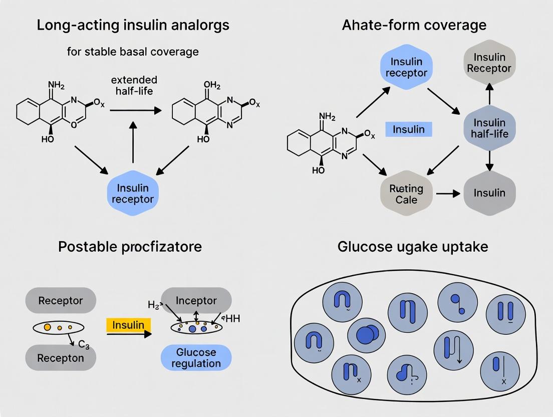
Molecular Mechanisms and Clinical Pharmacology: How Long-Acting Insulin Analogs Provide Sustained Basal Coverage
This article provides a comprehensive review for researchers, scientists, and drug development professionals on the molecular and pharmacological principles underpinning the stable, flat pharmacokinetic/pharmacodynamic (PK/PD) profiles of modern long-acting insulin...
Molecular Mechanisms and Clinical Pharmacology: How Long-Acting Insulin Analogs Provide Sustained Basal Coverage
Abstract
This article provides a comprehensive review for researchers, scientists, and drug development professionals on the molecular and pharmacological principles underpinning the stable, flat pharmacokinetic/pharmacodynamic (PK/PD) profiles of modern long-acting insulin analogs. It explores the foundational structural engineering of these analogs, the methodological advances in their formulation and delivery, key challenges and optimization strategies in their development, and their validation through comparative efficacy and safety data. The scope synthesizes current preclinical and clinical research to elucidate the translational journey from molecular design to stable glycemic control.
Beyond NPH: The Structural Engineering of Long-Acting Insulin Analogs for Basal Stability
Within the broader thesis on How do long-acting insulin analogs achieve stable basal coverage, the precise definition of a physiological basal profile is paramount. The physiological imperative is a basal insulin replacement that mimics the endogenous, non-prandial insulin secretion of a healthy pancreas: flat, stable, and peakless over a full 24-hour period. This whitepaper deconstructs the pharmacokinetic (PK) and pharmacodynamic (PD) parameters defining this profile and details the experimental paradigms used to quantify them in next-generation insulin analog research.
Defining the Target Profile: PK/PD Parameters
The ideal basal insulin profile is characterized by three core attributes, measurable through standardized clinical and preclinical experiments:
- Duration of Action (T>50% GIRmax): The time during which the glucose-lowering effect remains above 50% of the maximum effect. A true 24-hour coverage requires this to exceed 24h in the target population.
- Peak-to-Trough Ratio (PTR): The ratio of the maximum (GIR~max~) to minimum (GIR~min~) glucose infusion rate in a euglycemic clamp study. The ideal is 1.0 (completely flat). Ratios >2.0 indicate significant peaking.
- Intra-subject Variability (CV~AUC~ & CV~GIR~): The coefficient of variation in the area under the effect curve (AUC) and GIR profiles. Low variability (CV < 20-25%) is critical for predictable, stable daily coverage.
Table 1: Comparative PK/PD Parameters of Modern Long-Acting Analogs
| Insulin Analog | Mechanism of Protraction | Approx. Half-life (h) | Duration of Action (T>50% GIRmax, h) | Peak-to-Trough Ratio (PTR) | Intra-subject Variability (CV~AUC~, %) |
|---|---|---|---|---|---|
| Insulin Glargine U100 | Isoelectric precipitation | 12 | ~24 (varies) | ~1.5 - 2.0 | ~30-40 |
| Insulin Degludec | Multi-hexamer formation & albumin binding | 25+ | >42 | ~1.0 - 1.2 | ~20 |
| Insulin Glargine U300 | Enhanced precipitation | 19 | >24 | ~1.2 - 1.5 | ~20-30 |
Key Experimental Protocols for Assessment
The Euglycemic Glucose Clamp (Gold Standard PD)
Objective: To quantify the time-action profile of a basal insulin analog without confounding effects of endogenous insulin or counter-regulatory responses.
Detailed Protocol:
- Subject Preparation: Overnight fasted, diabetic (type 1) or pancreatectomized animal models to nullify endogenous insulin.
- Baseline Stabilization: A variable IV insulin infusion stabilizes blood glucose at target euglycemia (~5.5 mmol/L or 100 mg/dL).
- Test Insulin Administration: Subcutaneous injection of the investigational insulin analog at a standardized dose (e.g., 0.4 U/kg).
- Glucose Clamping: Frequent blood glucose monitoring (every 5-15 min) guides a variable IV infusion of 20% glucose. The glucose infusion rate (GIR, mg/kg/min) required to maintain euglycemia is precisely recorded.
- Data Collection: Continues for 24-36 hours post-dosing. The GIR profile directly mirrors the insulin's pharmacodynamic action.
- Analysis: Calculate GIR~max~, T~max~, AUC~GIR,0-24h~, and T>50% GIR~max~. PTR = GIR~max~ / GIR~trough~.
Pharmacokinetic Studies Using Tracer Methodology
Objective: To distinguish exogenous insulin PK from endogenous insulin, especially in non-diabetic models or early-phase human trials.
Detailed Protocol:
- Tracer Labeling: The investigational insulin analog is labeled with a stable, non-radioactive isotope (e.g., ^13^C, ^15^N, ^2^H).
- Co-administration: A microdose of the labeled insulin is administered simultaneously with the therapeutic dose of the unlabeled analog.
- Serial Sampling: Frequent plasma samples are collected over 24-48 hours.
- Mass Spectrometry Analysis: Samples are analyzed via liquid chromatography-tandem mass spectrometry (LC-MS/MS) to specifically quantify the labeled insulin fraction, excluding endogenous insulin and its metabolites.
- PK Modeling: Concentration-time data is analyzed using non-compartmental or compartmental modeling to derive AUC, C~max~, T~max~, half-life, and clearance.
Visualization of Mechanisms and Assessment
Title: Mechanism of Long-Acting Insulin Analog Protraction
Title: Euglycemic Glucose Clamp Experimental Workflow
The Scientist's Toolkit: Key Research Reagent Solutions
Table 2: Essential Materials for Basal Insulin Profile Research
| Research Reagent / Material | Function & Rationale |
|---|---|
| Clamp-Prepared T1D Animal Models (e.g., STZ-diabetic rats, diabetic dogs, pancreatectomized pigs) | Provides a consistent model devoid of endogenous insulin secretion, essential for clean PD assessment. |
| Stable Isotope-Labeled Insulin Analogs (^13^C/^15^N-full amino acid incorporation) | Enables specific PK tracing of the exogenous analog without interference from endogenous insulin in LC-MS/MS. |
| High-Sensitivity Human Insulin/Insulin Analog ELISA Kits (e.g., Mercodia, ALPCO) | For quantification of total immunoreactive insulin in plasma samples, though lacks analog specificity. |
| Automated Euglycemic Clamp Systems (e.g., Biostator, custom closed-loop systems) | Provides semi-automated glucose monitoring and infusion control, reducing operator variability in clamp studies. |
| Recombinant Human Albumin (rHA) | Critical for in vitro binding studies to assess albumin-binding kinetics of analogs like degludec. |
| Size-Exclusion Chromatography (SEC) & Analytical Ultracentrifugation (AUC) | Techniques to characterize the self-association state (monomer, hexamer, multi-hexamer) of insulin analogs in vitro. |
| Physiologically Buffered Subcutaneous Injection Simulants | Matches pH, hyaluronan, and collagen content of SC tissue to study dissolution and diffusion kinetics in vitro. |
Within the broader research thesis, "How do long-acting insulin analogs achieve stable basal coverage," two dominant molecular strategies have been engineered to protract insulin action and minimize pharmacokinetic (PK) and pharmacodynamic (PD) variability. These core strategies are: 1) Albumin Binding, which utilizes reversible binding to circulating albumin to create a circulating depot, and 2) Hexamer Stabilization, which enhances self-association in the subcutaneous depot to delay absorption. This whitepaper provides a technical dissection of these mechanisms, their experimental validation, and the resulting clinical profiles.
Mechanism of Action & Pharmacokinetic Principles
Albumin Binding Strategy: Analogs like insulin detemir and degludec are acylated with a fatty acid side chain. Following subcutaneous injection and hexamer disassociation, the modified monomers bind reversibly to albumin via the fatty acid moiety in the interstitial fluid and bloodstream. This binding creates a significant circulating reservoir, slowing diffusion to target tissues and reducing clearance. The dynamic equilibrium between bound and free insulin ensures a slow, continuous release of active monomer.
Hexamer Stabilization Strategy: Analog insulin glargine is engineered with alterations that shift its isoelectric point towards neutrality. In the acidic formulation (pH ~4), it is fully soluble. Upon injection into the neutral subcutaneous tissue, it precipitates, forming micro-precipitates that slowly dissolve. Insulin glargine U-300 (a more concentrated formulation) forms a stable subcutaneous depot with even slower dissolution. Similarly, insulin degludec, while also albumin-binding, employs a unique mechanism of multi-hexamer chain formation upon injection, facilitated by phenol and zinc in the formulation, creating a protracted subcutaneous depot.
Quantitative Pharmacokinetic/Pharmacodynamic Comparison
Table 1: Core Characteristics of Prototypical Analogs
| Parameter | Insulin Glargine U-100 (Hexamer Stabilization/Precipitation) | Insulin Detemir (Albumin Binding) | Insulin Degludec (Albumin Binding + Multi-hexamer) |
|---|---|---|---|
| Molecular Modification | A-chain: Gly21→Arg; B-chain: 2 Arg added to C-terminus | B29 Lys→myristoylated fatty acid | B29 Lys→hexadecandioyl-γ-Glu; Omits B30 Thr |
| Formulation pH | ~4.0 | ~7.4 | ~7.4 |
| Key Protraction Mechanism | Precipitation at neutral pH | Reversible albumin binding | Multi-hexamer formation + albumin binding |
| Albumin Binding (%) | Minimal | >98% | >99% |
| Reported t½ (h) | ~12 | 5-7 (dose-dependent) | ~25 |
| Duration of Action (h) | Up to 24 (with variability) | Up to 24 | >42 (steady-state) |
| CV for PK Endpoints | Higher | Moderate | Lowest (~20% GIR AUC) |
Table 2: Clinical Efficacy & Stability Metrics
| Metric | Glargine U-100 | Detemir | Degludec |
|---|---|---|---|
| Fasting Glucose Control (Δ vs. comparator) | Reference | Comparable | Superior in some studies |
| Hypoglycemia Rate (Nocturnal) | Reference | Lower in some | Significantly Lower |
| Within-subject PK/PD Variability | High | Lower | Lowest |
| Dosing Flexibility Window | ± 1 hour | Fixed timing | ± ~8 hours |
Detailed Experimental Protocols
4.1. Protocol for Assessing Albumin Binding Affinity (SPR/Biacore) Objective: Quantify the binding kinetics (Ka, Kd, KD) of acylated insulin analogs to human serum albumin (HSA). Materials: See "Scientist's Toolkit" below. Procedure:
- Surface Preparation: Immobilize HSA on a CM5 sensor chip using standard amine-coupling chemistry to achieve ~5000 RU.
- Running Buffer: Use HBS-EP+ (10 mM HEPES, 150 mM NaCl, 3 mM EDTA, 0.05% v/v Surfactant P20, pH 7.4).
- Analyte Preparation: Serially dilute the insulin analog (0.78 nM to 100 nM) in running buffer.
- Binding Cycle: Inject analyte for 180s at 30 μL/min, followed by a dissociation phase of 600s.
- Regeneration: Regenerate the surface with a 30s pulse of 10 mM Glycine-HCl, pH 2.0.
- Data Analysis: Double-reference sensorgrams. Fit data to a 1:1 Langmuir binding model to calculate association (ka) and dissociation (kd) rate constants. KD = kd/ka.
4.2. Protocol for Evaluating Subcutaneous Depot Formation (In Vitro Release) Objective: Model the dissolution/release rate from a stabilized hexamer or precipitate depot. Materials: USP apparatus 4 (flow-through cell), simulated interstitial fluid (pH 7.4), HPLC. Procedure:
- Depot Formation: Place 20 μL of insulin formulation (e.g., glargine U-100, degludec) into a small cavity in a cell filled with inert glass beads.
- Perfusion: Circulate pre-warmed (37°C) release medium (pH 7.4) through the cell at 8 mL/min.
- Sampling: Collect effluent fractions at predetermined timepoints (e.g., 0.5, 1, 2, 4, 8, 12, 24 h).
- Quantification: Analyze insulin concentration in each fraction by reverse-phase HPLC.
- Data Modeling: Plot cumulative release vs. time. Fit data to appropriate models (e.g., Higuchi, zero-order) to determine release kinetics.
4.3. Protocol for In Vivo Pharmacodynamic Assessment (Euglycemic Clamp in Rodents) Objective: Measure the time-action profile of long-acting insulin analogs. Materials: Cannulated rats/dogs, glucose infusion system, clamp software, insulin formulations. Procedure:
- Animal Preparation: Fast animals overnight. Insert catheters in jugular vein (for infusions) and carotid artery (for sampling).
- Basal Period: Infuse glucose as needed to establish stable baseline glycemia (~100 mg/dL).
- Insulin Dosing: Administer a single subcutaneous bolus of the test insulin analog at a standard dose (e.g., 6 nmol/kg).
- Clamp Phase: Initiate variable glucose infusion (GIR) to maintain euglycemia for up to 30 hours. Blood glucose is measured every 5-10 min.
- Endpoint: The Glucose Infusion Rate (GIR) over time is the primary PD readout. Calculate GIRmax, time to GIRmax, and GIR AUC for comparison.
Signaling Pathway & Experimental Workflow Visualizations
Diagram Title: Long-Acting Insulin Protraction Pathways (86 chars)
Diagram Title: Euglycemic Clamp Experimental Protocol (44 chars)
The Scientist's Toolkit: Key Research Reagent Solutions
Table 3: Essential Materials for Core Experiments
| Item/Category | Example/Supplier | Function in Research Context |
|---|---|---|
| Recombinant Human Insulins | Detemir, Glargine, Degludec (Sigma, Novo Nordisk) | Gold standards for comparative binding, release, and activity assays. |
| Human Serum Albumin (HSA) | Fatty acid-free, recombinant (Sigma) | Essential substrate for SPR/Biacore and equilibrium dialysis assays to measure binding affinity. |
| SPR/Biacore System | Cytiva Series S, Biacore T200 | Label-free real-time analysis of insulin-albumin binding kinetics (ka, kd, KD). |
| Simulated Interstitial Fluid | Custom buffer (pH 7.4, with electrolytes) | In vitro medium to model the subcutaneous environment for release/dissolution studies. |
| USP Apparatus 4 | Flow-through cell dissolution system (Sotax) | Standardized system for evaluating in vitro release from subcutaneous depot formulations. |
| Euglycemic Clamp System | Custom rodent/dog setup (BioDaq, Instech) | Integrated system for continuous glucose monitoring and infusion to measure in vivo PD profiles. |
| Stable Isotope Tracers | [6,6-²H₂]-Glucose (Cambridge Isotopes) | Used in advanced clamp studies to assess hepatic glucose production and peripheral uptake. |
| HPLC-MS/MS Systems | Triple quadrupole MS with UPLC (Waters, Sciex) | Ultra-sensitive quantification of insulin analogs and metabolites in plasma/tissue samples. |
Framing Thesis Context: This whitepaper details the physicochemical mechanism of insulin glargine’s protracted action. Understanding this mechanism is a cornerstone case study for the broader research thesis: How do long-acting insulin analogs achieve stable basal coverage? This investigation focuses on the deliberate, pH-dependent precipitation phenomenon that underlies glargine’s pharmacokinetic profile.
Core Mechanism of Action
Insulin glargine is a recombinant human insulin analog engineered for prolonged, peakless absorption following subcutaneous injection. The key modifications are:
- A21: Glycine replaces asparagine.
- B30: Two arginines are added to the C-terminus.
These alterations shift the isoelectric point (pI) from approximately pH 5.4 for human insulin to pH 6.7 for insulin glargine. The formulation is stabilized at an acidic pH of ~4.0, where the molecule is fully soluble. Upon injection into the subcutaneous tissue (pH ~7.4), the solution is neutralized. As the environmental pH approaches the molecule's pI, insulin glargine precipitates, forming hexameric and multimeric micro-precipitates. These solid-phase reservoirs dissolve slowly, providing a continuous, slow release of insulin monomers into the systemic circulation. The U300 formulation (300 units/mL) presents a higher concentration of insulin in a proportionally smaller injection volume, leading to a smaller initial precipitation surface area and contributing to an even more prolonged and stable release profile compared to U100.
Table 1: Physicochemical and Pharmacokinetic Properties of Insulin Glargine U100 vs. U300
| Property | Insulin Glargine U100 | Insulin Glargine U300 | Notes |
|---|---|---|---|
| Concentration | 100 U/mL (3.64 mg/mL) | 300 U/mL (10.91 mg/mL) | U300 is 3x more concentrated. |
| Formulation pH | ~4.0 | ~4.0 | Acidic to ensure solubility. |
| Isoelectric Point (pI) | ~6.7 | ~6.7 | Unchanged by concentration. |
| Injection Volume | Larger (e.g., 0.3 mL for 30 U) | Smaller (e.g., 0.1 mL for 30 U) | Key driver of PK differences. |
| Onset of Action | ~1-2 hours | ~1-2 hours | Similar onset. |
| Time to Peak (Tmax) | ~5-6 hours (mild peak) | Flatter profile | U300 demonstrates a more consistent concentration-time curve. |
| Duration of Action (TDD) | Up to 24 hours (often 22-24h) | >24 hours (often >30h) | U300 provides a longer and more stable tail. |
| GIRAUC (0-24h) Stability | Reference | +18% to +24% more stable | Glucose Infusion Rate AUC indicates smoother action for U300. |
Table 2: Key Findings fromIn VitroPrecipitation/Dissolution Studies
| Study Parameter | Observation for U100 | Observation for U300 | Implication |
|---|---|---|---|
| Precipitation Onset (at pH ~7.4) | Rapid formation of discrete microprecipitates. | Formation of a denser, more cohesive precipitate network. | Altered dissolution kinetics. |
| Dissolution Rate | Faster dissolution per unit mass. | Slower dissolution per unit mass due to reduced surface area-to-volume ratio. | Contributes to prolonged release. |
| Precipitate Morphology | Amorphous/crystalline aggregates. | More homogeneous, gel-like structure. | Modified release profile from depot. |
Experimental Protocols for Key Studies
Protocol 1:In VitroPrecipitation Kinetics Assay
Objective: To visualize and quantify the pH-dependent precipitation of insulin glargine formulations. Methodology:
- Prepare formulation samples (U100 and U300) in their native acidic buffer.
- Rapidly mix 100 µL of formulation with 900 µL of neutral phosphate buffer (PBS, pH 7.4) in a cuvette.
- Immediately place the cuvette in a spectrofluorometer or UV-Vis spectrophotometer equipped with a stirrer and temperature control (set to 32°C to mimic subcutaneous temperature).
- Monitor turbidity development by measuring optical density (OD) at 350 nm or 600 nm every 10 seconds for 60 minutes.
- Plot OD vs. time. The slope of the initial linear increase indicates precipitation rate. The plateau OD indicates final precipitate density.
- Correlate findings with parallel dynamic light scattering (DLS) measurements for particle size distribution.
Protocol 2:In VivoPharmacokinetic/Pharmacodynamic (PK/PD) Study in Diabetic Animal Model
Objective: To compare the absorption and action profiles of U100 and U300. Methodology:
- Use chronically catheterized diabetic (e.g., pancreatectomized or streptozotocin-induced) canines or pigs.
- After an overnight fast and achieving stable euglycemia via a basal insulin infusion (glucose clamp technique), administer a single subcutaneous bolus of insulin glargine (U100 or U300) at 0.6 U/kg in a randomized, crossover design.
- PK: Collect serial plasma samples over 30 hours. Measure serum insulin glargine concentrations via a specific ELISA (does not cross-react with endogenous insulin).
- PD: Maintain euglycemia using a variable glucose infusion. The glucose infusion rate (GIR) required over time is the primary PD endpoint, representing the drug's biological activity.
- Calculate key parameters: Time to 50% of max GIR (onset), GIRmax, time to GIRmax, and total glucose infused (AUCGIR, 0-24h).
Mechanism and Workflow Visualizations
The Scientist's Toolkit: Key Research Reagents & Materials
Table 3: Essential Materials for Insulin Glargine Precipitation Research
| Item | Function/Description | Example Supplier/Catalog |
|---|---|---|
| Recombinant Insulin Glargine (Lyophilized) | High-purity standard for preparing controlled formulation comparisons or calibration curves. | Sigma-Aldrich, Thermo Fisher Scientific. |
| Lantus & Toujeo (U100 & U300) | Commercial formulations for in vitro and ex vivo comparative studies. Critical for translational research. | Pharmacy-sourced. |
| Phosphate Buffered Saline (PBS), pH 7.4 | Standard neutral medium to simulate subcutaneous fluid for precipitation assays. | Various biological buffer suppliers. |
| Glycine-HCl or Acetate Buffer, pH 4.0 | Acidic buffer for reconstituting or diluting glargine to mimic its formulation vehicle. | Various biological buffer suppliers. |
| Spectrophotometer with Peltier & Stirrer | For kinetic turbidity measurements (OD at 350/600 nm) under controlled temperature. | Agilent, PerkinElmer. |
| Dynamic Light Scattering (DLS) Instrument | To measure particle size distribution and z-average diameter of forming precipitates. | Malvern Panalytical, Wyatt Technology. |
| Insulin Glargine-Specific ELISA Kit | For quantifying glargine concentrations in plasma/serum without cross-reactivity with endogenous insulin in PK studies. | Mercodia, ALPCO. |
| Euglycemic Clamp Apparatus | Integrated pump system for maintaining constant blood glucose during in vivo PD studies in animals or humans. | Biostator or modern clinical research systems. |
Thesis Context: This analysis is a component of a broader investigation into "How do long-acting insulin analogs achieve stable basal coverage?" This whitepaper examines the molecular design and pharmacokinetic principles of insulin detemir, focusing on its acylation-based mechanism for prolonged action.
Molecular Design and Mechanism of Action
Insulin detemir (Levemir) is a long-acting human insulin analog engineered for stable, peakless basal coverage. The core modification is the acylation of the insulin B-chain. Specifically, lysine at position B29 is conjugated with a myristic acid (C14 fatty acid) side chain. This single alteration confers two interconnected mechanisms for prolonged action:
- Reversible albumin binding: The fatty acid side chain facilitates binding to albumin, the most abundant protein in circulation, creating a circulating depot.
- Slower absorption from subcutaneous tissue: The albumin-binding property also slows diffusion from the injection site.
Upon subcutaneous injection, detemir binds to albumin present in interstitial fluid. Following absorption into the bloodstream, it binds to serum albumin (>98% bound). Only the free, unbound fraction is pharmacologically active at the insulin receptor. The equilibrium between bound and free states provides a slow, continuous release of active insulin.
Key Pharmacokinetic/Pharmacodynamic Data
Table 1: Comparative Pharmacokinetic Properties of Insulin Detemir vs. NPH Insulin
| Parameter | Insulin Detemir | NPH Insulin (Neutral Protamine Hagedorn) | Notes/Methodology |
|---|---|---|---|
| Time to Max Concentration (T~max~) | 6 - 8 hours | 4 - 6 hours | Measured via radioimmunoassay or ELISA following subcutaneous administration in clinical trials. |
| Half-life (t~1/2~) | 5 - 7 hours | 2 - 3 hours | Determined from venous blood sampling in pharmacokinetic studies. |
| Duration of Action | Up to 24 hours | 12 - 18 hours | Assessed by glucose infusion rate (GIR) in euglycemic clamp studies. |
| Intra-subject Variability (CV%) in Absorption | ~20-25% | ~40-50% | Lower variability attributed to albumin-based mechanism vs. crystal dissolution (NPH). Measured by AUC variability. |
| Albumin Binding (%) | >98% | 0% | Assessed using ultrafiltration or equilibrium dialysis with radiolabeled insulin. |
Table 2: Impact of Acyl Chain Characteristics on Pharmacokinetics
| Acyl Chain Feature | Rationale & Effect on PK/PD | Experimental Evidence |
|---|---|---|
| Chain Length (C14) | Optimal balance of albumin affinity and reversible dissociation. Shorter chains reduce duration; longer chains increase affinity, potentially reducing free fraction. | Structure-activity relationship (SAR) studies with C12, C14, C16, and C18 chains measuring serum half-life in animal models. |
| Attachment Site (B29 Lysine) | Position chosen to minimize interference with insulin receptor binding (mediated primarily via B24-B26 region). | Comparative assays of receptor binding affinity and mitogenic potential for analogs acylated at different residues. |
| Presence of Threonine at B30 | Native human insulin has threonine at B30; detemir lacks B30 (desB30). This omission slightly increases solubility and facilitates the acylation process. | Comparative stability and hexamer formation studies of desB30 human insulin. |
Detailed Experimental Protocols
Protocol 1: Assessing Albumin Binding Affinity (Equilibrium Dialysis)
- Objective: Quantify the percentage of insulin detemir bound to human serum albumin (HSA) under physiological conditions.
- Materials: ³H- or ¹²⁵I-labeled insulin detemir, unlabeled insulin detemir, purified HSA, isotonic phosphate buffer (pH 7.4), equilibrium dialysis chambers (e.g., DispoEquilibrium Dialyzer), dialysis membrane (MWCO 10 kDa), liquid scintillation counter or gamma counter.
- Method:
- Prepare a solution of HSA (40 g/L) in phosphate buffer to mimic physiological concentration.
- Spike the HSA solution with a trace amount of radiolabeled insulin detemir and a known concentration of unlabeled detemir (e.g., 100 nM).
- Load the HSA-detemir solution into one chamber (donor) and buffer only into the adjacent chamber (receiver). Separate chambers with the dialysis membrane.
- Incubate at 37°C with gentle agitation for 16-24 hours to reach equilibrium.
- Sample equal volumes from both chambers and measure radioactivity.
- Calculation: % Bound = (CPM~donor~ - CPM~receiver~) / CPM~donor~ × 100. The free fraction concentration is equal to the receiver chamber concentration.
Protocol 2: Euglycemic Clamp for Pharmacodynamic Profiling
- Objective: Measure the time-action profile of insulin detemir in vivo.
- Materials: Human or animal subjects, variable-rate intravenous insulin infusion, 20% glucose solution, insulin detemir for subcutaneous injection, automated glucose clamp system (e.g., Biostator) or manual setup, frequent blood glucose monitor.
- Method:
- After an overnight fast, establish a baseline. Initiate a primed, continuous intravenous insulin infusion to lower blood glucose to the target euglycemic level (e.g., 90 mg/dL).
- Administer a subcutaneous dose of insulin detemir.
- Maintain blood glucose at the target level for 24+ hours by adjusting the infusion rate of exogenous glucose based on frequent (e.g., every 5-10 min) glucose measurements.
- The Glucose Infusion Rate (GIR) required to maintain euglycemia is the primary endpoint, directly reflecting the insulin's biological activity over time.
- Plot GIR (mg/kg/min) vs. time to visualize onset, peak, and duration of action.
Visualization of Mechanism and Research Pathways
Diagram 1: Insulin Detemir Pharmacokinetic Pathway
Diagram 2: Structural Engineering of Insulin Detemir
The Scientist's Toolkit: Key Research Reagents & Materials
Table 3: Essential Research Reagents for Studying Insulin Detemir
| Reagent/Material | Function in Research | Key Application Example |
|---|---|---|
| Recombinant Insulin Detemir (High Purity) | The core analyte for in vitro and in vivo studies. | Used as reference standard in binding assays, receptor studies, and formulation development. |
| ³H- or ¹²⁵I-labeled Insulin Detemir | Radiolabeled tracer for sensitive quantification. | Essential for equilibrium dialysis, pharmacokinetic distribution studies, and cellular uptake assays. |
| Human Serum Albumin (HSA), Fatty Acid-Free | The primary binding partner in plasma. | Used in binding affinity assays (SPR, ITC) and to simulate physiological conditions in stability studies. |
| Anti-Insulin/Detemir Antibodies (ELISA Kit) | Immunoassay-based detection and quantification. | Measuring insulin detemir concentrations in serum/plasma samples from preclinical and clinical studies. |
| CHO/HEK Cells Overexpressing Human Insulin Receptor (IR) | Cellular model for receptor binding and downstream signaling. | Assessing in vitro potency, mitogenic potential, and phosphorylation cascades (e.g., AKT, MAPK). |
| Euglycemic Clamp System | Gold-standard in vivo pharmacodynamic assessment. | Determining the precise time-action profile (onset, peak, duration) in animal models or human subjects. |
| Surface Plasmon Resonance (SPR) Chip with Immobilized HSA | Label-free, real-time analysis of binding kinetics (ka, kd, KD). | Precisely measuring the association/dissociation rates of detemir and its analogs to albumin. |
This whitepaper examines the molecular and pharmacological mechanisms underlying the ultra-long duration of action of insulin degludec, within the broader thesis research of How do long-acting insulin analogs achieve stable basal coverage.
Core Molecular Mechanism
Insulin degludec is engineered by removing threonine at position B30 and conjugating a 16-carbon fatty diacid (hexadecanedioic acid) via a glutamic acid spacer to lysine at B29. Upon subcutaneous injection at therapeutic concentrations (600 µM, U100), it self-associates into stable dihexamers. In the subcutaneous tissue, phenol and zinc diffuse away, leading to the formation of multi-hexamer chains. These chains act as a soluble depot, from which monomers slowly and continuously dissociate into the bloodstream, providing a flat, stable pharmacokinetic (PK) profile.
Table 1: Key Physicochemical and Pharmacokinetic Parameters of Insulin Degludec
| Parameter | Value/Range | Experimental Context |
|---|---|---|
| Molecular Weight | 6103.97 g/mol | Calculated for C274H411N65O81S6 |
| Critical Self-Association Concentration | ~5 nM | In vitro dissociation constant (Kd) for dihexamer formation |
| Formulation Concentration | 600 µM (U100) | Therapeutic formulation with phenol and zinc. |
| Time to Steady-State (PK) | 2-3 days | In clinical studies after repeated dosing. |
| Terminal Half-life (t1/2) | ~25.1 hours | Subcutaneous administration in humans. |
| Duration of Action | >42 hours | Pharmacodynamic (glucose infusion rate) assessment. |
| Albumin Binding Affinity (Kd) | High (bound fraction >99%) | Due to fatty acid side chain; reversible binding. |
| Coefficient of Variation (PK) | 20% | For total exposure (AUC) at steady state, indicating low variability. |
Table 2: Comparison of Hexamer Stability Metrics (In Vitro)
| Insulin Analog | Dissociation Rate Constant (koff) for Monomer Release | Relative Chain Propagation Propensity | Key Structural Modifier |
|---|---|---|---|
| Human Insulin | High | None | N/A |
| Insulin Detemir | Moderate | Low (Dihexamer max) | C14 fatty acid (Myristic acid) at B29 |
| Insulin Glargine | Low (precipitate) | High (Microprecipitate) | Isoelectric point shift to ~6.7 |
| Insulin Degludec | Very Low | Very High (Soluble Multi-Hexamer Chains) | C16 fatty diacid chain at B29 via spacer |
Experimental Protocols for Key Studies
Protocol: Analysis of Multi-Hexamer Chain Formation by Size-Exclusion Chromatography (SEC)
Objective: To characterize the self-association state of insulin degludec in solution under varying conditions. Materials: Insulin degludec stock (600 µM), buffer (pH 7.4, 30 mM phosphate, 100 mM NaCl, with/without 0.2% phenol and 0.06 mM Zn2+), HPLC-SEC system with UV detection. Procedure:
- Prepare degludec samples in (a) formulation buffer (with phenol/Zn2+) and (b) physiological buffer (without phenol/Zn2+).
- Incubate at 37°C for 0, 6, 24 hours.
- Inject samples onto a calibrated Superdex 75 Increase 10/300 GL column.
- Elute with physiological buffer at 0.5 mL/min, monitor at 280 nm.
- Compare elution volumes to protein standards to determine molecular size/complex formation. Interpretation: Formulation buffer shows dihexamer peak. Physiological buffer shows high-molecular-weight aggregates/smear indicative of soluble multi-hexamer chains.
Protocol: Surface Plasmon Resonance (SPR) for Albumin Binding Kinetics
Objective: Quantify the binding affinity of insulin degludec to human serum albumin (HSA). Materials: Biacore T200 SPR system, CMS sensor chip, HSA, insulin degludec, running buffer (HBS-EP+, pH 7.4). Procedure:
- Immobilize HSA on a CM5 chip via amine coupling to ~5000 response units (RU).
- Use a flow cell without HSA as a reference.
- Inject a series of insulin degludec concentrations (0.1-100 nM) over HSA and reference surfaces at 30 µL/min for 120s association, followed by 600s dissociation.
- Regenerate surface with a 30s pulse of 10 mM glycine-HCl, pH 2.0.
- Fit the resulting sensograms to a 1:1 Langmuir binding model to determine association (kon) and dissociation (koff) rate constants, and calculate equilibrium dissociation constant (KD = koff/kon).
Protocol:In VivoPharmacokinetic/Pharmacodynamic (PK/PD) Study in Diabetic Animal Model
Objective: Assess the time-action profile of insulin degludec. Materials: Streptozotocin-induced diabetic pigs or dogs, insulin degludec, continuous glucose monitoring system, automated blood sampler, glucose infusion setup. Procedure:
- Cannulate animals for frequent blood sampling and glucose infusion.
- Administer a single subcutaneous dose of insulin degludec (0.4 U/kg) or comparator.
- Monitor plasma insulin concentration via specific ELISA (distinguishes bound and free fractions) for 48+ hours.
- Simultaneously, clamp blood glucose at euglycemic level (e.g., 5.5 mmol/L) via variable glucose infusion rate (GIR).
- Plot PK (plasma concentration) and PD (GIR) profiles over time. Calculate half-life, time to peak, and duration of action (time until GIR returns to baseline).
Visualizations
Diagram 1: Action mechanism of insulin degludec from injection to effect.
Diagram 2: SEC workflow for multi-hexamer chain analysis.
The Scientist's Toolkit: Research Reagent Solutions
Table 3: Essential Research Materials for Insulin Degludec Studies
| Item | Function/Application in Research | Key Characteristics |
|---|---|---|
| Recombinant Insulin Degludec (Lyophilized) | Core molecule for in vitro biophysical and in vivo studies. | High purity (>99%), fatty acid conjugation confirmed by mass spectrometry. |
| Human Serum Albumin (HSA), Fatty Acid Free | For albumin binding assays (SPR, equilibrium dialysis). | Essential to study reversible binding kinetics and plasma distribution. |
| Size-Exclusion Chromatography (SEC) Columns | To analyze self-association states (dihexamers, multi-hexamer chains). | High-resolution matrix (e.g., Superdex Increase series) for protein aggregates. |
| Insulin Degludec-Specific ELISA Kit | To measure low concentrations in plasma, distinguishing free from total drug. | High specificity, no cross-reactivity with endogenous insulin or albumin. |
| Phenol/Zinc-Free Buffer Systems | To mimic subcutaneous interstitial fluid and trigger chain formation in vitro. | Typically pH 7.4 phosphate or HEPES buffer. |
| Streptozotocin (STZ)-Induced Diabetic Animal Models | For in vivo PK/PD profiling. | Rodents or larger animals (pigs, dogs) provide predictive data for human action. |
| Surface Plasmon Resonance (SPR) Instrument | To quantify real-time kinetics of albumin binding. | Enables determination of kon, koff, and KD. |
This whitepaper provides an in-depth technical guide on leveraging pharmacokinetic/pharmacodynamic (PK/PD) correlates to rationally design long-acting insulin analogs that achieve stable basal coverage. The principles discussed are framed within the broader thesis of understanding how molecular engineering translates to predictable, flat, and prolonged time-action profiles.
The therapeutic goal of a basal insulin is to provide a consistent, peakless background of insulin activity that mimics physiological fasting insulin secretion. Achieving this requires a profound understanding of the PK/PD correlate—the quantitative link between a molecule's structural attributes, its resulting plasma concentration-time profile (PK), and the subsequent glucose-lowering effect (PD). Molecular design directly manipulates the PK profile; the PK profile dictates the PD response. For long-acting analogs, the primary objective is to decelerate absorption from the subcutaneous depot and/or delay receptor clearance to produce a stable plasma concentration plateau.
Molecular Design Strategies for Protraction
Current long-acting insulin analogs employ three primary mechanisms to prolong action, each with distinct PK/PD consequences.
Table 1: Molecular Design Strategies for Long-Acting Insulin Analogs
| Strategy | Molecular Modification | Primary PK Effect | Key PK/PD Parameter Impact | Example Analog |
|---|---|---|---|---|
| Self-Association | Substituents (e.g., fatty acids) enabling reversible hexamer-to-dimer/monomer dissociation and albumin binding. | Slows absorption from SC depot; albumin binding reduces free fraction and delays clearance. | Increases t~1/2~, flattens C~max~/C~min~ ratio. | Insulin detemir, degludec |
| Isoelectric Point Shift | Amino acid substitutions raising pI from ~5.4 to near-neutral (~7.0). | Precipitates in neutral pH SC tissue, forming a stable subcutaneous depot. | Markedly prolongs t~1/2~ and duration of action. | Insulin glargine |
| PEGylation & Fc Fusion | Conjugation to polyethylene glycol (PEG) or immunoglobulin Fc domain. | Increases hydrodynamic radius; Fc binds neonatal Fc receptor (FcRn) promoting recycling. | Dramatically reduces clearance, extends t~1/2~. | PEGylated lispro (not marketed), Fc-fusion proteins |
The PK/PD Modeling Framework
The relationship between plasma concentration (C~p~) and effect (E) for insulin is typically described by an Indirect Response Model, as glucose lowering is mediated through a complex cascade of insulin receptor signaling and subsequent metabolic processes.
Diagram Title: PK/PD Indirect Response Model for Insulin
The core equations for a simple inhibitory indirect response model (for hepatic glucose production) are:
- PK: C~p~(t) = f(Dose, k~a~, V~d~, CL) [Often bi-exponential for SC administration]
- PD: dR/dt = k~in~ * (1 - (I~max~ * C~p~) / (IC~50~ + C~p~)) - k~out~ * R Where R is the response variable (e.g., glucose production rate), k~in~ is the zero-order production rate, k~out~ is the first-order loss rate, I~max~ is the maximum inhibitory effect, and IC~50~ is the plasma concentration producing 50% of I~max~.
Table 2: Key PK/PD Parameters for Basal Insulin Assessment
| Parameter | Symbol | Definition | Target for Stable Coverage |
|---|---|---|---|
| Time to Max Concentration | T~max~ | Time from dosing to C~max~. | Longer T~max~ indicates slower absorption, desirable. |
| Peak-Trough Ratio | C~max~/C~min~ | Ratio of maximum to minimum concentration over dosing interval. | Closer to 1.0 indicates flatter profile. |
| Half-life | t~1/2~ | Time for plasma concentration to reduce by 50%. | Longer t~1/2~ supports once-daily dosing. |
| Duration of Action | - | Time glucose infusion rate (GIR) remains above a threshold (e.g., 50% of max). | Should cover ≥24h with minimal fluctuation. |
| GIR~AUC~/C~AUC~ Ratio | - | Measure of pharmacodynamic potency per unit exposure. | Reflects receptor affinity and signaling efficiency. |
Experimental Protocols for Characterizing the PK/PD Correlate
Euglycemic Glucose Clamp (Gold Standard PD Assay)
- Objective: Quantify the time-action profile of an insulin analog under steady-state glucose conditions.
- Protocol:
- Subject Preparation: Overnight-fasted, healthy or diabetic subjects are catheterized for insulin/glucose infusion and frequent blood sampling.
- Basal Period: A variable glucose infusion establishes a steady baseline blood glucose (~5.0 mmol/L or 90 mg/dL).
- Insulin Administration: A single subcutaneous dose of the test insulin analog is administered.
- Clamp Phase: Blood glucose is measured every 5-10 minutes. The glucose infusion rate (GIR) is adjusted dynamically to counteract insulin-induced glucose lowering and maintain the target euglycemia.
- Duration: Continues for 24-36 hours or until GIR returns to baseline.
- Primary Output: The GIR-time profile is the direct PD readout. Key metrics are derived: time to 50% max GIR, max GIR, GIR~AUC~, and duration of action.
Pharmacokinetic Sampling & Bioanalysis
- Objective: Determine the plasma concentration-time profile of the insulin analog.
- Protocol:
- Sample Collection: Frequent venous blood samples are drawn in parallel with the clamp study (e.g., pre-dose, then 0.5, 1, 2, 4, 6, 8, 10, 12, 16, 20, 24, 30, 36h post-dose).
- Sample Processing: Plasma is separated via centrifugation and stored at ≤-70°C.
- Quantification: Analyzed via specific immunoassays (ELISA) or liquid chromatography-tandem mass spectrometry (LC-MS/MS). Assays must distinguish the analog from endogenous insulin.
- PK Analysis: Concentration-time data are modeled using non-compartmental analysis (NCA) or compartmental modeling to estimate AUC, C~max~, T~max~, t~1/2~, and clearance.
Diagram Title: PK/PD Study Workflow for Insulin Analogs
The Scientist's Toolkit: Key Research Reagent Solutions
Table 3: Essential Research Reagents for Insulin PK/PD Studies
| Item | Function & Specificity | Example Application |
|---|---|---|
| Analog-Specific ELISA Kits | Quantify specific insulin analog in biological matrices without cross-reactivity with human insulin or other analogs. | Measuring plasma PK profiles in preclinical/clinical studies. |
| Human Insulin Receptor (hIR) Phosphorylation Assay | Measures phosphorylated tyrosine residues on hIR in cell lysates. | Assessing in vitro potency and signaling kinetics of analogs. |
| Stable GLUT4 Reporter Cell Lines | Cells expressing a tagged glucose transporter (GLUT4) to monitor translocation. | Quantifying downstream metabolic signaling potency. |
| SC Injection Site Mimetic Matrix | Synthetic or animal-derived hydrogel simulating subcutaneous tissue. | In vitro study of insulin analog hexamer/dissociation and precipitation kinetics. |
| Recombinant Human Serum Albumin (HSA) | High-purity, fatty-acid-free HSA. | Studying binding kinetics and affinity of albumin-binding analogs (detemir, degludec). |
| Liquid Chromatography-Mass Spectrometry (LC-MS/MS) Systems | High-sensitivity, high-specificity detection and quantification of insulins and metabolites. | Definitive bioanalysis, especially for novel analogs without immunoassays. |
| Automated Glucose Clamp Systems | Integrated systems for glucose measurement and variable infusion pump control. | Standardizing and improving reproducibility of euglycemic clamp studies. |
Case Study: Insulin Degludec's Ultra-Flat Profile
Insulin degludec employs a unique design: addition of a hexadecanedioic acid via a glutamic acid spacer at B29, and omission of B30 threonine. This enables the formation of multi-hexamers in the subcutaneous depot, which slowly dissociate into monomers for absorption. Furthermore, its strong albumin binding (>99%) following absorption creates a significant circulating depot. This dual-depot mechanism results in an exceptionally flat and stable PK profile, with a t~1/2~ of ~25 hours and a peak-trough ratio of <1.2 in steady state. Its GIR profile shows a smooth, peakless activity over 42+ hours, exemplifying the successful application of the PK/PD correlate in molecular design to achieve stable basal coverage.
From Bench to Bedside: Formulation Science and Delivery Optimization of Basal Analogs
Within the pursuit of stable basal glycemic control, the development of long-acting insulin analogs represents a pinnacle of protein engineering. While modifications to the insulin polypeptide chain (e.g., at positions B28, B29, B31, B32) are central to altering pharmacokinetics, the formulation excipients zinc, phenol, and m-cresol are critical, non-polypeptide components that dictate the stable, soluble depot formation required for protracted action. This whitepaper dissects the distinct and synergistic roles of these excipients in mediating hexamer stabilization, solubility, and controlled disassembly, framing their function within the core thesis of achieving stable basal insulin coverage.
The therapeutic goal of a basal insulin is to provide a steady, peakless, and reproducible concentration profile over approximately 24 hours. Achieving this requires a formulation that remains soluble at high concentration in the injection vial yet forms a delayed-release depot upon subcutaneous administration. Insulin analogs like insulin glargine, degludec, and detemir solve this through a combination of amino acid substitutions and carefully engineered formulation chemistry. The excipients zinc, phenol, and m-cresol are not mere preservatives or stabilizers; they are active directors of the insulin's assembly state and dissolution kinetics.
Mechanistic Roles of Critical Excipients
Zinc: The Architect of Hexamerization
Zinc ions (Zn²⁺) are integral to the native quaternary structure of insulin. In pharmaceutical formulations, zinc is added to stabilize insulin hexamers, which are too large for rapid capillary absorption.
Mechanism: Two Zn²⁺ ions bind coordinately to the His(B10) residues of six insulin monomers, forming a stable, symmetric R6 hexamer. This hexamer is the storage form in the vial.
Protraction Contribution: The subcutaneous dissociation sequence—hexamer → dimer → monomer—is rate-limited by the initial hexamer disassembly. Zinc-mediated hexamer stability directly slows this first step. In analogs like insulin glargine, the isoelectric point shift means precipitation occurs at physiological pH, and zinc further modulates the physical characteristics of the precipitate.
Phenol andm-Cresol: Allosteric Stabilizers and Antimicrobials
Phenol and its derivative m-cresol serve dual, essential functions.
Allosteric Hexamer Stabilization: These phenolic compounds bind to specific hydrophobic pockets at the hexamer interface, inducing a conformational change from the T-state (tense) to the R-state (relaxed). The R-state hexamer is significantly more stable. This allosteric stabilization further delays subcutaneous disassembly.
Solubility Modulation: Phenolic compounds enhance the solubility of insulin at the acidic pH of some formulations (e.g., glargine at pH ~4). Upon injection into neutral subcutaneous tissue, the dilution of phenol/m-cresol removes this solubilizing effect, contributing to the controlled precipitation of certain analogs.
Antimicrobial Preservation: At their employed concentrations (typically 0.1-0.3%), they provide essential bacteriostatic activity in multi-dose vials.
Synergistic Action for Protraction
The protraction mechanism is a concert: Zinc provides the structural core of the hexamer, while phenolic compounds "lock" it in a stable R-state. Upon subcutaneous injection, dilution and diffusion of phenolic compounds initiate hexamer destabilization. Subsequent dissociation and, for some analogs, precipitation are controlled by the remaining zinc ions and the altered solubility profile of the engineered protein. This multi-stage retardation creates the desired flat, prolonged absorption profile.
Table 1: Typical Concentration Ranges of Critical Excipients in Commercial Long-Acting Insulin Analogs
| Insulin Analog | Zinc (µg/mL) | m-Cresol (mg/mL) | Phenol (mg/mL) | Key Formulation pH | Primary Protraction Mechanism |
|---|---|---|---|---|---|
| Insulin Glargine | 30-80 | 2.7 | - | ~4.0 | Microprecipitate at neutral pH |
| Insulin Detemir | ~65 | 1.6 | 1.5 | ~7.4 | Albumin binding + Zn-phenol hexamer |
| Insulin Degludec | ~70 | 1.6 | 1.5 | ~7.4 | Multi-hexamer chain formation |
| Human NPH | 20-100 | 0.65 (or Phenol) | 0.60 (or m-cresol) | ~7.0 | Protamine crystallization |
Table 2: Impact of Excipient Removal on Key Pharmacokinetic Parameters (Model Study)
| Formulation Variant | Tmax (h) | t1/2 (h) | AUC0-24 | Relative Bioavailability |
|---|---|---|---|---|
| Full Commercial Formulation | 6-8 | 12-20 | 100% | 100% |
| Without Phenolic Compounds | 2-3 | 4-6 | ~85% | ~95% |
| With Reduced Zinc (<10 µg/mL) | 3-4 | 5-8 | ~90% | ~98% |
| Without Zn & Phenolics | 1-2 | 2-3 | ~80% | ~90% |
Experimental Protocols for Investigating Excipient Roles
Protocol 4.1: Size-Exclusion Chromatography (SEC) for Assembly State Analysis Objective: To determine the oligomeric state (monomer, dimer, hexamer) of an insulin formulation under varying excipient conditions. Methodology:
- Column: Use a high-resolution SEC column (e.g., Superdex 75 Increase 10/300 GL).
- Mobile Phase: Prepare buffers mimicking formulation (pH 4) and physiological (pH 7.4) conditions, with and without critical excipients. Include 150 mM NaCl to mimic ionic strength.
- Sample Preparation: Dilute insulin sample to 1 mg/mL in corresponding mobile phase. Incubate for 1 hour at 25°C.
- Run Conditions: Flow rate 0.5 mL/min, detection at 280 nm.
- Calibration: Use protein standards of known molecular weight to create a calibration curve. The elution volume identifies the dominant oligomeric state.
Protocol 4.2: Isothermal Titration Calorimetry (ITC) for Binding Affinity Objective: To quantify the binding affinity (Kd), stoichiometry (n), and thermodynamics (ΔH, ΔS) of phenol/zinc binding to insulin. Methodology:
- Sample Preparation: Thoroughly degas all solutions. Insulin solution (50 µM in monomer) is loaded into the sample cell. Titrant solutions contain zinc acetate (e.g., 500 µM) or phenol (e.g., 1 mM) in identical buffer.
- Titration: Perform automated injections (e.g., 19 injections of 2 µL) of titrant into the insulin solution with constant stirring.
- Data Analysis: Fit the raw heat signal vs. molar ratio data using a one-set-of-sites or two-set-of-sites binding model provided by the instrument software (e.g., MicroCal PEAQ-ITC Analysis Software) to extract binding parameters.
Protocol 4.3: In Vitro Release Kinetics via Franz Diffusion Cell Objective: To model the subcutaneous release profile of insulin from different formulations. Methodology:
- Membrane: Use a regenerated cellulose or polysulfone membrane with a molecular weight cutoff appropriate for insulin monomers/hexamers.
- Receptor Chamber: Fill with phosphate-buffered saline (PBS, pH 7.4) at 37°C, stirred continuously.
- Donor Chamber: Apply a small, precise volume (e.g., 50 µL) of the insulin formulation directly onto the membrane.
- Sampling: At predetermined time points (e.g., 0.5, 1, 2, 4, 8, 12, 24 h), withdraw aliquots from the receptor chamber and replace with fresh buffer.
- Analysis: Quantify insulin concentration in samples using HPLC-UV or ELISA. Plot cumulative release vs. time to generate release profiles.
Visualizing Pathways and Workflows
Diagram 1: Protraction Pathway of Long-Acting Insulin Analogs
Diagram 2: Excipient Mechanism Experimental Workflow
The Scientist's Toolkit: Key Research Reagent Solutions
Table 3: Essential Materials for Excipient Mechanism Research
| Item/Catalog (Example) | Function in Research | Critical Specification / Note |
|---|---|---|
| Recombinant Insulin Analog (e.g., custom synthesis) | The core active pharmaceutical ingredient for formulation studies. | High purity (>99%), defined amino acid sequence and modification. |
| Zinc Acetate Dihydrate (e.g., Sigma-Aldrich 383317) | Source of Zn²⁺ ions for controlled formulation. | Trace metal analysis grade to avoid confounding ions. |
| Phenol, for molecular biology (e.g., Thermo Fisher 178340250) | Allosteric stabilizer and preservative. | Highly purified, colorless crystals to avoid oxidation products. |
| m-Cresol, ≥98% (e.g., Merck 8.22272) | Alternative/complementary phenolic stabilizer. | Purified by redistillation for formulation use. |
| Size-Exclusion Chromatography Column (e.g., Cytiva 28989333) | Separates insulin monomers, dimers, and hexamers. | Superdex 75 Increase 10/300 GL for optimal resolution. |
| Isothermal Titration Calorimeter (e.g., Malvern MicroCal PEAQ-ITC) | Measures binding constants of Zn²⁺/phenols to insulin. | Requires high-sensitivity cell and precise temperature control. |
| Franz Diffusion Cell System (e.g., PermeGear 4-FDC-07) | Models subcutaneous release kinetics in vitro. | Standard 7 mL receptor volume, with jacketed cell for 37°C control. |
| HPLC System with UV Detector | Quantifies insulin concentration in release studies. | Compatible with SEC and reverse-phase columns. C18 column for quantification. |
| pH-Stable Buffer Salts (e.g., Citrate, Phosphate) | Maintains precise formulation and physiological pH conditions. | USP/Ph.Eur. grade for formulation work. |
Within the broader research thesis on How do long-acting insulin analogs achieve stable basal coverage, a critical hurdle is the pharmaceutical development phase. Achieving the requisite prolonged, peakless pharmacokinetic profile is not solely a function of molecular design (e.g., albumin binding, hexamer stability) but equally depends on overcoming significant formulation challenges. The translation of a stable insulin analog molecule into a commercially viable, patient-administered product necessitates a sophisticated balance of stability (chemical and physical), solubility, and syringability. This guide details the core challenges and methodologies in formulating long-acting insulin analogs for basal coverage.
Core Formulation Challenges
Stability
Stability encompasses both chemical integrity (prevention of deamidation, hydrolysis, covalent dimer/oligomer formation) and physical stability (prevention of fibrillation, aggregation, and precipitation).
- Chemical Stability: Insulin is prone to deamidation at AsnA21 and AsnB3. Long-acting analogs often modify these residues (e.g., glargine substitutes AsnA21 for Gly). Formulation pH is critical; glargine is formulated at pH 4.0 to ensure solubility, but upon subcutaneous injection (pH ~7.4), it precipitates, forming a depot. This acidic environment itself can accelerate chemical degradation pathways.
- Physical Stability: Insulin fibrillation is a nucleation-dependent process exacerbated by mechanical stress (shaking), thermal stress, and interfacial tension (air-liquid, solid-liquid). Fibrils can seed further aggregation, compromising efficacy and safety.
Solubility
Solubility must be precisely engineered for both product storage and in vivo performance.
- Product State: The formulation must maintain the drug in a stable, monomeric or hexameric state in a vial or cartridge. Excipients like phenolic compounds (e.g., m-cresol) are often used to stabilize hexamers.
- In Vivo Release: For long-acting analogs, solubility is deliberately modulated to control release. Glargine’s low solubility at neutral pH causes precipitation. Degludec is designed to form multi-hexamers upon injection in the presence of phenol and zinc, driven by fatty acid side-chain interactions.
Syringability
Syringability refers to the ease with which a formulation can be drawn into and expelled from a syringe or pen needle. It is governed by viscosity and injection force.
- High-Concentration Formulations: Modern analogs like degludec (U100, U200) and glargine (U300) are high-concentration protein solutions. Increased concentration raises viscosity, which can increase injection force, cause needle clogging, and affect patient adherence and dose accuracy.
- Formulation Excipients: Agents like glycerol are used to modulate viscosity and osmolarity, directly impacting syringability.
Experimental Protocols for Assessment
Protocol 1: Accelerated Stability Study for Fibrillation
Objective: To assess the physical stability of an insulin formulation under thermal stress. Methodology:
- Prepare samples of the insulin formulation in sealed vials (0.5 mL).
- Place samples in a shaking incubator at 37°C, agitating at 100 rpm.
- Withdraw samples at defined intervals (e.g., 0, 1, 2, 4, 8 weeks).
- Analyze samples by:
- Visual Inspection: For turbidity or visible particles.
- Size-Exclusion Chromatography (SEC-HPLC): Quantify soluble high molecular weight protein (HMWP) aggregates.
- Thioflavin T (ThT) Assay: Measure fluorescence (excitation ~440 nm, emission ~485 nm) to detect amyloid fibril formation.
- Plot % monomer remaining and ThT fluorescence against time.
Protocol 2: Viscosity and Injection Force Measurement
Objective: To characterize the syringability of a high-concentration insulin formulation. Methodology:
- Condition the insulin cartridge or vial to 25°C.
- Using a texture analyzer or force gauge equipped with a standard insulin pen needle (e.g., 32G, 4mm), program a plunger to expel a fixed volume (e.g., 0.05 mL) at a constant speed (e.g., 5 mm/s).
- Record the peak force (in Newtons) required to initiate and maintain expulsion.
- Perform at least 10 replicates per formulation.
- Measure dynamic viscosity using a micro-viscometer (e.g., cone-and-plate) at 20-25°C and a shear rate relevant to injection (~10,000 s⁻¹).
Data Presentation
Table 1: Comparative Formulation Parameters of Commercial Long-Acting Insulin Analogs
| Parameter | Insulin Glargine (Lantus) | Insulin Degludec (Tresiba) | Insulin Detemir (Levemir) |
|---|---|---|---|
| Concentration | 100 U/mL (U100), 300 U/mL (U300) | 100 U/mL (U100), 200 U/mL (U200) | 100 U/mL (U100) |
| pH | 4.0 | 7.4-8.0 | 7.4 |
| Phenolic Excipient | m-cresol | Phenol | m-cresol |
| Zinc Content (µg/mL) | ~30 (U100) | ~71 (U100) | ~65 |
| Mechanism of Protraction | Isoelectric precipitation at neutral pH | Multi-hexamer formation & albumin binding | Albumin binding & hexamer stabilization |
| Key Stability Challenge | Acid-induced deamidation; precipitation control | High-concentration viscosity; fibrillation | Fatty acid-mediated aggregation |
Table 2: Representative Stability Data from Accelerated Studies
| Formulation | Condition (37°C, agitation) | Time Point (Weeks) | % Monomer (by SEC-HPLC) | ThT Fluorescence (A.U.) | Visual Clarity |
|---|---|---|---|---|---|
| Insulin Degludec (U100) | Static | 0 | 99.8% | 10 | Clear |
| 4 | 99.5% | 12 | Clear | ||
| 8 | 99.0% | 15 | Clear | ||
| Agitated (100 rpm) | 0 | 99.8% | 10 | Clear | |
| 4 | 98.2% | 25 | Slight Opalescence | ||
| 8 | 96.5% | 105 | Opalescent | ||
| Prototype (High-Concentration) | Static | 8 | 95.1% | 180 | Opalescent |
Visualizations
Diagram Title: Formulation & In Vivo Release Pathways of Long-Acting Insulins
Diagram Title: Formulation Development & Stability Testing Workflow
The Scientist's Toolkit: Key Research Reagent Solutions
Table 3: Essential Materials for Insulin Formulation Research
| Reagent / Material | Function in Research | Example / Note |
|---|---|---|
| Size-Exclusion HPLC (SEC-HPLC) Columns | Quantification of monomeric insulin vs. high molecular weight aggregates (HMWP). Critical for stability assessment. | TSKgel G2000SWxl, Superdex 75 Increase. Use mobile phase with organic modifier (e.g., acetonitrile) to prevent non-specific binding. |
| Thioflavin T (ThT) | Fluorescent dye that binds specifically to amyloid fibrils (cross-β-sheet structure). Used to monitor fibrillation kinetics. | Prepare fresh stock solution. Measure fluorescence at λex 440 nm / λem 485 nm. |
| Dynamic Light Scattering (DLS) Instrument | Measures hydrodynamic particle size distribution. Detects sub-visible aggregates and changes in oligomeric state. | Essential for characterizing hexamer/multi-hexamer formation (e.g., for degludec prototypes). |
| Micro-Viscometer | Measures dynamic viscosity of low-volume, high-value protein solutions under high shear rates. | Cone-and-plate or capillary viscometers suitable for ~100 µL samples. |
| Controlled Stress Rheometer | Applies controlled shear stress/strain to study injectability and viscoelastic properties of formulations. | Can simulate injection shear rates and measure yield stress. |
| Phenol / m-Cresol | Phenolic preservatives that stabilize insulin hexamers and modulate pharmacokinetics. | Critical formulation component. Used in screening for optimal hexamer stability and release profile. |
| Polysorbate 20/80 | Non-ionic surfactants used to minimize surface-induced aggregation and fibrillation at air-liquid interfaces. | Typically used at low concentrations (0.01-0.05% w/v). |
| Zinc Chloride / Acetate | Source of Zn²⁺ ions, essential for insulin hexamer formation and stability. Concentration affects crystallization and release kinetics. | Must be precisely controlled; impacts physical stability and pharmacology. |
| Recombinant Human Albumin (rHA) | Used in binding studies to characterize the pharmacokinetics of albumin-binding analogs (detemir, degludec). | Preferable to animal-derived albumin for consistency. |
Thesis Context: This whitepaper examines the development of higher-strength insulin formulations within the broader research thesis: How do long-acting insulin analogs achieve stable basal coverage? It details the technological innovations that enable concentrated formulations to provide more predictable pharmacokinetic (PK) and pharmacodynamic (PD) profiles, thereby enhancing metabolic control.
Long-acting insulin analogs are engineered for stable, peakless basal coverage. The pursuit of flatter, more prolonged action profiles led to the development of higher-strength formulations (U200, U300, U500). These are not simple dilutions but involve reformulation to alter subcutaneous (SC) depot dynamics. Increased concentration reduces the injection volume for an equivalent dose, fundamentally changing the absorption process and leading to improved PK/PD stability.
Core Technology: From U100 to U300/U200
The shift from U100 (100 units/mL) to U300 (300 units/mL) insulin glargine or U200 (200 units/mL) insulin degludec involves complex pharmaceutical engineering. The key is maintaining molecular stability while modifying the injection depot's behavior.
- U100 Insulin Glargine: Forms a microprecipitate in the neutral SC tissue, from which monomers slowly dissolve.
- U300 Insulin Glargine (Gla-300): Has the same molecular structure but is formulated at three times the concentration. This creates a smaller, more compact precipitate with a reduced surface area, slowing dissolution and prolonging release.
- U200 Insulin Degludec (Deg-200): Forms multi-hexamers that self-associate into long, stable chains at the injection site. The higher concentration influences the kinetics of this chain formation and dissociation, contributing to an ultra-long and flat profile.
Table 1: Comparative PK/PD Profiles of Higher-Strength vs. U100 Formulations
| Parameter | Insulin Glargine U100 | Insulin Glargine U300 | Insulin Degludec U100 | Insulin Degludec U200 |
|---|---|---|---|---|
| Strength | 100 U/mL | 300 U/mL | 100 U/mL | 200 U/mL |
| Median t½ (h) | ~12 | ~19 | ~25 | ~25 |
| Duration of Action (h) | 24-36 | >24 (often up to 36) | >42 | >42 |
| GIRmax (mg/kg/min)* | ~10.5 | ~7.5 | ~6.5 | ~6.0 |
| Time to GIRmax (h)* | ~12 | ~12 | ~9 | ~9 |
| Fluctuation Index (Lower = Flatter) | 1.0 (Reference) | ~0.7 | ~0.5 | ~0.5 |
| Injection Volume (for 30 U dose) | 0.3 mL | 0.1 mL | 0.3 mL | 0.15 mL |
*GIR: Glucose Infusion Rate in euglycemic clamp studies. Values are approximate from pooled studies.
Key Experimental Methodologies for Assessing Stable Basal Coverage
Research into basal coverage stability relies on standardized, rigorous in vivo models.
Euglycemic Glucose Clamp Study
Objective: To precisely quantify the time-action profile of an insulin formulation. Protocol:
- Subject Preparation: Overnight fasted, diabetic or healthy subjects (under hyperinsulinemic conditions).
- Basal Period: Establish a target blood glucose level (e.g., 5.0 mmol/L or 90 mg/dL).
- Insulin Administration: Subcutaneous injection of a standardized dose (e.g., 0.4 U/kg) of the test insulin.
- Glucose Clamping: Frequent blood glucose monitoring (every 5-15 min). A variable intravenous glucose infusion (20% dextrose) is adjusted in real-time to "clamp" the subject's glucose at the target level despite the exogenous insulin's action.
- Duration: Typically lasts 24-36 hours for long-acting insulins.
- Data Output: The Glucose Infusion Rate (GIR) over time is the primary PD endpoint. The area under the GIR curve (AUCGIR), time to 50% of total AUC, GIRmax, and fluctuation indices are calculated. Parallel PK sampling determines serum insulin concentration.
In Vivo Subcutaneous Depot Imaging & Biopsy
Objective: To visualize and analyze the formation and resolution of the insulin depot. Protocol:
- Labeling: Insulin is fluorescently or radioactively labeled (e.g., with ¹²⁵I or a near-infrared fluorophore).
- Administration & Imaging: The labeled insulin is injected SC into an animal model (e.g., pig, which has similar skin properties to humans).
- Time-Course Analysis: Using SPECT/CT, MRI, or high-frequency ultrasound, the physical characteristics (size, shape, density) of the depot are monitored over 24-48 hours.
- Biopsy: At predetermined time points, the injection site is surgically excised. The tissue is analyzed via histology, mass spectrometry, or chromatography to quantify remaining insulin and assess local tissue reaction and precipitate morphology.
Signaling Pathways in Insulin Action Stability
The molecular signaling pathway is identical for all insulin analogs; the difference lies in the stability of receptor activation driven by sustained plasma levels.
Diagram Title: Insulin Signaling Pathway for Stable Basal Coverage
Experimental Workflow for Formulation Development
Diagram Title: High-Strength Insulin Formulation Development Workflow
The Scientist's Toolkit: Key Research Reagent Solutions
| Item | Function in Research |
|---|---|
| Recombinant Insulin Analogs (Native & Labeled) | The core test molecule for in vitro and in vivo studies. Fluorescent/radio-labeled versions enable depot tracking. |
| Euglycemic Clamp System | Integrated system of pumps, glucometers, and software for performing standardized glucose clamp studies in humans or large animals. |
| Human Insulin Receptor (hIR) Kinase Assay Kit | In vitro tool to measure the intrinsic receptor binding affinity and tyrosine kinase activation potency of new analogs. |
| Insulin ELISA/Kits (Human-specific) | For precise quantification of serum/plasma insulin concentrations in PK studies without cross-reactivity with endogenous animal insulin. |
| SC Tissue Model (e.g., Pig, Artificial Membrane) | Provides a physiologically relevant environment to study injection depot formation, dispersion, and absorption kinetics. |
| Analytical U/HPLC with SEC Columns | To assess formulation stability, monitor high molecular weight protein aggregates, and ensure monomeric insulin content. |
| Isothermal Titration Calorimetry (ITC) | Measures the thermodynamics of insulin self-association (hexamer/ multi-hexamer formation) critical for depot design. |
Within the critical research framework of "How do long-acting insulin analogs achieve stable basal coverage?", the engineering of delivery devices emerges as a paramount, yet often underappreciated, variable. While molecular modifications (e.g., acylation, formulation with zinc and phenolic preservatives) confer extended pharmacokinetic profiles, the consistent and accurate subcutaneous delivery of these often highly viscous, proteinaceous formulations is the final determinant of therapeutic success. This whitepaper provides an in-depth technical guide to the engineering principles and validation of pens and pumps designed to ensure consistent dosing of viscous biologics, directly impacting the stable basal coverage promised by long-acting insulin analogs and similar therapies.
Core Engineering Challenges for Viscous Formulations
Long-acting insulin analogs (e.g., insulin glargine U300, insulin degludec U200) are formulated at high concentrations, leading to viscosities significantly greater than standard insulin solutions. This presents distinct challenges:
- Increased Flow Resistance: Higher pressure is required to initiate and maintain flow through fine needles (e.g., 32G, 34G).
- Dose Accuracy: High plunger forces can cause component deflection (dose button, cartridge bung) leading to under-dosing.
- Prime/Re-Prime Behavior: Viscous fluids are more prone to leaving voids, requiring reliable priming mechanisms.
- Consistency Across Environmental Conditions: Viscosity is temperature-dependent; performance must be stable across a defined use range (typically 4°C to 30°C).
Device Archetypes: Technical Specifications and Performance Data
Pens (Mechanical and Digital)
Pen injectors are the primary delivery method for patient-administered basal insulins. Key engineering adaptations include high-force springs or geared drive trains to manage viscous drag.
Table 1: Comparative Performance of Viscous Formulation-Compatible Pens
| Feature / Model Type | Standard Insulin Pen | High-Viscosity/Optimized Pen | Critical Function |
|---|---|---|---|
| Typical Spring Force | 15-30 N | 35-60 N | Overcomes static & dynamic friction of viscous fluid. |
| Dose Accuracy (ISO 11608-1) | ±5% for doses ≥20 IU | Maintains ±5% for doses ≥10 IU | Ensures delivered volume matches dialed dose despite high back-pressure. |
| Max Plunger Travel Force | < 30 N | Up to 50-70 N | Prevents stalling during injection; requires ergonomic design. |
| Needle Gauge Compatibility | 29G to 32G | 29G to 34G | Maintains flow rate through ultra-fine needles. |
| Dose Speed | 5-10 IU/sec | 3-6 IU/sec | Slower, controlled delivery reduces pain and tissue back-pressure. |
Pumps (Patch and Tethered)
Pumps for viscous formulations require precise micro-motors, advanced pressure sensors, and fluid path designs that minimize flow resistance and prevent occlusion.
Table 2: Pump Specifications for Continuous Viscous Drug Delivery
| System Component | Standard Pump Specification | Viscous-Formulation Adaptation | Rationale |
|---|---|---|---|
| Motor Type & Torque | Standard stepper or DC motor. | High-torque micro-stepper motor. | Provides consistent rotational force to drive a lead screw against high back-pressure. |
| Reservoir Material | Standard flexible polymer. | Low-sorption, chemically inert polymer (e.g., COC, COP). | Prevents adsorption of concentrated drug; maintains formulation stability. |
| Flow Path Diameter | Standard ~2-3 mm lumen. | Optimized, tapered connectors; minimized restriction points. | Reduces laminar flow resistance (per Hagen–Poiseuille law: ΔP ∝ 1/r⁴). |
| Occlusion Detection | Motor stall detection. | Real-time pressure sensing with adaptive algorithms. | Early detection of flow restriction due to increased viscosity or needle kinking. |
| Basal Flow Rate Range | 0.025 - 30 µL/hr. | Capable of reliable delivery at minimum rates for concentrated drugs (e.g., 0.05 µL/hr for U500). | Ensures stable basal infusion without pulsing or stalls. |
Key Experimental Protocols for Device Validation
Ensuring consistent dosing requires rigorous, standardized testing.
Protocol 1: Dose Accuracy and Precision (Gravimetric Method)
- Objective: Quantify the mean delivered dose and variability across the device's dose range under controlled conditions.
- Methodology:
- Condition devices and formulation to 20±2°C.
- Prime the device per instructions.
- Dial a set dose (e.g., 5, 30, 60 IU).
- Fire the device, delivering into a pre-weighed vial or onto a moisture-trapping seal.
- Weigh the recipient on a microbalance (0.1 mg resolution).
- Convert mass to volume using the formulation's density.
- Repeat (n≥10 per dose point).
- Calculate mean delivered volume, standard deviation, and % deviation from target.
Protocol 2: Plunger Force Profile Analysis
- Objective: Characterize the force required to initiate and complete dose delivery.
- Methodology:
- Mount a primed device in a materials testing system (e.g., Instron) equipped with a force transducer.
- Align actuator to depress the dose button or directly engage the plunger rod.
- Program the actuator to simulate a human injection speed (e.g., 3-6 mm/sec).
- Record force vs. displacement throughout the full dose travel.
- Key metrics: Break-loose force (initial peak), Glide force (average during travel), and End force.
Protocol 3: Environmental Robustness (Temperature-Dependent Viscosity Impact)
- Objective: Assess dose accuracy across declared temperature operating range.
- Methodology:
- Stabilize devices and formulation at three temperatures: 5°C (cold), 20°C (room), 30°C (warm).
- At each temperature, perform Protocol 1 for low and high dose settings.
- Additionally, measure injection time (dose speed) automatically.
- Compare dose accuracy and injection time across temperatures. Performance must remain within ISO 11608-1 limits.
Visualization: Experimental Workflow and Critical Relationships
Title: Device Validation Experimental Workflow
Title: Engineering's Role in Achieving Stable Basal Coverage
The Scientist's Toolkit: Research Reagent Solutions
Table 3: Essential Materials for Device Performance Research
| Item | Function & Rationale |
|---|---|
| High-Precision Microbalance (0.01 mg resolution) | For gravimetric dose accuracy testing. Essential for converting delivered mass to volume with high precision. |
| Materials Testing System (e.g., Instron, Zwick) | Quantifies plunger/actuation force profiles (break-loose, glide force) to validate mechanical design. |
| Programmable Temperature Chamber | Controls environmental conditions (5°C, 20°C, 30°C) to study viscosity-dependent performance. |
| High-Speed Camera | Visualizes fluid flow during priming and injection to identify cavitation, stalling, or flow anomalies. |
| Simulated Skin/Subcutaneous Layer (e.g., ballistic gelatin at defined % w/v) | Provides a repeatable medium for testing needle insertion force and assessing subcutaneous dispersion. |
| Dynamic Viscosity Meter (Capillary or Cone-Plate Rheometer) | Precisely measures formulation viscosity under shear rates mimicking delivery through fine needles. |
| Prototype Fluid Path Components (Cartridges, needles, seals) | Custom or commercial components for iterative testing of different materials and geometries. |
| Data Acquisition (DAQ) System with Pressure Sensors | Integrated into fluid paths of pump prototypes to monitor real-time pressure during infusion. |
This whitepaper provides a technical guide for assessing the protraction and action profiles of long-acting insulin analogs during development, framed within the broader thesis of understanding how these analogs achieve stable basal coverage.
The primary therapeutic goal of a long-acting basal insulin is to provide a flat, peakless, and reproducible pharmacokinetic (PK) and pharmacodynamic (PD) profile over approximately 24 hours or longer. This minimizes glycemic variability and hypoglycemia risk. In vitro and preclinical models are critical for deconvoluting the molecular and physiological mechanisms driving protraction, which generally fall into two categories:
- Delayed Absorption: Achieved via solubility shifts (e.g., hexamer stabilization, precipitation at neutral pH).
- Albumin Binding: Utilizing reversible binding to circulating albumin to delay receptor engagement.
Core In Vitro Assays and Models
Receptor Binding Kinetics
Protocol: Surface Plasmon Resonance (SPR) or Isothermal Titration Calorimetry (ITC) is used.
- SPR Protocol: The human insulin receptor (hIR-A or hIR-B) ectodomain is immobilized on a sensor chip. Analogs are flowed over at varying concentrations (typically 0.1 nM – 1 µM) in HBS-EP buffer (pH 7.4) at 25°C. Association (kon) and dissociation (koff) rates are derived from sensograms using a 1:1 Langmuir binding model. Equilibrium dissociation constant (KD) = koff/kon.
- Function: Quantifies affinity and binding kinetics. Albumin-binding analogs often show reduced receptor kon.
Table 1: Representative In Vitro Binding Data for Insulin Analogs
| Analog | Mechanism of Protraction | hIR-A KD (nM) | Albumin KD (µM) | Assay Type |
|---|---|---|---|---|
| Insulin Glargine | Precipitation at neutral pH | 1.2 ± 0.2 | Not Applicable | SPR |
| Insulin Detemir | Fatty Acid-acylated, Albumin Binding | 18.5 ± 3.1 | 0.42 ± 0.05 | SPR |
| Insulin Degludec | Multi-hexamer formation, Albumin Binding | 2.1 ± 0.4 | 0.11 ± 0.02 | ITC |
| Human Insulin | Reference | 0.9 ± 0.1 | Not Applicable | SPR |
Cellular Signaling & Mitogenic Potential
Protocol: Phospho-Akt ELISA/Cell-Based Assay.
- Methodology: Serum-starved recombinant cell lines (e.g., CHO-hIR) or physiologically relevant cells (e.g., L6 myoblasts, HepG2 hepatocytes) are stimulated with insulin analogs (0.1-100 nM) for 10-20 minutes. Cells are lysed, and phospho-Akt (Ser473) is quantified via ELISA or MSD assay. Data is normalized to total Akt and expressed as % of maximal human insulin response (EC50 calculated).
- Function: Measures canonical metabolic signaling potency and time-course.
Protocol: Thymidine Incorporation or BrdU Assay.
- Methodology: Serum-starved, responsive cells (e.g., Saos-2/B-10 osteosarcoma) are treated with analogs (1-1000 nM) for 18-24 hours. [³H]-thymidine is added for the final 4-6 hours, or BrdU is measured via ELISA. Incorporation indicates DNA synthesis as a surrogate for mitogenic activity.
- Function: Assesses mitogenic signaling risk profile.
Hexamer Stability & Solubility Profiling
Protocol: Size-Exclusion Chromatography (SEC) with Multi-Angle Light Scattering (MALS).
- Methodology: Analog solutions (0.6 mM in formulation buffer, pH 4) are injected onto an SEC column equilibrated in phosphate buffer (pH 7.4, 37°C). Elution profiles are monitored by UV, refractive index, and MALS. MALS directly determines absolute molecular weights of eluting species (monomer, hexamer, multi-hexamers).
- Function: Directly characterizes hexamer dissociation kinetics and multi-hexamer formation propensity under physiological conditions.
Key Preclinical In Vivo Models
Pharmacokinetic/Pharmacodynamic (PK/PD) in Diabetic Rodents
Protocol: Euglycemic Clamp in Streptozotocin (STZ)-Induced Diabetic Rats.
- Animal Model: Male Sprague-Dawley rats (300-350g) rendered diabetic via STZ (55-65 mg/kg, i.v.). Studies commence 5-7 days post-induction (fasting glucose >300 mg/dL).
- Clamp Procedure: Catheters are implanted in jugular vein (infusion) and carotid artery (sampling). After recovery, fasted animals receive a subcutaneous bolus of insulin analog (5-20 nmol/kg). A variable glucose infusion (20% solution) is initiated to maintain euglycemia (~100 mg/dL). Blood glucose is monitored every 5-10 minutes; glucose infusion rate (GIR) is the primary PD endpoint. Serial blood samples are taken for analog concentration measurement via specific ELISA.
- Function: Gold-standard for deriving time-action profiles (onset, Tmax, duration).
Table 2: Representative PK/PD Parameters from Euglycemic Clamp Studies in Diabetic Rats
| Analog | Dose (nmol/kg) | Tmax, GIR (h) | GIRmax (% of basal) | Duration (h, GIR >50% max) | Terminal t½ (h) |
|---|---|---|---|---|---|
| Insulin Glargine | 12 | 5.2 ± 1.1 | 450 ± 75 | 15.8 ± 2.3 | 7.5 ± 1.5 |
| Insulin Detemir | 12 | 3.0 ± 0.8 | 320 ± 60 | 10.5 ± 1.8 | 4.2 ± 0.9 |
| Insulin Degludec | 12 | 8.5 ± 1.5 | 480 ± 80 | >24 | 19.8 ± 3.5 |
| NPH Insulin | 12 | 2.5 ± 0.5 | 380 ± 65 | 8.2 ± 1.2 | 3.0 ± 0.7 |
Subcutaneous Depot Morphology
Protocol: Histological Analysis of Injection Site Depots.
- Methodology: Analog is injected s.c. in diabetic or normal rodents. At predetermined times (1h, 6h, 24h, 72h), the injection site is excised, fixed, and paraffin-embedded. Sections are stained with H&E or specific antibodies (anti-insulin, albumin). Microscopy (brightfield, confocal) characterizes depot shape, cellular infiltration, and analog distribution.
- Function: Visualizes depot formation, dissolution, and local tissue reaction.
The Scientist's Toolkit: Key Research Reagent Solutions
Table 3: Essential Materials for Protraction Profile Studies
| Item | Function & Application | Example/Supplier Note |
|---|---|---|
| Recombinant hIR Ectodomain | Essential for SPR/ITC binding studies to measure direct analog-receptor affinity. | Purified isoform A or B (e.g., Sino Biological). |
| Fatty Acid-Free BSA or HSA | Critical for evaluating albumin-binding kinetics and performing assays in physiologically relevant protein-containing buffers. | Sigma-Aldrich, essentially globulin-free. |
| Phospho-Specific Antibodies (pAkt, pErk) | For quantifying cellular signaling pathway activation via Western blot or ELISA. | Cell Signaling Technology, validated for specific species. |
| Species-Specific Insulin Analog ELISA | Quantifies analog concentration in complex biological matrices (serum, tissue homogenates) for PK studies. | Must not cross-react with endogenous insulin or other analogs (e.g., Mercodia). |
| STZ (Streptozotocin) | Beta-cell cytotoxin for creating insulin-deficient diabetic rodent models. | Sigma-Aldrich, stored dessicated at -20°C, prepared fresh in citrate buffer. |
| Radioactive [³H]-2-deoxy-D-glucose | Tracer for measuring in vivo glucose uptake in specific tissues during clamp studies. | PerkinElmer, requires radiation safety protocols. |
| Insulin-Sensitive Cell Lines | For in vitro potency and signaling assays. | L6 rat myotubes (glucose uptake), 3T3-L1 adipocytes, CHO/hIR overexpressors. |
| SEC-MALS System | Analytical system for characterizing oligomeric state and stability in solution. | Wyatt Technology Dawn series detectors coupled to HPLC. |
This whitepaper explores the principles of translational pharmacology, with a specific focus on its application in the development and optimization of long-acting insulin analogs. The core thesis under examination is: "How do long-acting insulin analogs achieve stable basal coverage?" This question is fundamentally answered through an integrated pharmacokinetic/pharmacodynamic (PK/PD) approach, where preclinical data on absorption, distribution, and receptor signaling is quantitatively scaled to predict stable, flat, and prolonged glycemic control in patients with diabetes.
Foundational PK/PD Principles of Long-Acting Insulins
The stability of basal insulin coverage is a function of its pharmacokinetic (PK) profile—specifically, a slow, predictable absorption from the subcutaneous depot and a long terminal half-life—and its pharmacodynamic (PD) response—the duration and flatness of glucose-lowering effect. Key molecular engineering strategies include:
- Acylation: Attaching a fatty acid side chain (e.g., insulin detemir, degludec) promotes reversible binding to albumin, delaying absorption and distribution.
- Formulation Manipulation: Creating hexamers and di-hexamers in a formulation with phenol and zinc (e.g., insulin glargine U100) that precipitate at physiological pH, forming a subcutaneous depot.
- Protein Engineering: Modifying amino acid sequences (e.g., glycine deletion, threonine substitution in insulin degludec) to promote multi-hexamer chain formation upon injection, creating a soluble depot.
The translational challenge lies in quantitatively predicting the human in vivo PK/PD profile from in vitro receptor binding data and in vivo animal studies.
Key Experimental Protocols in Preclinical Development
Protocol 1: In Vitro Insulin Receptor (IR) Binding and Affinity Assay
- Objective: Determine the binding affinity (Kd) and potency of novel analogs for the human insulin receptor A- and B-isoforms.
- Methodology: Use a radioligand (e.g., ¹²⁵I-labeled insulin) or fluorescence-based binding assay with cells overexpressing human IR-A or IR-B. Perform competitive binding curves with increasing concentrations of the unlabeled insulin analog. Data is fit to a one-site competition model to calculate IC₅₀, which is then used to derive Kd.
- Key Output: Relative receptor affinity compared to native human insulin.
Protocol 2: Subcutaneous Pharmacokinetics in Rodent Models
- Objective: Characterize the absorption rate and terminal half-life after subcutaneous (SC) administration.
- Methodology: Administer a single SC dose of the insulin analog to diabetic (e.g., STZ-induced) or non-diabetic rats. Collect serial blood samples over 24-48 hours. Measure serum analog concentration using a validated specific immunoassay (ELISA) or LC-MS/MS. Perform non-compartmental analysis (NCA) to determine Cmax, Tmax, AUC, and t₁/₂.
Protocol 3: Euglycemic Clamp Study in Canine or Porcine Model
- Objective: Establish the explicit PK/PD relationship—the time course of glucose infusion rate (GIR) required to maintain euglycemia relative to plasma drug concentration.
- Methodology: In conscious, catheterized animals, administer a single SC dose of the insulin analog. Initiate a glucose clamp at the target level (e.g., 90 mg/dL). Frequently measure blood glucose and adjust the exogenous glucose infusion rate (GIR) to counteract the insulin's effect. Simultaneously, collect plasma for PK analysis. The resulting GIR-time profile is the direct PD endpoint.
- Key Output: A PK/PD model linking plasma insulin concentration to the glucose-lowering effect, often described by an Indirect Response Model (see Diagram 1).
Table 1: Comparative Preclinical PK/PD Parameters of Long-Acting Analogs (Representative Data)
| Parameter | Human Insulin (Reference) | Insulin Glargine (U100) | Insulin Detemir | Insulin Degludec |
|---|---|---|---|---|
| Receptor Affinity (Relative %) | 100% | ~80-90% | ~20% (Serum) | ~70-80% |
| SC tmax (h, Rat) | ~0.5-1.5 | ~2-4 | ~4-6 | ~6-12 |
| Terminal t1/2 (h, Rat) | ~1-2 | ~5-7 | ~4-6 | ~12-16 |
| Duration of Action (h, Dog Clamp) | 6-10 | ~24 | 12-20 | >24 |
| Key PK Driver | Monomer diffusion | Precipitation & slow dissolution | Albumin binding | Multi-hexamer formation & albumin binding |
| Clinical Dosing Interval | N/A | Once daily | Once/Twice daily | Once daily |
Table 2: Translational Scaling Output for Clinical Prediction
| Scaling Step | Method | Application to Insulin Analogs | Predicted Human Outcome |
|---|---|---|---|
| Allometric Scaling (PK) | Use animal CL and Vd, scale by body weight (exponent ~0.75-0.85). | Predicts human clearance and volume to estimate half-life. | Degludec's prolonged t1/2 in animals predicts ~25h half-life in humans. |
| PK/PD Model Translation | Fit animal clamp data to an Indirect Response (IDR) Model. Assume similar in vivo receptor sensitivity (EC₅₀) in humans. | Transfer the structural PK/PD model; scale PK parameters allometrically, keep PD parameters system-specific. | Predicts flat, stable GIR-time profile in humans at doses of 0.2-0.4 U/kg. |
| Clinical Dose Prediction | Simulate human PK using allometric PK parameters. Drive PD model to achieve target glucose lowering (e.g., 40% suppression of EGP). | Target steady-state trough concentration (Css,min) that provides adequate basal suppression without peak hypoglycemia. | Starting dose prediction: ~10 units degludec provides stable 24h coverage. |
Visualizing Core Concepts
Diagram 1: PK/PD Indirect Response Model for Insulin Action
Diagram 2: Translational Workflow from Bench to Clinical Dose
The Scientist's Toolkit: Essential Research Reagents & Materials
Table 3: Key Research Reagent Solutions for Insulin Translational Pharmacology
| Item | Function/Application | Example/Note |
|---|---|---|
| Human Insulin Receptor (IR) Isoform Cell Lines | Stable cell lines (e.g., CHO) overexpressing human IR-A or IR-B for binding and signaling assays. | Critical for assessing isoform-specific affinity changes due to protein engineering. |
| Radioiodinated ([¹²⁵I]) Human Insulin | Tracer for competitive receptor binding assays to determine IC₅₀ and Kd of novel analogs. | Requires specific activity and purification; handled in licensed facilities. |
| Species-Specific Insulin Analog ELISA Kits | Quantification of analog concentration in biological matrices (serum, plasma) from preclinical species. | Must exhibit no cross-reactivity with endogenous insulin or other analogs. |
| Euglycemic Clamp Apparatus | Integrated system for automated or semi-automated glucose monitoring and infusion in conscious animals. | Includes pumps, glucose analyzer, and specialized cages/catheters for dogs/pigs. |
| Pharmacokinetic/Pharmacodynamic Modeling Software | Platform for data fitting, model development, and simulation (e.g., Phoenix WinNonlin, NONMEM, R/PKPDsim). | Essential for deriving parameters from animal data and performing human simulations. |
| Recombinant Human Serum Albumin (HSA) | Used in in vitro assays to characterize albumin-binding kinetics of acylated analogs (e.g., detemir, degludec). | Helps predict the impact of albumin binding on distribution and clearance. |
Navigating Development Hurdles: Variability, Safety, and Manufacturing of Long-Acting Analogs
Addressing Intra- and Inter-Individual Variability in Absorption and Response
Achieving stable basal glycemic control remains a central challenge in diabetes management. A primary source of this challenge is the significant intra- (within) and inter- (between) individual variability in the pharmacokinetics (PK) and pharmacodynamics (PD) of insulin. Within a single individual, day-to-day fluctuations in absorption rate can lead to unpredictable glucose-lowering effects. Between individuals, differences in physiology, body composition, and injection technique can result in varied dose requirements and therapeutic outcomes. This whitepaper details the mechanistic basis of this variability and the experimental approaches used to characterize it, framed within the research thesis: "How do long-acting insulin analogs achieve stable basal coverage?" Understanding variability is fundamental to designing next-generation insulins with improved profiles.
The journey of subcutaneously injected insulin to its site of action is complex and influenced by multiple physiological and physicochemical factors.
Intra-Individual Variability Drivers:
- Injection Site & Depth: Variations in local blood flow, lymphatic drainage, and subcutaneous fat structure between the abdomen, thigh, and arm.
- Local Factors: Temperature, massage, or exercise at the injection site affecting local perfusion.
- Day-to-Day Physiological States: Fluctuations in stress hormones, inflammation, or menstrual cycle phases.
Inter-Individual Variability Drivers:
- Body Composition: Mass and type of subcutaneous adipose tissue (e.g., lipohypertrophy).
- Demographics: Age, sex, BMI, and ethnicity.
- Disease States: Renal or hepatic impairment, obesity, and the presence of insulin antibodies.
- Injection Technique: Consistent use of correct needle length and injection practices.
Experimental Protocols for Quantifying Variability
Standardized methodologies are essential for robust comparison of insulin formulations.
Euglycemic Clamp Study (Gold Standard PD/PK Assessment)
This is the definitive technique for assessing the time-action profile of insulin.
- Objective: To quantify the glucose-lowering effect (PD) and serum concentration (PK) of an insulin formulation over time under standardized conditions.
- Protocol:
- Preparation: Overnight fasted subjects are admitted. Intravenous lines are placed for insulin/glucose infusion and frequent blood sampling.
- Basal Period: Target blood glucose (e.g., 5.5 mmol/L or 100 mg/dL) is established and maintained.
- Insulin Dosing: A standardized subcutaneous dose of the test insulin is administered.
- Clamp Phase: Blood glucose is measured every 5-10 minutes. A variable-rate intravenous glucose infusion (20% dextrose) is adjusted dynamically to "clamp" blood glucose at the target level despite the ongoing action of the subcutaneous insulin.
- Duration: Continues for 24-36 hours for long-acting analogs.
- Primary Outputs:
- GIR(t): Glucose Infusion Rate (mg/kg/min) over time = direct measure of insulin action (PD).
- Serum Insulin Concentration: PK profile.
- Variability Metrics: Coefficient of variation (CV%) for key PK/PD endpoints like
AUC_GIR(0-24h),GIR_max, andInsulin_Concentration_tmax.
Pharmacokinetic Study with Stable Isotope Labeling
- Objective: To precisely track the absorption and distribution of a specific insulin dose without interference from endogenous insulin.
- Protocol: Administer insulin analog labeled with non-radioactive stable isotopes (e.g., ^13C, ^15N). Use liquid chromatography-tandem mass spectrometry (LC-MS/MS) to specifically quantify the labeled insulin in serial plasma samples, generating a pure exogenous PK profile.
Radiolabeled Insulin Absorption Study (Preclinical)
- Objective: To visualize and quantify the subcutaneous depot fate.
- Protocol: Insulin is radiolabeled (e.g., with ^125I). After subcutaneous injection in an animal model, the residual radioactivity at the injection site is measured over time using a gamma counter or imaging, generating an absorption curve.
Quantitative Data on Variability
Table 1: Comparative PK/PD Variability of Basal Insulins (Representative Clamp Data)
| Insulin Formulation | Mechanism of Protraction | CV% of AUC_GIR(0-24h) (PD) |
CV% of AUC_INS(0-24h) (PK) |
T50% Variability (Hours) |
|---|---|---|---|---|
| NPH Insulin | Zinc suspension, crystal dissolution | 25-40% | 30-50% | High (2-8h) |
| Insulin Glargine U100 | Microprecipitation at neutral pH | 20-30% | 25-35% | Moderate (5-12h) |
| Insulin Detemir | Albumin binding, hexamer stabilization | 15-25% | 20-30% | Low (4-8h) |
| Insulin Degludec | Multi-hexamer chain formation | 10-20% | 10-20% | Very Low (~12h) |
Note: T50% = time for 50% of the glucose-lowering effect to dissipate. CV% = Coefficient of Variation. Data synthesized from Heise et al., *Diabetes Obes Metab (2018), Diabetes Care (2021).*
Table 2: Factors Contributing to Absorption Rate Variability
| Factor | Impact on Absorption Rate (↑ = Faster) | Primary Mechanism |
|---|---|---|
| Local Exercise/Massage | ↑↑ | Increased blood flow and lymphatic drainage |
| Higher Temperature | ↑ | Vasodilation and increased diffusion |
| Deep Intramuscular Injection | ↑↑ | Greater vascularity of muscle vs. fat |
| Lipohypertrophy | ↓↓ (Erratic) | Altered tissue structure, poor diffusion |
| High BMI (Adipose Tissue) | ↓ | Longer diffusion path, lower vascular density |
Molecular Mechanisms of Protraction and Reduced Variability
Modern analogs are engineered to minimize variability by controlling the rate of dissociation into absorbable monomers.
Diagram 1: The Absorption Cascade & Variability Leverage Point
Diagram 2: How Ultra-Long-Acting Design Buffers Variability
The Scientist's Toolkit: Research Reagent Solutions
Table 3: Essential Materials for Insulin Variability Research
| Item | Function/Application | Key Consideration |
|---|---|---|
| Recombinant Human Insulin & Analogs | Reference standards for PK/PD studies, in vitro receptor binding. | Source from certified suppliers (e.g., NIST SRM) for assay calibration. |
| Stable Isotope-Labeled Insulin | (^13C, ^15N) Enables precise tracing of exogenous insulin without endogenous interference in LC-MS/MS. | High isotopic purity (>99%) is critical. |
| LC-MS/MS System | Gold-standard for specific quantification of insulin analogs and labeled insulins in plasma. | Requires optimization of chromatography to separate analogs and sensitive MS detection. |
| Human Insulin Receptor (hIR) ELISA/Kinase Assay Kits | Measure in vitro receptor binding affinity and downstream signaling potency (PD correlate). | Cell-based assays account for differential signaling kinetics (Akt vs. MAPK pathways). |
| Radioimmunoassay (RIA) / ELISA Kits | Traditional immunoassays for total insulin concentration in clamp studies. | Potential for cross-reactivity with analogs and proinsulin; analog-specific assays preferred. |
| ClampMaster/Glucose Clamp Systems | Automated systems for precise glucose measurement and variable glucose infusion rate control. | Reduces operator error, crucial for multi-hour studies and inter-site reproducibility. |
| Subcutaneous Tissue Simulants (e.g., hydrogel matrices) | In vitro models to study insulin diffusion and release kinetics from the depot. | Should mimic the viscosity and diffusion barriers of human subcutaneous tissue. |
Within the broader thesis investigating "How do long-acting insulin analogs achieve stable basal coverage," a critical subtopic is the direct relationship between pharmacokinetic (PK) and pharmacodynamic (PD) profile flatness and the mitigation of nocturnal hypoglycemia. This whitepaper provides an in-depth technical analysis of this link, examining the molecular and clinical evidence that establishes flat, peakless, and predictable insulin action as a cornerstone of reduced hypoglycemic events during sleep.
Core Concepts: Defining "Flatness"
Profile flatness refers to the time-action characteristic of a basal insulin that exhibits minimal peak and low within- and between-subject variability. The ideal profile mirrors endogenous basal insulin secretion.
Table 1: Key PK/PD Metrics Defining Profile Flatness
| Metric | Definition | Ideal Target for Flat Profile | Clinical Implication |
|---|---|---|---|
| GIRmax / GIRAUC Ratio | Ratio of maximum glucose infusion rate to total area under the GIR curve. | Low ratio (<0.015 h⁻¹) indicates absence of a pronounced peak. | Predictable action; reduces post-absorption hypoglycemia risk. |
| Time to GIRmax (Tmax) | Time to reach maximum effect. | Extended (>12h) or no clear Tmax. | Delayed or absent peak minimizes nighttime risk window. |
| GIRCV at Steady State | Coefficient of variation (%) of GIR over the dosing interval at steady state. | Low variability (<20%). | Consistent day-to-day action, enhancing safety. |
| Duration of Action | Time during which GIR remains >50% of GIRmax. | ≥24 hours (true once-daily). | Prevents waning of effect before next dose. |
Molecular Mechanisms Enabling Flat Profiles
The flat profile of modern analogs (e.g., insulin glargine U300, insulin degludec) is engineered through molecular modifications that alter subcutaneous depot formation and absorption kinetics.
Table 2: Molecular Design and Resultant PK/PD Properties
| Insulin Analog | Molecular Modification | Depot Formation Mechanism | Key PK/PD Outcome |
|---|---|---|---|
| Insulin Glargine U100 | Glycine at A21, two arginines added to B-chain. | Precipitation at neutral pH; slow dissolution. | Flatter profile than NPH, but minor peak at 4-6h. |
| Insulin Glargine U300 | Higher concentration (300 U/mL). | Smaller surface-area-to-volume ratio of precipitated depot. | Flatter profile, longer duration vs U100. |
| Insulin Degludec | Removal of B30 threonine, addition of 16-C fatty diacid side-chain. | Multi-hexamer chain formation; slow monomer dissociation. | Ultra-flat, stable profile with lowest variability. |
Signaling Pathway of Insulin Receptor Activation from a Stable Depot
Title: Stable Insulin Depot to Metabolic Effect Pathway
Experimental Protocols for Assessing Flatness
Euglycemic Clamp Study (Gold Standard PD Assessment)
Objective: Quantify the time-action profile of a basal insulin analog. Methodology:
- Preparation: Subjects (healthy or T1D) are fasted overnight. Two intravenous cannulas are inserted (one for insulin/glucose infusion, one for frequent blood sampling).
- Basal Period: Blood glucose (BG) is stabilized at target euglycemia (~90-100 mg/dL).
- Intervention: A single subcutaneous dose of the investigational basal insulin is administered.
- Clamp Procedure: BG is measured every 5-10 minutes. A variable rate intravenous glucose infusion (GIR) is adjusted to maintain BG at the target level, counteracting the exogenous insulin's effect.
- Duration: Typically continues for 24-36 hours post-dose.
- Primary Output: The GIR curve over time. Flatness is quantified by calculating the GIRAUC for 0-24h, GIRmax, Tmax, and within-subject CV of the GIR profile.
Pharmacokinetic Study Using Stable Isotope Tracers
Objective: Precisely measure the appearance rate of exogenous insulin in circulation. Methodology:
- Labeling: The test insulin is produced with a stable isotopic label (e.g., 13C6-Phe).
- Administration: The labeled insulin is administered subcutaneously.
- Sampling: Frequent plasma samples are taken.
- Analysis: Samples are subjected to LC-MS/MS to distinguish and quantify labeled (exogenous) vs. unlabeled (endogenous) insulin.
- Primary Output: A concentration-time (PK) curve for the exogenous insulin. Flatness is assessed by calculating Cmax, Tmax, and fluctuation index.
Table 3: Experimental Workflow for PK/PD Profiling
| Stage | Technique | Primary Measures | Outcome for Flatness Assessment |
|---|---|---|---|
| In Vivo PD | Euglycemic Glucose Clamp | GIR over time, AUCGIR, GIRmax, Tmax | Direct measure of biological action profile. |
| In Vivo PK | Stable Isotope Tracer + LC-MS/MS | Insulin concentration over time, Cmax, Tmax | Direct measure of absorption kinetics. |
| In Vitro | Surface Plasmon Resonance (SPR) | Binding affinity (KD) to insulin receptor & IGF-1R. | Predicts on-target potency and off-risk profile. |
| Variability | Cross-over Clamp Studies | Intra-subject CV of GIRAUC (0-24h). | Quantifies predictability. |
Workflow for Hypoglycemia Risk Assessment
Title: From Clamp to Hypoglycemia Correlation
The Scientist's Toolkit: Key Research Reagent Solutions
Table 4: Essential Materials for Basal Insulin Research
| Item / Reagent | Function in Research | Example/Supplier Note |
|---|---|---|
| Stable Isotope-Labeled Insulin Analogs (13C, 15N) | Allows precise discrimination of administered insulin from endogenous insulin in PK studies. | Custom synthesis required (e.g., from Bachem, ChinaPeptides). |
| Human Insulin Receptor (IR) & IGF-1R Proteins | For in vitro binding affinity and kinase activation assays to assess specificity. | Recombinant extracellular domains (e.g., Sino Biological, R&D Systems). |
| Phospho-Specific Antibodies (pYIR, pAKT, pMAPK) | Detect activation of downstream signaling pathways in cell-based assays. | Available from Cell Signaling Technology, Abcam. |
| Glucose Oxidase/Hexokinase Assay Kits | Accurate, high-frequency glucose measurement during clamp studies. | Automated clinical analyzers (YSI, Roche). |
| Continuous Glucose Monitoring (CGM) Systems | Assess real-world glycemic variability and nocturnal hypoglycemia events in ambulatory trials. | Dexcom G7, Medtronic Guardian, Abbott Libre. |
| Subcutaneous Tissue Simulants (Hydrogels, Collagen Matrices) | In vitro models to study insulin analog diffusion and depot formation. | Matrigel, synthetic PEG hydrogels. |
Data Synthesis: Clinical Evidence Linking Flatness to Reduced Nocturnal Events
Quantitative data from head-to-head clamp studies and clinical trials substantiate the mechanistic link.
Table 5: Comparative Clinical Data of Long-Acting Analogs
| Parameter | Insulin Glargine U100 | Insulin Glargine U300 | Insulin Degludec | NPH Insulin (Reference) |
|---|---|---|---|---|
| GIRmax / AUC0-24 (h⁻¹) | 0.018 | 0.015 | 0.012 | 0.035 |
| Intra-subject CV of GIRAUC(0-24) (%) | ~30% | ~20% | <20% | >50% |
| Duration of Action (h) | ~24 | >24 | >42 | 12-16 |
| Rate of Nocturnal Hypoglycemia in Phase 3 Trials (events/patient-year) | 1.5 - 2.0 | ~1.2 (25% reduction vs U100) | ~1.0 (40% reduction vs Glargine U100) | 3.5 - 4.5 |
The synthesis of advanced molecular engineering, rigorous PK/PD characterization via clamp studies, and large-scale clinical outcomes confirms that the flatness and low variability of a basal insulin profile are primary determinants of its safety, particularly at night. This direct link is a fundamental pillar of the thesis on achieving stable basal coverage, guiding the development of next-generation therapies toward even more predictable physiological insulin replacement.
Managing Injection Site Reactions and Lipohypertrophy with New Formulations
The development of long-acting insulin analogs represents a cornerstone in diabetes management, aimed at achieving stable, peakless, and reproducible basal insulin coverage to mimic physiological fasting insulin secretion. The broader research thesis examines how molecular engineering of insulin peptide chains and formulation chemistry—utilizing mechanisms like hexamer stabilization, albumin binding, and solubility modulation—achieves this prolonged, stable pharmacokinetic (PK) and pharmacodynamic (PD) profile. However, the physicochemical properties enabling extended action and the necessitated frequent subcutaneous (SC) administration introduce the risk of local tissue complications, namely Injection Site Reactions (ISRs) and Lipohypertrophy (LH). This whitepaper provides an in-depth technical analysis of the pathogenesis of these complications and evaluates how next-generation formulations aim to mitigate them without compromising the stable basal coverage that is the hallmark of modern analog therapy.
Pathogenesis: ISRs and Lipohypertrophy
Injection Site Reactions (ISRs): These are localized inflammatory responses, ranging from mild erythema and itching to painful nodules. In the context of insulin therapy, they are often immune-mediated (Type I or Type IV hypersensitivity) or related to the irritant properties of the formulation excipients (e.g., phenolic preservatives, surfactants like polysorbate 20/80, or the acidic pH of some formulations).
Lipohypertrophy (LH): This is a pathological expansion of adipose tissue at injection sites, characterized by adipocyte hypertrophy and hyperplasia. The leading hypothesis posits that the repeatedly injected insulin, a potent growth factor, acts locally via the insulin and IGF-1 receptors, promoting adipocyte lipid filling and proliferation. Improper injection rotation exacerbates the issue, creating a cycle where LH alters insulin absorption, leading to erratic glycemic control and prompting further injections into the affected, less painful area.
Quantitative Data on Incidence and Impact
Recent clinical studies and meta-analyses provide quantitative evidence on the prevalence and metabolic consequences of these complications, particularly with long-acting analogs.
Table 1: Incidence of Lipohypertrophy and ISRs with Common Long-Acting Analogs
| Insulin Formulation | Reported LH Incidence (Range) | Common ISR Incidence (Range) | Key Contributing Formulation Factors |
|---|---|---|---|
| Insulin Glargine U100 | 20% - 48% (1,2) | 1% - 5% (3) | Acidic pH (~4.0), presence of zinc, m-cresol. |
| Insulin Detemir | 10% - 20% (1) | 5% - 15% (3) | Myristic acid side chain, phenolic preservatives, neutral pH. |
| Insulin Degludec U100/U200 | 2% - 8% (4,5) | 0.5% - 4% (5) | Phenol, phenol/ meta-cresol mix, high concentration (U200), multi-hexamer chain formation. |
| Insulin Glargine U300 | 3% - 10% (6) | 1% - 3% (6) | Higher concentration, modified subcutaneous precipitate morphology vs. U100. |
(Sources synthesized from latest meta-analyses and post-marketing surveillance. 1: Blanco et al., 2013; 2: Deng et al., 2021; 3: Mistry et al., 2022; 4: Heise et al., 2012; 5: CHMP Assessment Report; 6: Becker et al., 2015)
Table 2: Impact of Lipohypertrophy on Glycemic Control and Insulin Use
| Parameter | Finding in Patients with LH vs. Without LH | Study Design |
|---|---|---|
| HbA1c | Increased by 0.5% - 1.0% (7) | Cross-sectional, n>500 |
| Glycemic Variability (GV) | Significantly higher (Mean Amplitude of Glycemic Excursion +40%) (8) | Continuous Glucose Monitoring study |
| Total Daily Insulin Dose | Increased by 20% - 30% (7) | Observational cohort |
| Unexplained Hypoglycemia | 2.5x higher odds ratio (9) | Case-control |
(7: Famulla et al., 2016; 8: Conget et al., 2016; 9: Kalra et al., 2010)
Formulation Strategies to Mitigate Complications
New formulations target reduction of local tissue burden through three primary strategies:
A. Reduced Injection Frequency: Ultra-long-acting profiles (e.g., insulin icodec, weekly basal) drastically reduce the annual number of SC insults from ~365 to ~52, inherently lowering LH and ISR risk.
B. Excipient Optimization: Reformulating to remove or reduce irritating agents. This includes using alternative preservative systems (e.g., glycerol, tromethamine buffers) or eliminating phenolic compounds entirely in single-use devices.
C. Molecular Engineering for Lower Local Bioactivity: Designing molecules with high self-association and slow systemic dissociation but reduced direct signaling through the insulin receptor at the injection site depot. The hypothesis is that the molecule is largely "inactive" while in the SC depot and only becomes fully active upon systemic dispersion or slow release.
Experimental Protocols for Assessing ISR and LH Potential
Protocol 1: In Vitro Adipocyte Hypertrophy/Hyperplasia Assay
- Objective: Quantify the direct proliferative and lipogenic effects of insulin formulations on adipocytes.
- Methodology:
- Culture 3T3-L1 preadipocytes or primary human adipocytes.
- Differentiate cells into mature adipocytes using standard cocktail (IBMX, dexamethasone, insulin).
- Serum-starve cells for 12-24 hours.
- Treat with experimental formulations (e.g., insulin glargine, degludec, icodec, vehicle controls) at clinically relevant SC concentrations (e.g., 1-100 nM) for 72 hours.
- Assay Endpoints:
- Proliferation: Cell counting kit-8 (CCK-8) or EdU incorporation assay.
- Lipid Accumulation: Oil Red O staining, quantified via spectrophotometry.
- Signaling Activation: Phospho-Akt (Ser473) and phospho-ERK1/2 Western blot at early time points (15, 30, 60 min) to assess acute signaling potency.
Protocol 2: In Vivo Repeat Injection Study in Diabetic Rodent Model
- Objective: Evaluate the long-term tissue response to repeated SC injections.
- Methodology:
- Use Zucker Diabetic Fatty (ZDF) rats or STZ-induced diabetic mice.
- Establish two injection sites on the dorsum (shaved). One site receives daily SC injections of the test insulin formulation (dose normalized to U/kg). The contralateral site receives vehicle control.
- Continue injections for 4-8 weeks to model chronic use.
- Tissue Analysis (at endpoint):
- Gross Morphology & Histology: Excise injection site tissue. Measure subcutaneous fat pad thickness. Perform H&E staining for inflammatory infiltrate and adipocyte size measurement.
- Immunohistochemistry: Stain for macrophage markers (F4/80, CD68), fibrosis (Masson's Trichrome, α-SMA), and adipocyte proliferation (Ki67).
- Gene Expression: qPCR of excised tissue for inflammatory cytokines (TNF-α, IL-6), adipogenic markers (PPARγ, aP2), and fibrotic markers (TGF-β, collagen I).
Signaling Pathways in Lipohypertrophy Pathogenesis
The following diagram illustrates the hypothesized local signaling cascade leading to adipocyte growth from a persistent SC insulin depot.
Diagram 1: Local insulin signaling in adipocyte growth.
Experimental Workflow for Formulation Safety Screening
Diagram 2: Preclinical safety screening workflow.
The Scientist's Toolkit: Key Research Reagents & Materials
Table 3: Essential Reagents for Investigating Insulin Formulation Effects
| Reagent / Material | Function in Research | Example Product / Vendor |
|---|---|---|
| 3T3-L1 Cell Line | Standard murine preadipocyte model for differentiation into adipocytes to study lipid accumulation and hormonal response. | ATCC CL-173 |
| Human Primary Preadipocytes | More relevant model for human-specific signaling and metabolic responses. | Lonza, PromoCell |
| Phospho-Specific Antibodies (p-Akt Ser473, p-ERK1/2) | Detect acute activation of insulin signaling pathways via Western Blot or ELISA. | Cell Signaling Technology |
| Oil Red O Stain | Stains neutral lipids (triglycerides) in adipocytes for quantitative analysis of lipid accumulation. | Sigma-Aldrich (O0625) |
| Mouse/Rat Insulin ELISA Kits | Accurately measure serum and tissue insulin levels from in vivo studies. | Mercodia, ALPCO |
| Continuous Glucose Monitoring System (Animal) | Monitor glycemic variability and control in rodent models during chronic studies. | DEXCOM G6 Pro, Abbott Libre |
| Tissue Clearing Kit (e.g., CUBIC) | Enables 3D imaging of entire injection site tissue to visualize depot morphology and inflammation. | Tokyo Chemical Industry |
| Multiplex Cytokine Assay Panel | Quantify a broad profile of inflammatory cytokines/chemokines from tissue homogenates. | Bio-Plex Pro (Bio-Rad), MSD |
| Zucker Diabetic Fatty (ZDF) Rats | Genetic model of type 2 diabetes with insulin resistance and hyperinsulinemia, relevant for LH studies. | Charles River Laboratories |
| Polysorbate 20/80, Phenol, m-cresol | Common formulation excipients used as controls or for developing mitigation strategies. | Sigma-Aldrich |
The pursuit of stable basal insulin coverage through long-acting analogs is intrinsically linked to the challenge of local tissue tolerability. Next-generation formulations are being actively designed with a dual mandate: to extend pharmacokinetic profiles and to minimize the local tissue burden that leads to ISRs and LH. This requires a sophisticated research approach that integrates molecular pharmacology (reducing local bioactivity), advanced formulation science (optimizing excipients), and rigorous preclinical models capable of predicting clinical tissue outcomes. Success in this endeavor will improve both the efficacy and safety of basal insulin therapy, enhancing patient adherence and long-term metabolic health.
Thesis Context: This technical guide is framed within the ongoing research to understand How do long-acting insulin analogs achieve stable basal coverage? A core aspect of this inquiry involves surmounting significant analytical hurdles in characterizing the unique pharmacokinetic (PK) profiles and the stable hexameric structures that underpin prolonged action.
Long-acting insulin analogs are engineered for delayed absorption and flat, prolonged pharmacokinetic profiles to mimic basal insulin secretion. This is primarily achieved through formulations that promote stable hexamer formation and albumin binding. Analyzing these properties presents distinct challenges: deconvoluting complex PK curves with multiple absorption phases and quantitatively assessing hexamer stability in physiological conditions.
Characterizing Complex Pharmacokinetics
The PK profile of analogs like insulin glargine, degludec, and icodec is not a simple mono-exponential decline. It features a protracted, often multi-phasic, absorption process from the subcutaneous depot.
Key PK Parameters & Analytical Challenges
The primary challenge is accurately measuring the low, steady concentrations over extended periods (often >24 hours) and modeling the delayed absorption phase, which is influenced by formulation precipitation (glargine), multi-hexamer chain formation (degludec), or strong albumin binding (icodec).
Table 1: Comparative PK Parameters of Select Long-Acting Insulin Analogs
| Parameter | Insulin Glargine | Insulin Degludec | Insulin Icodec | Analytical Challenge |
|---|---|---|---|---|
| t½ (h) | ~12 | ~25 | ~196 | Requires ultra-sensitive assays for accurate terminal phase quantification. |
| tmax (h) | ~6-8 | ~9 | ~16 | Demands frequent sampling in preclinical/clinical studies to capture peak. |
| Duration (h) | 24+ | >42 | ~168 | Necessitates prolonged study design with low limit of quantification (LLOQ). |
| Key Mechanism | Precipitation at neutral pH | Multi-hexamer chains at injection site | Strong, reversible albumin binding | PK modeling must account for complex dissociation/absorption kinetics. |
| Coefficient of Variation (CV%) for Absorption | ~30% | ~20% | <20% | High intersubject variability complicates bioequivalence studies. |
Experimental Protocol: Pharmacokinetic Study with Tandem Mass Spectrometry (LC-MS/MS)
Objective: To quantify plasma concentrations of a novel long-acting insulin analog over 168 hours post-dose in a preclinical model.
Methodology:
- Sample Collection: Serial blood samples are collected pre-dose and at defined intervals post-dosing (e.g., 0.5, 1, 2, 4, 8, 12, 24, 36, 48, 72, 96, 120, 144, 168h). Plasma is separated via centrifugation (1500 x g, 15 min, 4°C).
- Sample Preparation (Solid Phase Extraction - SPE): Internal standard (stable isotope-labeled insulin) is added to plasma. Proteins are precipitated with acidified ethanol. The supernatant is loaded onto a mixed-mode SPE cartridge, washed, and the insulin analog is eluted with an organic solvent (e.g., acetonitrile with 1% ammonium hydroxide).
- LC-MS/MS Analysis: The extract is chromatographically separated using a reversed-phase C18 column with a gradient of water/acetonitrile containing 0.1% formic acid. Detection is via a triple-quadrupole mass spectrometer in positive electrospray ionization (ESI+) mode, monitoring specific precursor-to-product ion transitions (MRM).
- Data Analysis: Peak areas are quantified. A calibration curve (1-1000 pM) is constructed using blank plasma spiked with the analog. PK parameters (AUC, Cmax, tmax, t½) are calculated using non-compartmental analysis (NCA) software (e.g., Phoenix WinNonlin).
Title: Workflow for LC-MS/MS Based Insulin PK Study
Analyzing Stable Hexamer Formation
The self-association of insulin into stable hexamers is a critical determinant of delayed dissolution and absorption. Analytical methods must assess hexamer stability in formulation and under dilution in the subcutaneous space.
Key Techniques and Challenges
The challenge lies in studying hexamers at physiological concentrations (nM-pM), where rapid dissociation occurs, and in distinguishing between different oligomeric states (monomer, dimer, hexamer).
Table 2: Techniques for Hexamer Stability Analysis
| Technique | Application | Measurable Parameter | Key Limitation |
|---|---|---|---|
| Analytical Ultracentrifugation (AUC) | Gold standard for in-solution oligomerization. | Sedimentation coefficient; direct quantification of oligomeric distribution. | Low throughput; requires high sample concentration (mg/mL). |
| Size Exclusion Chromatography (SEC) with Multi-Angle Light Scattering (MALS) | Online separation and size determination. | Hydrodynamic radius (Rh) and absolute molecular weight. | Potential for on-column dissociation due to dilution. |
| Native Mass Spectrometry (MS) | Direct detection of oligomeric ions. | Mass of intact oligomers under non-denaturing conditions. | Can be disruptive to non-covalent complexes; specialized instrumentation. |
| Circular Dichroism (CD) Spectroscopy | Monitoring structural changes. | Secondary/tertiary structure; conformational stability. | Indirect measure; cannot directly quantify oligomer ratio. |
Experimental Protocol: Assessing Hexamer Stability via AUC-SV
Objective: To determine the oligomeric state distribution of insulin degludec in formulation buffer under near-physiological conditions.
Methodology:
- Sample Preparation: The insulin analog is dialyzed exhaustively into the desired buffer (e.g., phosphate buffer with phenol and zinc). Sample is loaded at concentrations relevant to the formulation (e.g., 600 µM).
- AUC Experiment: Samples are loaded into dual-sector centerpieces. Sedimentation velocity (SV) experiments are performed in an analytical ultracentrifuge (e.g., Beckman Optima AUC) at 50,000 rpm, 20°C. Absorbance (280 nm) and/or interference data are collected continuously.
- Data Analysis: Data are analyzed using software like SEDFIT. A continuous c(s) distribution model is applied to calculate sedimentation coefficient distributions. Peaks corresponding to monomers (~1 S), dimers (~2 S), and hexamers (~5 S) are identified and integrated to determine relative populations.
Title: From Stable Hexamer to Prolonged Pharmacokinetics
The Scientist's Toolkit: Research Reagent Solutions
Table 3: Essential Reagents and Materials for Characterization Studies
| Item | Function / Application |
|---|---|
| Stable Isotope-Labeled Insulin Internal Standard (¹³C, ¹⁵N) | Essential for accurate quantification in LC-MS/MS bioanalysis, correcting for matrix effects and recovery losses. |
| Zinc Chloride (ZnCl₂) & Phenol/M-Cresol | Critical formulation excipients that promote and stabilize hexamer formation. Required for in vitro stability studies. |
| Recombinant Human Albumin (HSA) | For studying albumin-binding kinetics of analogs like icodec via techniques like Surface Plasmon Resonance (SPR) or equilibrium dialysis. |
| SEC-MALS Columns (e.g., silica-based with wide pore size) | For separating insulin oligomers with minimal non-specific interaction before light scattering detection. |
| Reference Standard for Insulin Oligomers | Well-characterized monomeric, dimeric, and hexameric insulin preparations for calibrating analytical methods (SEC, AUC). |
| Physiological Buffer Kits (for AUC/Native MS) | Pre-formulated buffers at precise pH and ionic strength to mimic subcutaneous interstitial fluid conditions. |
This technical guide examines the scale-up and manufacturing challenges for complex protein-excipient systems, specifically within the context of developing long-acting insulin analogs. The stable basal coverage achieved by these therapeutics is not solely a function of molecular design but is critically dependent on the consistent, large-scale production of a precisely formulated protein-excipient matrix. This document details the methodologies and controls required to ensure batch-to-batch consistency in these sophisticated systems.
Core Challenges in Scale-Up
The transition from lab-scale formulation to commercial manufacturing introduces variables that can disrupt the delicate equilibrium of protein-excipient interactions. For long-acting insulin analogs like insulin glargine, insulin detemir, and insulin degludec, this equilibrium governs the predictable subcutaneous depot formation and subsequent release rate. Key scale-up challenges include:
- Mixing Dynamics: Achieving homogeneous dispersion of excipients (e.g., zinc, phenol, m-cresol, polysorbates, trehalose) without inducing shear stress or interfacial denaturation.
- Stoichiometric Precision: Maintaining exact molar ratios of protein to modifying agents (e.g., fatty acid chains for albumin binding) across large volumes.
- Crystallization Control: For analogs like insulin glargine, ensuring consistent nanocrystal size and morphology, which directly impacts release kinetics.
- Container Closure Interactions: Managing protein adsorption and excipient leaching at increased surface-area-to-volume ratios in holding tanks and filling lines.
Key Experimental Protocols for Consistency Assurance
Protocol for Assessing Formulation Homogeneity
Objective: To quantify the distribution of protein and critical excipients throughout a manufactured lot. Methodology:
- Sampling: Utilize a validated stratified sampling plan from a representative mixing vessel or fill batch (e.g., samples from top, middle, bottom, and discharge port).
- Protein Concentration Assay: Analyze samples via reversed-phase high-performance liquid chromatography (RP-HPLC) with UV detection. Calculate relative standard deviation (RSD) across all samples.
- Excipient Analysis: For preservatives (phenol/m-cresol), use HPLC. For zinc, employ inductively coupled plasma mass spectrometry (ICP-MS).
- Acceptance Criteria: RSD for all critical quality attributes (CQAs) must be ≤ 2.0%.
Protocol for Characterizing Insulin Nanocrystal Morphology
Objective: To ensure consistent particle size distribution (PSD) of insulin analog crystals, a critical determinant of release profile. Methodology:
- Sample Preparation: Dilute the drug product suspension appropriately in its formulation buffer to prevent Ostwald ripening.
- Dynamic Light Scattering (DLS): Perform for an initial assessment of hydrodynamic diameter and polydispersity index (PDI).
- Laser Diffraction: Utilize a wet dispersion method (e.g., Malvern Mastersizer) to obtain a volume-based PSD.
- Microscopy: Use scanning electron microscopy (SEM) for qualitative assessment of crystal shape and aggregation state.
- Data Analysis: Report Dv(10), Dv(50), Dv(90) and span value [(Dv(90)-Dv(10))/Dv(50)].
Protocol for In Vitro Release Kinetics under Biorelevant Conditions
Objective: To predict in vivo basal release performance and detect batch variations. Methodology:
- Apparatus: Use a dialysis membrane or a flow-through cell apparatus.
- Release Medium: Phosphate-buffered saline (PBS) at pH 7.4, 37°C, with or without the addition of human serum albumin (HSA) at physiological concentrations (40 mg/mL) to simulate binding.
- Procedure: Place a precise volume of formulation in the donor compartment. Continuously agitate and maintain sink conditions. Sample the receptor compartment at predetermined time points over 24-48 hours.
- Analysis: Quantify insulin analog concentration in sampled medium using a stability-indicating HPLC method.
- Modeling: Fit release data to relevant kinetic models (e.g., zero-order, Higuchi). The time to release 50% (T~50%) is a key comparator.
Data Presentation: Comparative Analysis of Long-Acting Insulin Analogs
Table 1: Key Formulation Components and Their Functions
| Excipient/Component | Insulin Glargine | Insulin Detemir | Insulin Degludec | Primary Function |
|---|---|---|---|---|
| Zinc (µg/mL) | 30 | 65.4 | 30.6 | Promotes hexamer stability and crystallization (glargine); modulates solubility. |
| m-Cresol (mg/mL) | 2.7 | 1.50 | 1.50 | Antimicrobial preservative; stabilizes protein conformation. |
| Phenol (mg/mL) | - | - | 1.50 | Antimicrobial preservative. |
| Polysorbate 20 (mg/mL) | - | 1.72 | - | Surfactant; minimizes surface-induced aggregation. |
| Glycerol (mg/mL) | - | 16.0 | 19.6 | Tonicity adjuster; minor stabilization effect. |
| Trehalose (mg/mL) | - | - | 3.15 | Stabilizer, cryoprotectant. |
| Disodium Phosphate | Yes | Yes | Yes | Buffering agent, pH control. |
| Mechanism of Protraction | pH-induced precipitation & nanocrystal formation | Albumin binding via fatty acid acylation | Multi-hexamer chain formation & albumin binding | Determines release kinetics. |
Table 2: Critical Quality Attributes (CQAs) for Manufacturing Control
| CQA | Target Range (Example) | Analytical Method | Impact on Performance |
|---|---|---|---|
| Protein Purity / Potency | ≥ 98.0% by HPLC | RP-HPLC, Bioassay | Directly affects pharmacological activity. |
| High Molecular Weight Proteins (HMWP) | ≤ 1.0% | Size-Exclusion HPLC (SE-HPLC) | Indicator of aggregation; impacts immunogenicity. |
| Nanocrystal Size Dv(50) | 200 - 500 nm (glargine-specific) | Laser Diffraction, DLS | Controls dissolution rate and PK profile. |
| Preservative Content | 90.0-110.0% of label | HPLC | Ensures sterility and formulation stability. |
| Zinc Content | 90.0-110.0% of label | ICP-MS / AAS | Critical for self-association and stability. |
| Sub-Visible Particles (≥10µm) | As per USP <788> | Light Obscuration / Microscopy | Safety and consistency indicator. |
| pH | 7.0 - 8.0 (varies by product) | Potentiometry | Affects solubility, stability, and injection site comfort. |
Signaling Pathways & Workflow Visualizations
Title: Manufacturing CQAs Impact Insulin PK/PD
Title: Manufacturing Consistency Verification Workflow
The Scientist's Toolkit: Key Research Reagent Solutions
| Item | Function / Role in Development |
|---|---|
| Stability-Indicating HPLC Methods | Essential for quantifying intact protein, related proteins, and degradation products (deamidation, oxidation) under stress and shelf-life conditions. |
| Forced Degradation Standards | Chemically or thermally stressed protein samples used to validate analytical methods and identify potential degradation pathways. |
| Biorelevant Release Media | PBS with HSA (40 mg/mL) to simulate physiological subcutaneous interstitial fluid conditions for in vitro release testing. |
| Reference Standard (WHO/NIBSC) | Internationally recognized standard for insulin analogs (e.g., NIBSC code) for calibrating potency assays and ensuring data comparability. |
| Sub-Visible Particle Counters | Light obscuration or micro-flow imaging instruments to characterize and control particulate matter, critical for suspension products. |
| Isothermal Titration Calorimetry (ITC) | Used to measure binding affinities (e.g., between insulin analog and excipient like zinc or albumin) to optimize stoichiometry. |
| Surface Plasmon Resonance (SPR) | Label-free technique to study real-time kinetics of protein-excipient or protein-protein interactions relevant to formulation stability. |
| DSC Microcalorimeters | Differential scanning calorimetry determines the thermal unfolding temperature (Tm), a key indicator of conformational stability in the chosen formulation. |
Achieving stable basal coverage with long-acting insulin analogs is a function of precision at both the molecular and manufacturing scales. Consistency in the complex protein-excipient system is non-negotiable, as variations in crystal morphology, excipient ratios, or aggregate levels can directly alter the pharmacokinetic profile. A rigorous, QbD-driven approach—defining CQAs, establishing a design space, and implementing continuous process verification—is essential to ensure that every manufactured unit delivers the intended therapeutic performance. The protocols and controls detailed herein provide a framework for navigating the scale-up challenges inherent to these life-saving therapies.
The pursuit of stable basal insulin coverage necessitates the engineering of long-acting insulin analogs with prolonged pharmacokinetic profiles. This is achieved through specific structural modifications, such as fatty acid acylation (e.g., insulin degludec) or amino acid substitutions altering isoelectric points (e.g., insulin glargine). However, these modifications can create novel epitopes or alter the molecule's tertiary structure, potentially increasing its immunogenic risk. Immunogenicity, the unwanted induction of anti-drug antibodies (ADAs), can impact pharmacokinetics, pharmacodynamics, efficacy, and safety. This whitepaper examines how structural modifications in long-acting insulin analogs influence antibody formation, providing a technical guide for researchers in therapeutic protein development.
Mechanisms of Immunogenicity and Structural Triggers
Immunogenicity arises from the breaking of immune tolerance. Key factors include:
- Sequence Foreignness: Introduction of non-human or altered human sequences.
- Aggregation: Modified proteins may exhibit altered stability, leading to aggregates that are potent immunogenic triggers.
- Altered Processing: Modifications can change how antigen-presenting cells (APCs) process and present peptide fragments on MHC II.
- Post-Translational Modifications (PTMs): Novel glycosylation or chemical conjugation sites (e.g., from acylation) can be recognized as neo-epitopes.
Diagram 1: Immunogenicity Pathway Triggered by Protein Modifications
Comparative Analysis of Long-Acting Insulin Analogs
The table below summarizes key structural modifications, their intended functional role, and their associated immunogenicity profile based on clinical data.
Table 1: Structural Modifications and Immunogenicity Profile of Long-Acting Insulin Analogs
| Insulin Analog | Core Structural Modification | Primary Functional Purpose | Reported Incidence of ADA in Pivotal Trials (%) | Neutralizing Antibody Incidence | Key Immunogenicity Risk Factor |
|---|---|---|---|---|---|
| Insulin Glargine | Glycine for Asn(A21), Arg/Arg extension on B31/B32 | Shift isoelectric point → precipitation at neutral pH | 0-6.8% (type 1), 0-3.5% (type 2) | Low (<2%) | Altered hexamer stability; potential for subvisible aggregate formation over time. |
| Insulin Detemir | Fatty acid (myristic acid) attached to Lys(B29), omission of Thr(B30) | Albumin binding via fatty acid | 22-74% (binding ADA), type 1 DM | High binding, low neutralizing (~1%) | Fatty acid conjugate as a potential neo-epitope; high-affinity binding antibodies common. |
| Insulin Degludec | Fatty acid (hexadecanedioic acid) attached via linker to Lys(B29), omission of Thr(B30) | Multi-hexamer chain formation & albumin binding | ~1% (type 1), ~0.6% (type 2) | Very low | Stable, consistent formulation with low aggregate levels; conjugate shielded in albumin-binding state. |
| Insulin Glargine U300 | Higher concentration formulation of glargine | Altered precipitation kinetics → longer, flatter profile | Comparable to U100 | Low | Similar to glargine U100; injection volume reduction may alter local protein behavior. |
Data synthesized from EMA/FDA assessment reports and peer-reviewed publications (2015-2023). ADA = Anti-Drug Antibodies; PK/PD = Pharmacokinetics/Pharmacodynamics.
Key Experimental Protocols for Immunogenicity Assessment
In Silico Epitope Prediction
Purpose: To predict T-cell and B-cell epitopes introduced by structural modifications. Protocol:
- Obtain the 3D structure of the modified analog (PDB file) and the native human insulin.
- Use T-cell epitope prediction tools (e.g., NetMHCIIpan) to scan for novel peptides with high affinity to common HLA-DR alleles.
- Use B-cell epitope prediction tools (e.g., DiscoTope-3.0) to map potential conformational epitopes altered by the modification.
- Compare predictions for the analog vs. native sequence to flag "neo-epitopes."
Biophysical Characterization for Aggregation Propensity
Purpose: To assess stability and aggregation, a key risk factor for immunogenicity. Protocol (Size-Exclusion Chromatography with Multi-Angle Light Scattering - SEC-MALS):
- Sample Preparation: Prepare solutions of the insulin analog at relevant formulation concentrations (e.g., 100 U/mL). Stress samples via thermal (e.g., 40°C for 2 weeks) or mechanical agitation.
- Chromatography: Inject sample onto an analytical SEC column (e.g., TSKgel G3000SWxl) equilibrated with formulation buffer.
- Detection: Use an inline UV detector (280 nm), MALS detector, and refractive index (RI) detector.
- Analysis: Determine absolute molecular weight across the elution peak using MALS/RI data. Quantify the percentage of high molecular weight species (HMWs > insulin hexamer).
Cell-Based Antigen Presentation Assay
Purpose: To experimentally validate in silico T-cell epitope predictions. Protocol:
- Isolate monocytes from human donor PBMCs and differentiate into immature dendritic cells (DCs) using IL-4 and GM-CSF over 5-7 days.
- Pulse DCs with the native insulin or the modified analog (10 µg/mL) for 24 hours.
- Co-culture pulsed DCs with autologous CD4+ T-cells (isolated via magnetic sorting) at a 1:10 ratio (DC:T-cell) for 6 days.
- Measure T-cell activation via flow cytometry (CD69+, CD25+ expression) or ELISA for IFN-γ in supernatant.
- A significant increase in T-cell activation for the analog-pulsed DCs indicates a novel T-cell epitope.
Diagram 2: Cell-Based ADA & Epitope Validation Workflow
The Scientist's Toolkit: Key Research Reagent Solutions
Table 2: Essential Reagents for Immunogenicity Risk Assessment of Insulin Analogs
| Category | Item/Reagent | Function & Rationale |
|---|---|---|
| Reference Standards | WHO International Standard for Human Insulin; Native human recombinant insulin. | Critical positive controls for assays; baseline for comparing modified analog behavior. |
| Cell Culture & Assay | Human PBMCs from multiple HLA-typed donors; GM-CSF & IL-4 cytokines; Anti-human CD4, CD14, CD25, CD69 antibodies (flow cytometry). | Enables donor-specific immune response assessment; DC differentiation and T-cell activation measurement. |
| Biophysical Analysis | SEC-MALS columns (e.g., TSKgel); Static/Dynamic Light Scattering instrument; Microcalorimetry (ITC/DSC) system. | Quantifies aggregates (size/distribution) and measures conformational stability (melting temperature, binding affinity). |
| Immunoassays | Bridging Electrochemiluminescence (ECL) ADA assay kits (e.g., Meso Scale Discovery); Anti-idiotypic antibodies specific to the insulin modification. | Gold-standard for sensitive ADA detection; specific reagents to confirm ADA binding to the novel epitope. |
| In Silico Tools | MHC-II prediction software (NetMHCIIpan, IEDB tools); Molecular dynamics simulation software (GROMACS, AMBER). | Predicts theoretical immunogenic risk from sequence/structure; models conformational changes due to modifications. |
Head-to-Head and Real-World Evidence: Validating the Clinical Superiority of Modern Basal Insulins
This whitepaper, framed within a broader thesis on how long-acting insulin analogs achieve stable basal coverage, provides a technical comparison of the pharmacokinetic (PK) and pharmacodynamic (PD) profiles of insulin glargine U300, insulin degludec, and insulin detemir. Emphasis is placed on the mechanisms driving profile flatness and duration, which are critical for consistent glycemic control in diabetes therapy.
The evolution of basal insulin aims to mimic physiological insulin secretion—providing a stable, peakless, and prolonged action profile. Achieving this requires molecular and formulation engineering to modify subcutaneous absorption kinetics.
Molecular Mechanisms & PK/PD Drivers
Key Mechanisms of Protraction
- Insulin Glargine U300: Upon injection into the neutral subcutaneous tissue, the acidic solution (pH ~4) is neutralized, leading to the formation of microprecipitates. The higher concentration (300 U/mL) creates a smaller injection volume and a larger surface area of precipitate, resulting in a more gradual dissolution and prolonged, flatter release compared to Glargine U100.
- Insulin Degludec: In formulation, exists as dihexamers bound to phenol and zinc. After injection, phenol rapidly diffuses away, leading to the formation of long, multi-hexamer chains. These slowly dissociate at the tissue-capillary interface, releasing monomers into the bloodstream. This provides an ultra-long and stable action profile.
- Insulin Detemir: Acylation of the insulin molecule with a myristic acid side chain allows reversible binding to albumin in the subcutaneous tissue and plasma. This albumin-binding creates a circulating reservoir, delaying action and distribution.
Signaling Pathway & Action
The downstream signaling of all three analogs is mediated through binding to the endogenous insulin receptor, triggering the canonical metabolic pathways for glucose uptake and inhibition of hepatic gluconeogenesis.
Diagram Title: Mechanism of Action from Injection to Cellular Effect
Comparative PK/PD Data from Key Studies
Data synthesized from recent euglycemic clamp studies in individuals with type 1 or type 2 diabetes.
Table 1: Summary of Key PK/PD Parameters (Steady-State)
| Parameter | Insulin Glargine U300 | Insulin Degludec | Insulin Detemir |
|---|---|---|---|
| Time to Max Concentration (Tmax, h) | 12-16 | 9-12 (fasting) | 6-8 |
| Half-life (t½, h) | ~19 | ~25 | ~7 |
| Duration of Action (h)* | ≥36 | ≥42 | ~24 |
| GIRAUC (0-24h) %CV | ~20% | ~20% | ~30% |
| GIRmax / GIRavg | ~1.2 | ~1.1 | ~1.4 |
| Onset of Action (h) | 1-2 | 1-2 | 1-2 |
GIR: Glucose Infusion Rate; CV: Coefficient of Variation (measure of profile flatness, lower = flatter). *Duration defined as time until blood glucose rises >8.3 mmol/L after stopping clamp.
Table 2: Euglycemic Clamp Study Outcomes (Example 24h Profile)
| Study Outcome | Glargine U300 | Degludec | Detemir |
|---|---|---|---|
| Mean GIR (mg/kg/min) | 1.8 - 2.2 | 1.9 - 2.3 | 1.7 - 2.1 |
| Within-subject PK Variability | Low | Lowest | Moderate |
| Peak-to-Trough Fluctuation | Low | Very Low | Moderate-High |
Experimental Protocols for Clamp Studies
Standardized Euglycemic Clamp Protocol (Steady-State)
Objective: To quantify the pharmacodynamic action profile of basal insulins under steady-state conditions.
Methodology:
- Preparation: Participants (T1D or T2D) discontinue personal insulin 48h prior. Fasted overnight.
- Basal Insulin Infusion: An intravenous insulin infusion is initiated overnight to establish target euglycemia (~5.5 mmol/L or 100 mg/dL).
- Test Insulin Administration: At t=0h, the assigned subcutaneous basal insulin (Glargine U300, Degludec, or Detemir) is administered at a clinically relevant dose (e.g., 0.4 U/kg).
- Glucose Clamping: The overnight IV insulin is stopped. A variable-rate 20% glucose infusion is adjusted based on frequent (every 5-10 min) arterialized venous blood glucose measurements to maintain the target glucose level for 24-36 hours.
- Data Collection: The Glucose Infusion Rate (GIR) required to maintain euglycemia is the primary PD endpoint, plotted over time to generate an action profile. Frequent blood sampling for serum insulin concentration (PK) is performed.
- Analysis: PK parameters (AUC, Cmax, tmax) and PD parameters (GIRAUC, GIRmax, time to 50% GIRAUC, duration) are calculated. Flatness is assessed via GIRAUC %CV and GIRmax/GIRavg ratio.
Diagram Title: Euglycemic Clamp Study Workflow
Study Designs for Comparative Assessment
- Double-blind, randomized, two-period crossover design is the gold standard.
- Each participant receives two different insulins in separate study periods, separated by a washout.
- Primary Endpoint: Often the area under the GIR curve for the last 24 hours of a steady-state clamp (GIRAUC,0-24h,ss).
The Scientist's Toolkit: Key Research Reagent Solutions
Table 3: Essential Materials for In Vivo PK/PD Studies
| Item | Function/Application | Example/Note |
|---|---|---|
| Human Insulin-Specific ELISA/RIA | Quantifies serum levels of the administered analog without cross-reacting with endogenous insulin or C-peptide. | Mercodia Iso-Insulin ELISA, specific for Glargine, Degludec, or Detemir. |
| Stable Isotope-Labeled Insulin Internal Standards | Enables precise and accurate quantification of insulin analogs in biological matrices using LC-MS/MS. | 13C6,15N-labeled versions of the analog for mass spec. |
| Glycated Hemoglobin (HbA1c) Point-of-Care Analyzer | Monitors long-term glycemic control of participants during long-term trials. | DCA Vantage Analyzer. |
| High-Performance Glucose Analyzer | Provides immediate, accurate plasma glucose readings for real-time clamp decisions. | Yellow Springs Instruments (YSI) 2900 Series. |
| Standardized Clamp Software | Integrates glucose readings and calculates the required glucose infusion rate (GIR) using a validated algorithm. | e.g., Biostator GCIIS software. |
| Human Albumin (Fatty Acid-Free) | Critical for in vitro studies of insulin detemir binding kinetics and formulation buffers. | Sigma-Aldrich A3782. |
| Hexamer-Stabilizing Agents (Phenol, m-Cresol, Zn²⁺) | Used in formulation stability studies and to investigate the dissociation kinetics of degludec multi-hexamers. | Pharmaceutical grade. |
The pursuit of stable basal insulin coverage is central to modern diabetes management. This whitepaper examines two critical efficacy endpoints, Glycated Hemoglobin (HbA1c) and Time-in-Range (TIR), in the context of evaluating long-acting insulin analogs (LAIAs). The core thesis investigates how next-generation LAIAs achieve stable basal coverage by optimizing pharmacokinetic (PK) and pharmacodynamic (PD) profiles to minimize glycemic variability. HbA1c provides a measure of long-term average glucose, while TIR, derived from continuous glucose monitoring (CGM), offers a dynamic, short-term assessment of glycemic stability. Understanding the relationship and comparative utility of these endpoints is essential for researchers designing clinical trials and interpreting the real-world performance of novel basal insulins.
Endpoint Definitions and Physiological Basis
Glycated Hemoglobin (HbA1c): Formed via a non-enzymatic reaction between glucose and the N-terminal valine of the hemoglobin beta-chain. It reflects average plasma glucose over the preceding 8-12 weeks, weighted toward the most recent 2-4 weeks. It is a validated predictor of long-term microvascular complications.
Time-in-Range (TIR): Defined as the percentage of time an individual spends within a target glucose range, typically 70-180 mg/dL (3.9-10.0 mmol/L), as measured by CGM. It provides a direct measure of glycemic variability and stability, capturing both hyperglycemic and hypoglycemic excursions. TIR is increasingly recognized as a predictor of both micro- and macrovascular risk.
Methodological Protocols for Endpoint Assessment
Protocol for HbA1c Measurement in Clinical Trials
- Sample Collection: Venous blood drawn into EDTA tubes.
- Analysis Method: High-performance liquid chromatography (HPLC) is the gold standard. Point-of-care (POC) devices may be used for monitoring but are less common in pivotal trials.
- Timing: Baseline, at regular intervals (e.g., 12, 26, 39 weeks), and at study endpoint. Timing should be standardized relative to dose.
- Central Lab: Use a single, certified central laboratory for all samples in a trial to minimize assay variability.
- Reporting: Reported as a percentage (%) or in mmol/mol (IFCC units).
Protocol for TIR Assessment via Continuous Glucose Monitoring
- Device: Use regulatory-body-approved professional or personal CGM systems (e.g., Dexcom G7, Abbott Freestyle Libre 3).
- Blinding: For objective endpoint assessment, use professional ("blinded") CGM where the participant cannot see readings.
- Wear Period: Minimum of 14 consecutive days is recommended for reliable estimation. Aligns with sensor life.
- Data Sufficiency: Require ≥70% of CGM data available over the wear period for analysis validity.
- Metrics Calculated: Primary: % TIR (70-180 mg/dL). Secondary: Time Below Range (<70 mg/dL, <54 mg/dL), Time Above Range (>180 mg/dL), Glycemic Variability (Coefficient of Variation, %CV <36% is stable).
Comparative Analysis of Endpoints in LAIA Trials
The following table summarizes quantitative data from recent Phase 3 clinical trials of next-generation LAIAs, illustrating the relationship between HbA1c reduction and TIR improvement.
Table 1: Efficacy Endpoints in Recent Long-Acting Insulin Analog Trials
| Insulin Analog (Comparator) | Study Duration | Δ HbA1c (% vs Comp) | Achieved TIR (% vs Comp) | Δ TIR (%-points vs Comp) | Key PK/PD Feature Enabling Stability |
|---|---|---|---|---|---|
| Insulin Icodec (Insulin Glargine U100) | 52 weeks | -0.3 to -0.5 %* | ~71% vs ~66% | +5 to +8 %-points | Ultra-long half-life (~196 hrs), low fluctuation. |
| Insulin Degludec (Insulin Glargine U100) | 52 weeks | Non-inferior | ~60% vs ~57% | +2 to +4 %-points | Multi-hexamer formation, >24-hr duration. |
| Insulin Glargine U300 (Insulin Glargine U100) | 26 weeks | Non-inferior | ~65% vs ~62% | +3 %-points | Concentrated formulation, flatter PD profile. |
*Statistically significant. Data synthesized from ONWARDS, BRIGHT, and CONCLUDE trials.
Table 2: Strengths and Limitations of HbA1c vs. TIR as Endpoints
| Parameter | HbA1c | Time-in-Range (CGM-derived) |
|---|---|---|
| Measurement | Single point, laboratory-based. | Continuous, ambulatory. |
| Reflects | Long-term (2-3 month) average glucose. | Real-time glycemic fluctuations & stability. |
| Predictive Value | Strong for microvascular complications. | Emerging for both micro- and macrovascular risk. |
| Limitations | Insensitive to hypoglycemia & glycemic variability. Influenced by hemoglobinopathies. | Requires patient compliance with device wear. Data analysis complexity. |
| Role in LAIA Development | Primary endpoint for registration (historical). Confirms glucose-lowering potency. | Co-primary or key secondary endpoint. Demonstrates stability & safety (reduced hypoglycemia). |
Visualizing the Role of Endpoints in LAIA Development
Diagram 1: Efficacy endpoints in LAIA development pathway.
Diagram 2: How stable coverage drives HbA1c and TIR.
The Scientist's Toolkit: Research Reagent Solutions
Table 3: Essential Reagents and Materials for In Vitro/In Vivo LAIA Studies
| Item | Function in Research | Example/Supplier |
|---|---|---|
| Human Insulin Receptor (IR) Isoform A & B | For binding affinity assays (SPR, ELISA) to assess analog-target interaction. | Recombinant proteins (Sino Biological, R&D Systems). |
| Phospho-specific Antibodies (pY1158/1162/1163 IR, pAKT, pERK) | To measure downstream signaling potency and kinetics in cell-based assays. | ELISA/Kits (Cell Signaling Technology). |
| Artificial Plasma/Albumin Solutions | To study insulin analog hexamer dissociation and albumin binding kinetics in physiologically relevant media. | In-house formulation or commercial serum substitutes (Sigma-Aldrich). |
| Clamped Euglycemic/Hyperinsulinemic Animal Model | The gold-standard in vivo PD model to measure glucose infusion rate (GIR) profiles over 24+ hours, defining flatness and duration. | Rodent or canine models with surgical catheterization. |
| Radio-labeled Insulin Analogs (³H, ¹²⁵I) | For detailed tissue distribution, metabolic clearance, and receptor binding studies in vivo. | Custom synthesis required. |
| Continuous Glucose Monitoring (CGM) Systems | For longitudinal glycemic assessment in preclinical (large animal) and clinical studies to calculate TIR and variability. | Dexcom, Abbott, Medtronic (adapted for species). |
| Liquid Chromatography-Mass Spectrometry (LC-MS/MS) | For high-sensitivity, specific quantification of insulin analog concentrations in PK studies, distinguishing analog from endogenous insulin. | Requires specific method development per analog. |
Thesis Context: This analysis is framed within ongoing research into how long-acting insulin analogs achieve stable basal coverage, with a primary focus on their safety profile as measured by hypoglycemia rates in pivotal clinical trials. Understanding these benchmarks is critical for evaluating the mechanistic success of next-generation analogs in providing predictable, peakless, and durable action.
Long-acting insulin analogs represent a cornerstone of modern diabetes management, designed to mimic physiological basal insulin secretion. The development of these agents—from first-generation (e.g., insulin glargine U100, insulin detemir) to next-generation (e.g., insulin glargine U300, insulin degludec, insulin icodec)—has been driven by the goal of achieving flatter, more stable pharmacokinetic (PK) and pharmacodynamic (PD) profiles. This whitepaper synthesizes hypoglycemia rate data, with particular emphasis on nocturnal hypoglycemia, from major registration and head-to-head trials. These rates serve as the key safety benchmark, directly reflecting the success of molecular engineering in minimizing off-target effects and providing consistent coverage.
Quantitative Safety Benchmark: Hypoglycemia Rates in Key Trials
The following tables summarize severe and nocturnal hypoglycemia event rates from pivotal Phase 3 clinical trials. Rates are typically expressed as events per patient-year of exposure.
Table 1: Hypoglycemia Rates in Basal Insulin vs. Standard of Care Trials
| Trial (Year) | Intervention (I) | Comparator (C) | Duration | Severe Hypoglycemia Rate (I vs C) | Nocturnal Hypoglycemia Rate (I vs C) | Comments |
|---|---|---|---|---|---|---|
| ORIGIN (2012) | Insulin Glargine (U100) | Standard Care | ~6.2 yrs | 0.97 vs 0.85 /100 py | 2.12 vs 1.56 /100 py* | *Nocturnal event rates were low overall; increased risk with glargine vs standard care. |
| DEVOTE (2017) | Insulin Degludec | Insulin Glargine U100 | ~2 yrs | 0.60 vs 0.70 /100 py | 1.86 vs 2.39 /100 py* | *Degludec showed a significant 40% lower rate of nocturnal hypoglycemia (p<0.001 for superiority). |
Table 2: Hypoglycemia Rates in Head-to-Head Trials of Next-Generation Analogs
| Trial (Year) | Intervention (I) | Comparator (C) | Duration | Severe Hypoglycemia Rate (I vs C) | Nocturnal Hypoglycemia Rate (I vs C) | Key Finding |
|---|---|---|---|---|---|---|
| EDITION 2 (2014) | Glargine U300 | Glargine U100 | 6 mo | Comparable | 1.39 vs 1.73 /pt-yr | 21% lower risk of nocturnal hypoglycemia (95% CI: 0.62-0.99) with U300. |
| BRIGHT (2018) | Degludec | Glargine U300 | 24 wk | Very low & comparable | 0.45 vs 0.58 /pt-yr* | *No statistically significant difference in nocturnal hypoglycemia rates between two next-gen analogs. |
| ONWARDS 1 (2023) | Insulin Icodec | Glargine U100 | 78 wk | 0.30 vs 0.16 /100 py | 0.53 vs 0.31 /100 py* | *Icodec had a higher observed rate of level 2 nocturnal hypoglycemia (<54 mg/dL). |
Experimental Protocols for Assessing Hypoglycemia & Stable Coverage
The safety data in Section 2 are derived from standardized, large-scale clinical trial designs. Key methodological components include:
Protocol: Randomized, Controlled, Treat-to-Target Trial
- Objective: To compare the efficacy and safety of two basal insulin regimens, with hypoglycemia as a primary or key secondary safety endpoint.
- Design: Multicenter, open-label or double-blind, parallel-group.
- Population: Adults with type 1 (T1D) or type 2 diabetes (T2D) inadequately controlled on current therapy.
- Intervention: Titration to a predefined fasting glucose target (e.g., 80-100 mg/dL) using a forced titration algorithm.
- Key Safety Measurements:
- Self-Measured Blood Glucose (SMBG): Documented episodes of hypoglycemia.
- Severe Hypoglycemia: An event requiring external assistance (ADA definition).
- Nocturnal Hypoglycemia: An event occurring between 00:01 and 05:59 (trial-defined) while the patient is asleep.
- Continuous Glucose Monitoring (CGM): Used in modern trials to provide additional metrics (e.g., time below range [TBR], % <70 mg/dL).
- Statistical Analysis: Hypoglycemia rates are analyzed using a negative binomial regression model, accounting for treatment period and presented as events per patient-year.
Protocol: Euglycemic Clamp Study
- Objective: To directly characterize the PK/PD profile of a basal insulin, quantifying its duration of action and glucose-lowering effect over time.
- Design: Single-dose, double-blind, randomized, crossover study in healthy volunteers or patients with diabetes.
- Procedure:
- Baseline Stabilization: Insulin infusion is used to lower blood glucose to the target clamp level (~90-100 mg/dL).
- Test Insulin Administration: A subcutaneous dose of the investigational insulin is administered.
- Glucose Clamping: A variable-rate intravenous glucose infusion is adjusted based on frequent (e.g., every 5-10 min) blood glucose measurements to maintain the target euglycemia for up to 36 hours.
- Data Collection: The glucose infusion rate (GIR) is recorded continuously, creating a GIR-over-time curve. Blood samples are taken for serum insulin concentration (PK).
- Outcomes: Key PD parameters include GIRmax, time to GIRmax, and GIR-AUC. A flat, stable GIR profile with minimal peak correlates with lower risk of nocturnal hypoglycemia in clinical practice.
Visualizing the Mechanisms of Stable Coverage
Title: From Insulin Design to Hypoglycemia Risk
Title: Hypoglycemia Data Collection in RCT Workflow
The Scientist's Toolkit: Key Research Reagents & Materials
Table 3: Essential Materials for Basal Insulin Research
| Item / Reagent | Primary Function in Research |
|---|---|
| Human Insulin Receptor (hIR) Isoform B | Recombinant protein for in vitro binding affinity assays (SPR, ELISA) to quantify analog-receptor interaction kinetics. |
| Radio-labeled Insulin Analogs (e.g., ¹²⁵I) | Tracer molecules used in subcutaneous absorption studies in animal models and for tissue distribution profiling. |
| Euglycemic Clamp Apparatus | Integrated system of variable IV glucose pump, frequent blood sampler, and glucose analyzer for precise PD profiling in human clinical pharmacology studies. |
| Subcutaneous Microdialysis Catheter | For sampling interstitial fluid in the SC depot to measure local insulin concentration and metabolic changes post-injection. |
| Stable Isotope Tracers (e.g., [6,6-²H₂]-glucose) | Used in advanced metabolic studies to measure endogenous glucose production and assess hepatic vs. peripheral insulin action. |
| Next-Gen CGM Systems | Provides high-resolution, continuous interstitial glucose data for calculating hypoglycemia metrics like LBGI and time <70 mg/dL in clinical trials. |
Within the broader thesis investigating how long-acting insulin analogs achieve stable basal coverage—through mechanisms such as reversible albumin binding, altered isoelectric points, and sustained depot formation—a critical evaluation of their long-term clinical safety is paramount. Cardiovascular (CV) and renal outcome trials (CVOTs) have become a regulatory cornerstone for novel antidiabetic therapies, including newer insulin analogs. This whitepaper provides a technical analysis of CVOT data for these agents, focusing on the methodological frameworks and mechanistic insights relevant to their safety profiles.
Core CVOT Trial Designs and Protocols
Standardized CVOT Methodology
CVOTs for diabetes therapies are typically event-driven, randomized, double-blind, placebo-controlled trials. Key experimental protocols include:
- Primary Endpoint Composition: Time to first occurrence of a Major Adverse Cardiovascular Event (MACE), a composite typically comprising CV death, non-fatal myocardial infarction (MI), and non-fatal stroke. Some trials include hospitalization for unstable angina.
- Patient Population: High-risk patients with established CV disease (secondary prevention) or multiple risk factors (primary prevention), often with an estimated glomerular filtration rate (eGFR) criterion.
- Non-Inferiority Design: The primary analysis is a non-inferiority test against a pre-specified margin (usually a hazard ratio upper bound of 1.3 or 1.8) to rule out excess CV risk.
- Hierarchical Testing: Following confirmation of non-inferiority, a formal superiority test for CV benefit may be conducted. Secondary and exploratory endpoints include hospitalization for heart failure (HHF), renal composite outcomes, and all-cause mortality.
- Follow-up & Adjudication: Long-term follow-up (2-5 years) with all potential endpoint events blindly adjudicated by a Clinical Events Committee (CEC).
The following table summarizes quantitative outcomes from major CVOTs for newer long-acting insulin analogs.
Table 1: Cardiovascular and Renal Outcomes from Key Insulin Analog CVOTs
| Trial Name (Analogue) | Patient Population (N) | Median Follow-up | Primary MACE Outcome (HR; 95% CI) | Key Secondary Outcomes (HR; 95% CI) | Renal Composite Outcome (HR; 95% CI) |
|---|---|---|---|---|---|
| DEVOTE (Insulin degludec vs. glargine U100) | T2D at high CV risk (7,637) | 2.0 years | Non-fatal MI, non-fatal stroke, CV death: 0.91 (0.78-1.06); p<0.001 for non-inferiority | Severe Hypoglycemia: 0.60 (0.48-0.76) | Not a pre-specified endpoint |
| ORIGIN (Insulin glargine U100 vs. standard care) | Dysglycemia + high CV risk (12,537) | 6.2 years | CV death, non-fatal MI, non-fatal stroke: 1.02 (0.94-1.11) | Microvascular Outcomes: 0.97 (0.89-1.06) | New Microalbuminuria: 0.86 (0.79-0.93) |
| BRIGHT (Insulin degludec vs. glargine U100 in CAD) | T2D + Coronary Artery Disease (1,494) | 1.1 years | Not an event-driven CVOT | Nocturnal Hypoglycemia Rate Ratio: 0.43 (0.28-0.66) | Not assessed |
Mechanistic Pathways and Safety Signal Investigation
The stable pharmacodynamics of newer analogs like degludec and glargine U300 may influence CV/renal safety through indirect pathways, primarily via hypoglycemia risk mitigation. The following diagram illustrates the hypothesized mechanistic relationship between analog pharmacology and clinical outcomes.
Diagram Title: Mechanistic link of stable coverage to CV/renal safety
The Scientist's Toolkit: Key Research Reagent Solutions
Table 2: Essential Materials for Investigating Insulin Analog Pharmacology & Safety
| Reagent / Material | Function in Research |
|---|---|
| Recombinant Human Serum Albumin (rHSA) | Used in in vitro binding assays to quantify the albumin-binding affinity of insulin analogs (e.g., degludec), a key determinant of protracted action. |
| Isothermal Titration Calorimetry (ITC) Kit | Measures the thermodynamics of protein-ligand interactions; directly quantifies the binding constants (Kd) of insulin analogs to albumin or the insulin receptor. |
| Clamped Euglycemic Glucose Infusion Model | The gold-standard in vivo protocol in canines or humans to establish pharmacodynamic (PD) time-action profiles and within-subject variability. |
| Human Insulin Receptor (IR) Isoform B Phosphorylation Assay | Cell-based assay using HepG2 or other IR-expressing lines to compare post-receptor signaling potency and kinetics of analogs versus native insulin. |
| Specific Radioimmunoassays (RIAs) or ELISAs | For precise measurement of low levels of circulating insulin analog concentrations during pharmacokinetic (PK) studies, distinguishing them from endogenous insulin. |
| Hypoglycemic Clamp with Radiolabeled Tracers | Advanced protocol to assess counter-regulatory hormone response (glucagon, epinephrine) and glucose turnover during induced hypoglycemia with different analogs. |
| Cultured Human Podocyte or Proximal Tubule Cell Lines | In vitro models to investigate direct renal cellular effects of insulin analogs under normoglycemic and hyperglycemic conditions. |
Cost-Effectiveness and Value Assessments in Diabetes Management
The evaluation of cost-effectiveness in diabetes management is inextricably linked to therapeutic efficacy and pharmacokinetic/pharmacodynamic (PK/PD) profiles. This analysis is framed within the broader thesis on how long-acting insulin analogs achieve stable basal coverage. For researchers, the "value" of these agents is measured by their ability to mimic physiological basal insulin secretion—minimizing hypoglycemia risk and glycemic variability—which directly translates into long-term clinical and economic outcomes. Value assessments must therefore integrate molecular design, experimental PK/PD data, and real-world health economic models.
Core PK/PD Principles of Long-Acting Analogs and Economic Impact
Stable basal coverage is achieved through molecular modifications that delay absorption and prolong action. This stability is the primary driver of cost-effectiveness, reducing complications.
Table 1: Molecular Strategies and PK/PD Outcomes of Long-Acting Insulin Analogs
| Analog (Example) | Molecular Strategy | Key PK Parameter (Duration) | Key PD Outcome (Hypoglycemia Risk vs NPH) |
|---|---|---|---|
| Insulin Glargine U100 | Isoelectric point shift (precipitation in subcutaneous tissue) | ~24 hours | Significant reduction in nocturnal hypoglycemia |
| Insulin Detemir | Fatty acid acylation (albumin binding) | 12-24 hours (dose-dependent) | Moderate reduction in overall hypoglycemia |
| Insulin Degludec | Multi-hexamer formation (slow dissociation) | >42 hours (ultra-long) | Pronounced reduction in nocturnal hypoglycemia |
| Insulin Glargine U300 | Increased concentration, smaller injection volume depot | ~24-36 hours (flatter profile) | Reduction in severe hypoglycemia |
Key Experimental Methodologies for Evaluating Stable Coverage
The assessment of basal stability relies on standardized experimental protocols.
Protocol 1: The Euglycemic Clamp Study
- Objective: Quantify the time-action profile of a basal insulin.
- Methodology:
- Subject Preparation: Overnight fasted subjects (healthy or T1DM) are brought to a target euglycemic level (~5.5 mmol/L).
- Basal Insulin Administration: A standardized dose of the test insulin is administered subcutaneously.
- Glucose Clamping: A variable-rate intravenous glucose infusion is adjusted every 5-10 minutes based on frequent plasma glucose measurements to maintain the target level.
- Data Collection: The glucose infusion rate (GIR) is the primary PD endpoint, plotted over time (often 24-36 hours). The area under the GIR curve (GIR-AUC), peak GIR, and coefficient of variation (CV) of GIR are calculated.
- Analysis: A flatter, more stable GIR profile with lower CV indicates more physiological basal coverage.
Protocol 2: Continuous Glucose Monitoring (CGM) in Clinical Trials
- Objective: Assess glycemic variability and hypoglycemia in a real-world setting.
- Methodology:
- Study Design: Randomized controlled trial (RCT) comparing two basal insulins over 24+ weeks.
- Intervention: Subjects wear a blinded or unblinded CGM device measuring interstitial glucose every 1-5 minutes.
- Endpoints: Primary: Time-in-Range (TIR, 3.9-10.0 mmol/L). Key Secondary: Time Below Range (TBR, <3.9 mmol/L & <3.0 mmol/L), Glycemic Variability (Standard Deviation, CV%).
- Statistical Analysis: Mixed models for repeated measures to compare treatments. Lower TBR and CV% directly correlate with stable coverage and inform cost models by predicting complication avoidance.
Protocol 3: Pharmacokinetic Modeling for Dose-Response
- Objective: Develop population PK/PD models to predict insulin action.
- Methodology:
- Data Source: Serial plasma insulin (PK) and clamp GIR (PD) data from Phase I studies.
- Model Structure: Fit data to a compartmental model (e.g., two-compartment PK linked to an effect compartment PD model).
- Parameter Estimation: Estimate key parameters like absorption half-life, distribution volume, and insulin sensitivity.
- Simulation: Use the finalized model to simulate action profiles for different patient populations or dosing regimens, informing trial design and value propositions.
Visualizing Mechanisms and Methodologies
Molecular Action of Insulin Glargine
Euglycemic Clamp Experimental Workflow
The Scientist's Toolkit: Key Research Reagent Solutions
Table 2: Essential Materials for Basal Insulin Research
| Reagent / Material | Function in Research |
|---|---|
| Recombinant Human Insulin & Analogs | Gold standard comparators for in vitro receptor binding and in vivo studies. |
| INS-1 833/13 Cell Line | Clonal rat insulinoma cell line used to study insulin secretion mechanisms and beta-cell function. |
| Human Insulin Receptor (hIR) ELISA Kit | Quantifies insulin receptor binding affinity and autophosphorylation of novel analogs. |
| Phospho-Akt (Ser473) Antibody | Key downstream marker in insulin signaling pathway; Western blot analysis indicates insulin action potency. |
| Hyperinsulinemic-Euglycemic Clamp Kit (Rodent) | Standardized reagents for performing preclinical clamp studies in diabetic animal models. |
| Liquid Chromatography-Mass Spectrometry (LC-MS) | High-sensitivity method for quantifying insulin analog concentrations in pharmacokinetic studies. |
| Continuous Glucose Monitoring (CGM) System | Provides high-resolution glycemic data (TIR, TBR, GV) for long-term efficacy and safety trials. |
| Population PK/PD Modeling Software (e.g., NONMEM) | Industry-standard for analyzing sparse clinical data to predict dose-response relationships. |
Quantitative Data for Value Assessment
Table 3: Clinical & Economic Outcomes of Long-Acting Analogs
| Outcome Measure | NPH Insulin (Reference) | Long-Acting Analogs (Pooled/Example) | Source / Key Trial |
|---|---|---|---|
| Severe Hypoglycemia Rate (events/pt-yr) | 1.0 - 2.5 | 0.3 - 1.2 (~40-60% reduction) | Cochrane Review, 2020 |
| HbA1c Reduction (%) | ~7.5% (baseline dependent) | Comparable or marginally superior (~0.1-0.3% advantage) | Meta-analyses |
| Cost per QALY Gained vs. NPH | - | $15,000 - $50,000 (generally cost-effective at <$100K/QALY threshold) | Country-specific HTA reports (e.g., NICE, ICER) |
| Annual Drug Cost per Patient | Low ($100 - $500) | High ($2,000 - $6,000) | US Red Book, 2023 |
| Cost Driver Offset | High complication costs | Reduced costs from avoided hypoglycemia & long-term complications | EAGLE Study (Diabetes Care, 2018) |
For drug development professionals, the pathway from stable basal coverage to proven cost-effectiveness is clear. Demonstrating a flat, predictable PK/PD profile via clamp studies correlates directly with reduced hypoglycemia and improved Time-in-Range in CGM trials. These hard endpoints must be robustly modeled in health economic analyses, translating molecular stability into long-term quality-adjusted life years (QALYs) and justifying the premium of next-generation analogs within value-based healthcare systems. Future assessments will demand even more granular data from advanced CGM metrics and real-world evidence studies.
This technical guide explores next-generation basal insulin technologies, framed within the ongoing research thesis: How do long-acting insulin analogs achieve stable basal coverage? Current analogs (e.g., insulin glargine U300, degludec) achieve stability through mechanisms like subcutaneous depot formation and reversible albumin binding. The future extends this paradigm toward ultra-long-acting weekly formulations and dynamically responsive "smart" insulins, aiming to improve pharmacokinetic/pharmacodynamic (PK/PD) profiles and reduce hypoglycemia risk.
Core Technological Platforms
Ultra-Long-Acting Weekly Insulins
These insulins aim for a plasma half-life exceeding 7 days, requiring novel molecular and formulation strategies to prolong absorption and circulation.
Key Design Strategies:
- Fc-Fusion Proteins: Fusion of an insulin analog to the Fc region of immunoglobulin G (IgG), leveraging the neonatal Fc receptor (FcRn) recycling pathway to extend plasma half-life.
- Albumin-Binding Moieties: Covalent attachment of fatty acid chains or albumin-binding protein domains to facilitate strong, reversible binding to serum albumin.
- Polymer Conjugation: PEGylation or conjugation to other hydrophilic polymers (e.g., XTEN polypeptides) to increase hydrodynamic radius, reducing renal clearance and delaying subcutaneous absorption.
- Crystalline Depot Formulations: Development of highly stable, slow-dissolving subcutaneous crystalline suspensions.
Glucose-Responsive "Smart" Basals
These are closed-loop-mimicking molecular systems whose activity is modulated by physiological glucose concentrations.
Key Mechanistic Approaches:
- Glucose-Binding Protein Conjugates: Insulin conjugated to concanavalin A (ConA) or other lectins, where competitive binding of glucose triggers insulin release.
- pH-Sensitive Formulations: Insulin encapsulated in polymers (e.g., phenylboronic acid-based) that swell and degrade in response to the acidic byproducts of glucose metabolism.
- Enzyme-Responsive Systems: Use of glucose oxidase (GOx); elevated glucose generates gluconic acid, lowering local pH and triggering insulin release from a carrier matrix.
- Competitive Binding Hydrogels: Hydrogel networks where insulin is bound via glucose-sensitive linkers; increasing glucose competitively displaces insulin, enabling diffusion.
Table 1: Comparison of Current Long-Acting and Next-Generation Basal Insulin Candidates
| Parameter | Insulin Degludec (Current) | Icodec (Weekly, Phase 3) | LAPSInsulin-115 (Smart, Preclinical) | Fc-Fusion Insulin (Preclinical) |
|---|---|---|---|---|
| Half-life (hr) | ~25 | ~196 | Glucose-Dependent | ~120 |
| Duration of Action | >42 hours | ~7 days | Glucose-Dependent | ~4-5 days |
| Molecular Strategy | Fatty acid di-hexamer | Albumin-binding + 3 amino acid alterations | PBA-based polymer coating | Insulin analog fused to IgG4-Fc |
| Key PK Metric (AUC) | Steady-state, flat profile | Cumulative exposure over week | >95% release at 400 mg/dL vs <10% at 80 mg/dL | Linear, dose-proportional PK |
| Clinical Status | Approved | Phase 3 complete | In vivo proof-of-concept | Lead optimization |
Table 2: Key In Vitro Assays for Characterizing "Smart" Insulins
| Assay | Purpose | Key Readout | Typical Benchmark |
|---|---|---|---|
| Glucose-Responsive Release | Quantify insulin release vs. [glucose] | % Insulin released at 100 vs 400 mg/dL glucose | >50% differential release ratio |
| Reversibility/Cycling | Test system's ability to repeatedly respond | Release rate over 3-5 glucose on/off cycles | <20% attenuation per cycle |
| Albumin Binding Affinity (Kd) | Measure strength of albumin interaction for long-acting variants | Dissociation Constant (nM) | <200 nM for weekly action |
| FcRn Binding (SPR) | Affinity to FcRn at pH 6.0 vs 7.4 for Fc-fusions | Binding response units | >10-fold higher affinity at pH 6.0 |
Experimental Protocols
Protocol:In VitroGlucose-Responsive Release Kinetics
Objective: To characterize the dynamic insulin release profile of a smart insulin formulation in response to varying glucose concentrations.
Methodology:
- Dialysis Setup: Place a known quantity (e.g., 5 mg) of the smart insulin formulation into a dialysis bag (MWCO 100 kDa). Submerge in 50 mL of release buffer (PBS, pH 7.4, 0.1% BSA) at 37°C under gentle agitation.
- Glucose Stimulation Cycles: At predetermined time points, replace the entire external buffer with fresh buffer containing either low glucose (100 mg/dL) or high glucose (400 mg/dL) to simulate physiological cycles.
- Sampling: Collect aliquots (500 µL) from the external buffer at frequent intervals (e.g., every 30 min for 8h). Replenish with equal volume of corresponding fresh buffer.
- Quantification: Measure insulin concentration in aliquots using a validated Human Insulin ELISA kit.
- Data Analysis: Calculate cumulative insulin release. Plot release rate versus time and glucose concentration. Calculate the Release Ratio (RR) = (AUC Release at 400mg/dL) / (AUC Release at 100mg/dL).
Protocol:In VivoPharmacodynamic Profile in Diabetic Rodent Model
Objective: To assess the duration of action and glucose-responsive behavior of a weekly or smart insulin candidate.
Methodology:
- Model Induction: Use male STZ-induced diabetic Sprague-Dawley rats (blood glucose >350 mg/dL).
- Dosing: Administer a single subcutaneous dose of the test insulin or vehicle control. For smart insulins, include a standard long-acting analog as a non-responsive control.
- Glucose Challenge: At regular intervals post-dose (e.g., 24h, 72h, 120h), fast animals for 6h and perform an intraperitoneal glucose tolerance test (IPGTT, 2g/kg glucose).
- Monitoring: Measure blood glucose via tail nick at t= -30, 0, 15, 30, 60, 90, and 120 minutes relative to glucose injection.
- Analysis: Calculate the area under the blood glucose curve (AUC) for each IPGTT. The primary endpoint is the Glucose-Lowering Activity (GLA) defined as the reduction in AUC compared to vehicle, plotted over time to determine duration of action. For smart insulins, also measure hypoglycemia incidence during fasting periods.
Signaling Pathways and Workflows
Diagram 1: PK/PD Pathway of Weekly Basal Insulin Analogs
Diagram 2: Glucose-Responsive Insulin Release Mechanism
The Scientist's Toolkit: Research Reagent Solutions
Table 3: Essential Materials for Next-Gen Insulin Research
| Item / Reagent | Function in Research | Example Supplier/Cat # (Representative) |
|---|---|---|
| Human Serum Albumin (HSA), Fatty Acid-Free | Critical for in vitro binding studies to simulate plasma conditions and assess albumin-binding kinetics. | Sigma-Aldrich, A3782 |
| Surface Plasmon Resonance (SPR) Chip (CM5) | Gold-standard for real-time, label-free analysis of insulin-albumin or insulin-FcRn binding kinetics. | Cytiva, BR100530 |
| Recombinant Human FcRn Protein | Essential for characterizing the PK of Fc-fusion insulin candidates via binding assays at different pH levels. | Sino Biological, 10377-H08H |
| Glucose Oxidase (GOx) from Aspergillus niger | Key enzyme for constructing glucose-responsive "smart" insulin systems that operate via the pH-change mechanism. | Sigma-Aldrich, G7141 |
| Phenylboronic Acid (PBA) Functionalized Monomers | Building blocks for synthesizing glucose-sensitive hydrogel polymers that reversibly bind insulin. | TCI Chemicals, D4455 |
| STZ-Induced Diabetic Rodent Model | In vivo model for evaluating the duration of action and glucose-lowering efficacy of new insulin formulations. | Charles River Laboratories |
| Human Insulin ELISA Kit (High Sensitivity) | Quantifies low levels of insulin released in in vitro assays or present in serum/plasma samples. | Mercodia, 10-1113-01 |
| Dialysis Membranes (Various MWCO) | For in vitro release studies; MWCO must be selected to retain the insulin formulation while allowing free insulin diffusion. | Spectrum Labs, 132676 |
| Size-Exclusion Chromatography (SEC) Columns (e.g., Superdex 200) | Analyzes the oligomeric state (monomer, hexamer, aggregate) of insulin analogs in formulation buffers. | Cytiva, 17517501 |
Conclusion
The achievement of stable basal insulin coverage represents a triumph of rational drug design, where targeted molecular modifications and advanced formulation science directly address the pharmacokinetic limitations of earlier insulins. From foundational albumin-binding strategies to novel hexamer dynamics, each long-acting analog provides a distinct solution to prolonging action and flattening the profile, thereby reducing hypoglycemia risk. Methodological advances in high-concentration formulations and delivery systems have further optimized clinical utility. While development challenges related to variability and manufacturing persist, robust comparative validation confirms the clinical benefits of improved PK/PD profiles. The future trajectory points toward even longer-acting weekly formulations and the nascent field of glucose-responsive insulins, promising to further refine basal replacement therapy and personalize diabetes management. For researchers and developers, this field underscores the critical interplay between protein engineering, translational pharmacology, and clinical outcomes in creating next-generation biologics.