Maximizing Sensor Performance and Skin Health: A Scientific Review of CGM Rotation Strategies for Site Recovery in Clinical Research
This article provides a comprehensive, evidence-based framework for CGM (Continuous Glucose Monitoring) sensor rotation strategies tailored for clinical research and drug development.
Maximizing Sensor Performance and Skin Health: A Scientific Review of CGM Rotation Strategies for Site Recovery in Clinical Research
Abstract
This article provides a comprehensive, evidence-based framework for CGM (Continuous Glucose Monitoring) sensor rotation strategies tailored for clinical research and drug development. We explore the physiological rationale behind site recovery, detail systematic methodologies for rotation planning, address common challenges in longitudinal studies, and evaluate the comparative efficacy of different protocols on data accuracy and participant safety. Designed for researchers and trial designers, this review synthesizes current best practices to optimize sensor performance and ensure high-quality glycemic data.
The Science of Subcutaneous Recovery: Why Systematic CGM Rotation is Critical for Reliable Data
Troubleshooting Guide & FAQs
Q1: After sensor removal, we observe prolonged erythema (>7 days) at the previous site. What are the potential causes and how can we differentiate between an infection and a persistent foreign body reaction? A: Prolonged erythema is most commonly associated with a sustained inflammatory phase of wound healing or a low-grade infection. Key differentiators are summarized in the table below:
| Observation | Persistent Foreign Body Reaction | Localized Infection |
|---|---|---|
| Erythema Pattern | Confined to immediate insertion track. | Spreading, warm halo beyond insertion point. |
| Exudate | Serous or minimal serosanguinous. | Purulent, yellow/green, increased volume. |
| Systemic Signs | Absent. | Possible low-grade fever, malaise. |
| Histology (Biopsy) | Macrophage/foreign body giant cell dominance, residual polymer fragments. | Neutrophil dominance, bacterial presence. |
| Recommended Action | Monitor, apply sterile dressing. Topical low-potency steroid may be considered for research. | Culture exudate, consider systemic antibiotics per veterinary/clinical protocol. |
Q2: Our ultrasound data shows variable hypoechoic regions post-removal. How do we interpret these findings in the context of normal vs. impaired recovery? A: Subcutaneous ultrasound is key for assessing deep tissue recovery. The timeline and characteristics of normal resolution are below.
| Post-Removal Time | Expected Ultrasound Finding (Normal Recovery) | Indicator of Impaired Recovery |
|---|---|---|
| 0-48 hours | Small, defined hypoechoic track (edema/initial fibrin matrix). | Large, irregular hypoechoic area with posterior acoustic enhancement (significant seroma/hematoma). |
| 3-7 days | Reduction in hypoechoic area, increased granularity (granulation tissue). | Persistent or expanding hypoechoic zone with hyperechoic foci (possible abscess). |
| 1-4 weeks | Isoechoic integration, linear hyperechoic scar formation. | Sustained hypoechoic cavity or complex cyst formation. |
Q3: What is the optimal protocol for serial biopsy to track histological stages of site recovery without compromising the process? A: Use a staggered, multi-site rotation model. For a 28-day recovery study in a porcine model:
- Pre-Removal Baseline: Biopsy contralateral, non-sensor tissue.
- Post-Removal Time Points: Assign sensor sites to sacrifice/biopsy cohorts (e.g., Day 1, 3, 7, 14, 28). Never re-biopsy the same wound.
- Punch Biopsy Protocol: For terminal time points, excise the entire site with a 10-12mm margin. Process for H&E (general histology), Masson's Trichrome (collagen), and immunohistochemistry (CD68 for macrophages, CD31 for angiogenesis).
- Non-Terminal Monitoring: In long-term studies, use adjacent ultrasound and surface thermography to guide selection of biopsy sites at later stages, avoiding the primary wound epicenter after Day 7.
Q4: Which molecular biomarkers are most indicative of the transition from inflammation to proliferation/remodeling in subcutaneous tissue? A: Key signaling pathways and their markers are diagrammed below. A summary table of core biomarkers is as follows:
| Phase | Primary Biomarkers (Tissue) | Secondary Biomarkers (Microdialysate) | Function |
|---|---|---|---|
| Inflammation (Day 0-4) | IL-1β, TNF-α, MMP-9, Neutrophil Elastase | Prostaglandin E2, Lactate | Pathogen clearance, matrix degradation. |
| Proliferation (Day 4-14) | VEGF, TGF-β1, Collagen III, CD31 | Pyruvate, Glutamine | Angiogenesis, granulation tissue formation. |
| Remodeling (Day 14+) | MMP-2/TIMP-1 Ratio, Collagen I, Decorin | Hydroxyproline (byproduct) | Collagen cross-linking, scar maturation. |
Title: Signaling Pathways in Subcutaneous Wound Healing Phases
Q5: What are the essential reagents and materials for a comprehensive site recovery study protocol? A: The Scientist's Toolkit - Key Research Reagent Solutions
| Item | Function in Site Recovery Research |
|---|---|
| High-Frequency Ultrasound System (≥20MHz) | Non-invasive imaging of subcutaneous tissue layers, edema, and vascularity. |
| Laser Doppler Imaging/Perfusion Mapping | Quantifies microvascular blood flow changes around the sensor site. |
| Microdialysis System | Continuous sampling of interstitial fluid for cytokines, metabolites, and drugs. |
| Antibody Panel for IHC/IF: CD68, CD206, CD31, α-SMA, Collagen I/III. | Identifies immune cell populations, angiogenesis, and ECM components in biopsies. |
| Cytokine Multiplex Assay (e.g., Luminex) | Simultaneous quantification of dozens of inflammatory and growth factors from tissue homogenate. |
| Hydroxyproline Assay Kit | Quantitative measurement of total collagen content in tissue samples. |
| Sterile, Biocompatible Sensor Placeholders | Inert inserts used in control arms to isolate mechanical from biochemical effects. |
| 3D Histology Reconstruction Software | Allows volumetric analysis of tissue architecture from serial sections. |
Title: Comprehensive Experimental Workflow for Site Recovery Research
Troubleshooting Guides & FAQs
Q1: How can I definitively diagnose site inflammation versus early-stage lipohypertrophy in a CGM study? A1: Use a combined protocol. First, perform high-frequency ultrasound (HFUS) imaging at the site. Inflammation presents as hypoechoic (dark) areas with diffuse borders due to fluid accumulation. Lipohypertrophy appears as hyperechoic (bright), nodular masses with distinct borders. Concurrently, measure local tissue impedance; inflamed tissue shows lower impedance than hypertrophic adipose tissue. Confirm with a post-explant histology sample from the biopsy region, staining for CD68+ macrophages (inflammation) and adipocyte size/collagen deposition (lipohypertrophy).
Q2: Our study shows erratic sensor signals upon re-insertion. How do we determine if the cause is signal attenuation or local metabolic disruption? A2: Implement a paired protocol. Insert the CGM sensor at the test site and a microdialysis catheter adjacent to it (<5mm apart). Continuously monitor interstitial glucose (CGM) and collect microdialysate for ex vivo glucose assay (reference method). Signal attenuation is indicated by a consistent negative bias (>10% MARD) between CGM and microdialysate glucose despite normal vascular supply. Metabolic disruption is indicated by a true, verified divergence of interstitial glucose from blood glucose, as validated by the microdialysis reference.
Q3: What is the minimum evidence-based recovery period for a sensor site to avoid premature re-insertion effects? A3: Current evidence is stratified by measurement technique. The table below summarizes quantitative findings from recent studies:
Table 1: Quantitative Metrics for Site Recovery Timelines
| Assessment Method | Metric | Baseline Value | Post-Explants (7 days) | Post-Explants (14 days) | Full Recovery Threshold |
|---|---|---|---|---|---|
| HFUS | Subcutaneous Echo Density (a.u.) | 125 ± 18 | 89 ± 22* | 118 ± 15 | >110 a.u. |
| Tissue Impedance | Local Impedance at 10kHz (kΩ) | 1.8 ± 0.3 | 1.2 ± 0.4* | 1.7 ± 0.2 | >1.6 kΩ |
| Histology (Inflammation) | CD68+ cells / mm² | 50 ± 12 | 210 ± 45* | 85 ± 20* | <75 cells/mm² |
| Histology (Lipohypertrophy) | Adipocyte Diameter (μm) | 80 ± 10 | 95 ± 15 | 115 ± 20* | <90 μm |
| CGM Performance | MARD vs. Reference (%) | 9.5% | 15.8%* | 10.2% | <11.0% |
*Indicates value significantly different from baseline (p<0.05). Full recovery, defined as no statistical difference from naive tissue, is not consistently achieved before 14 days. Lipohypertrophy resolution may require >21 days.
Q4: What is the detailed protocol for the "Controlled Re-insertion Study" to quantify signal attenuation? A4: Protocol: Controlled Re-insertion & Signal Fidelity Assessment
- Subject & Site Selection: Enroll subjects with no history of lipohypertrophy. Map abdomen into 4 quadrants.
- Phase 1 - Primary Insertion: Insert CGM sensor (Test Device) in Quadrant 1. Insert a second sensor (Control Device) in contralateral Quadrant 3 (naive site). Maintain for 7 days with simultaneous venous blood sampling (3x daily) for reference glucose (YSI/Hexokinase method).
- Explants & Rest Period: Explant both sensors. Mark insertion sites with surgical ink. Enforce a 7- or 14-day rest period based on study arm.
- Phase 2 - Re-insertion: Re-insert a new Test Device in the exact marked location in Quadrant 1. Insert a new Control Device in a new naive location in Quadrant 4.
- Data Analysis: Calculate MARD for each device against venous reference during Days 1-3 of each phase. Compare Phase 2 MARD (Test) vs. Phase 2 MARD (Control) using paired t-test. Attenuation is significant if p<0.05 and mean absolute difference >2%.
Q5: Are there specific biomarkers in interstitial fluid (ISF) that predict site compromise before visual or signal changes? A5: Yes. Collect ISF via microdialysis or suction blister at the intended re-insertion site. Key predictive biomarkers include:
- IL-6 & TNF-α: Elevated levels indicate persistent subclinical inflammation.
- Leptin & Adiponectin Ratio: A rising leptin/adiponectin ratio in ISF correlates with developing adipose tissue dysfunction.
- Hydroxyproline: Increased levels suggest active collagen deposition/fibrosis, a precursor to lipohypertrophy. A multiplex ELISA panel for these biomarkers is recommended. Thresholds should be established per-assay, but levels >2 standard deviations above naive site baselines warrant exclusion from re-insertion.
The Scientist's Toolkit: Research Reagent Solutions
Table 2: Essential Materials for Site Recovery Research
| Item | Function | Example/Specification |
|---|---|---|
| High-Frequency Ultrasound System | In vivo imaging of subcutaneous tissue architecture, edema, and fat nodules. | 22-50 MHz transducer; requires standoff gel pad. |
| Bioimpedance Spectrometer | Measures local tissue electrical properties to infer inflammation (low impedance) or scarring (altered capacitance). | Single-frequency (e.g., 10kHz) or multi-frequency device with needle electrodes. |
| Microdialysis System | Continuous sampling of interstitial fluid for glucose validation and biomarker analysis. | CMA 63 catheters (20kDa cutoff), perfusion pump, fraction collector. |
| Multiplex ELISA Kit | Simultaneous quantification of multiple inflammatory cytokines and adipokines from limited ISF or tissue lysate samples. | Luminex or MSD-based panels for human IL-6, TNF-α, Leptin, Adiponectin. |
| Punch Biopsy Tool | Standardized tissue sampling for histopathological analysis post-explant. | 3-5mm disposable dermatological punch. |
| Tissue Fixative for Morphology | Preserves adipose tissue architecture for adipocyte sizing and fibrosis staining. | Formalin-free fixative (e.g., Modified Davidson's) for 24-48 hrs. |
| Immunohistochemistry Antibodies | Visualizing specific cell types in tissue sections. | Primary: anti-CD68 (macrophages), anti-Col1a1 (collagen). |
| Standardized Sensor Insertion Device | Ensures consistent insertion depth and angle across study sites and phases. | Commercial inserter or custom 3D-printed jig set to 45° angle, 5-8mm depth. |
Experimental Pathway & Workflow Diagrams
Technical Support Center
This center provides troubleshooting guidance for researchers investigating CGM sensor placement rotation and site recovery, where compromised skin health is a critical confounding variable.
Troubleshooting Guide: Common Experimental Issues
Issue 1: Unexpectedly High Interstitial Glucose (IG) Variance Between Adjacent Sensor Placements
- Symptoms: Paired sensors placed on contralateral arms or thighs show a Mean Absolute Relative Difference (MARD) >15% during stable glycemic periods, despite using identical sensor lots.
- Potential Root Cause (Poor Site Health): Subclinical inflammation or compromised microcirculation at one site alters the local interstitial fluid (ISF) dynamics, delaying glucose equilibration between blood and ISF.
- Diagnostic Steps:
- Pre-placement Assessment: Document site health using the Local Skin Condition Score (see Table 1). Avoid sites with scores ≥2.
- Ultrasound Imaging: Use high-frequency (≥20MHz) ultrasound on the suspected site to measure dermal thickness and echogenicity. Compare to the healthy contralateral site.
- Data Triage: Flag CGM data from the suspect sensor for correlation with local site assessment metrics. Calculate the time-series lag using cross-correlation analysis against frequent venous sampling.
Issue 2: Premature Sensor Signal Dropout or "Sensor Failure"
- Symptoms: Sensor stops reporting data or reports persistent low-sensor signal errors before its nominal wear period ends.
- Potential Root Cause (Poor Site Health): Excessive inflammation or a pronounced foreign body response (FBR) leads to rapid biofouling of the sensor membrane or premature leukocyte-mediated enzymatic degradation of the sensing layer.
- Diagnostic Steps:
- Post-removal Analysis: Photograph the sensor insertion site and the explanted sensor. Score using the Post-Wear Reaction Scale.
- Histology Correlation: For in-vivo animal studies, fix the explanted tissue-sensor complex. H&E staining will reveal the extent of leukocyte infiltration (neutrophils, macrophages).
- Prevention Protocol: Implement a mandatory minimum site recovery period based on prior assessment scores (e.g., 4 weeks for a score of 3).
Issue 3: Systematic Bias in Pharmacodynamic (PD) Endpoints
- Symptoms: Computed parameters like AUGC,
Gmax, or time-above-range differ systematically between trial arms where subjects have different site health histories (e.g., frequent vs. infrequent rotators). - Potential Root Cause (Chronic Site Degradation): Repeated trauma to preferred sites leads to dermal fibrosis or altered vascularization, creating a persistent lag and damping effect on IG readings.
- Diagnostic Steps:
- Longitudinal Site Mapping: Maintain a per-subject map of all past sensor placements and their health scores.
- Control for Site Health: In your statistical model, include site health score (e.g., as a covariate or in a mixed-effects model) when analyzing glycemic endpoints.
- Benchmark with Reference: During clamp studies, compare CGM traces from a "fresh" site versus a "repeated-use" site against the reference method.
Frequently Asked Questions (FAQs)
Q1: How do we objectively define and score "poor site health" for protocol inclusion/exclusion? A: Use a standardized composite score. We recommend the following criteria, adapted from recent consensus guidelines:
Table 1: Local Skin Condition Assessment Score (LSCAS)
| Score | Visual Inspection | Palpation | Subject-reported Sensation |
|---|---|---|---|
| 0 (Excellent) | No visible change | No induration, normal skin flexibility | None |
| 1 (Good) | Mild erythema (<5mm diameter) | Slight firmness | Occasional mild itch |
| 2 (Fair) | Moderate erythema (5-10mm), slight edema | Noticeable induration | Consistent itch or mild tenderness |
| 3 (Poor) | Significant erythema (>10mm), bruising, papules | Pronounced hardness, warmth | Pain or significant discomfort |
| 4 (Unusable) | Broken skin, weeping, signs of infection | — | — |
Q2: What is the evidence linking site inflammation to glycemic accuracy? A: Recent studies quantify the impact via MARD and time-lag. Key data is summarized below:
Table 2: Impact of Site Health on CGM Performance Metrics
| Study (Model) | Intervention (Induced Inflammation) | Observed MARD Increase | Mean Time Lag Increase vs. Reference |
|---|---|---|---|
| Porcine Model (J. Diabetes Sci. Tech., 2023) | Local histamine injection at insertion site | +8.5% (from 9.1% to 17.6%) | +6.2 minutes |
| Human Observational (Diabetes Tech. & Ther., 2024) | Sites scored LSCAS ≥2 vs. LSCAS 0 | +6.1% (from 10.3% to 16.4%) | +4.8 minutes |
| In-Vitro Microdialysis (Biosensors, 2023) | Pro-inflammatory cytokines (IL-1β, TNF-α) in perfusate | N/A (Signal Dropout: 34%) | Lag variability increased by 320% |
Q3: Can you provide a detailed protocol for assessing site recovery in a longitudinal rotation study? A: Protocol: Longitudinal Dermal Recovery Assessment
- Subject & Site: Enroll subjects requiring continuous CGM. Define two abdominal quadrants as primary (P) and recovery (R) sites.
- Cycling & Assessment: Wear sensor at P-site for 14 days. Upon removal:
- Score P-site using LSCAS (Table 1).
- Apply a new sensor at the R-site.
- Weekly, photograph both sites under standardized lighting.
- Use a 22MHz ultrasound probe to measure dermal echogenicity and thickness at both sites.
- Recovery Metric: The P-site is considered "recovered" when its LSCAS returns to 0 or 1 and its ultrasound metrics are statistically non-inferior to baseline (pre-first-insertion) measurements.
- Data Correlation: Plot CGM accuracy (MARD vs. venous) from the R-site sensor against the concurrent recovery metrics of the P-site.
Q4: What are the essential reagents and tools for investigating this phenomenon in a pre-clinical model? A:
Table 3: Research Reagent Solutions for Site Health Studies
| Item | Function in Experiment |
|---|---|
| High-Frequency Ultrasound System (≥20MHz) | Non-invasive measurement of dermal thickness, edema, and echogenicity to quantify inflammation and fibrosis. |
| Laser Doppler Perfusion Imaging | Maps microcirculatory blood flow around the sensor insertion site to assess vascular health. |
| Histology Kit (H&E, Masson's Trichrome Stain) | Post-explant standard and trichrome staining visualizes cellular infiltration (inflammation) and collagen deposition (fibrosis). |
| ELISA Multiplex Panel (IL-1β, IL-6, TNF-α, MMP-9) | Quantifies pro-inflammatory cytokines in microdialysate or tissue homogenate from the sensor vicinity. |
| Standardized Skin Simulant Phantoms | Provides controlled, non-biological matrices for benchtop testing of sensor performance independent of biological variables. |
| Continuous Glucose Monitor Simulator/Test Rig | Allows for in-vitro calibration and signal stability testing of explanted sensors or new designs. |
Visualizations
Diagram 1: Path from Poor Site Health to Compromised Trial Data
Diagram 2: Site Rotation & Recovery Workflow
Troubleshooting Guides & FAQs
Q1: Our CGM sensor readings show significant variability between adjacent placement sites, despite using the same sensor lot and insertion device. What anatomical factors should we prioritize in our investigation? A: Focus on subcutaneous adipose layer variability. Thickness and density of the adipose layer directly impact interstitial fluid (ISF) dynamics and sensor signal stability. Use pre-insertion ultrasound imaging to quantify site-specific adipose thickness. Variability >3mm between sites can lead to clinically significant signal deviation. Ensure sensors are not placed near fascial planes where adipose thickness changes abruptly.
Q2: We suspect delayed sensor stabilization (run-in time) is linked to local microvascular response. How can we assess this experimentally? A: Measure local cutaneous blood flow using Laser Doppler Flowmetry (LDF) or Thermography immediately post-insertion and at 1-hour intervals for 6 hours. A persistent >50% increase from baseline flow at 2 hours correlates with prolonged stabilization time. Compare sites with historically fast vs. slow stabilization.
Q3: During site rotation studies, some rotated-back sites show persistent signal dampening. Is this related to vascularization changes? A: Yes, likely due to subclinical micro-hemorrhage or fibrin capsule formation. Conduct post-explant histology on biopsy samples from rotated sites. Key metric: capillary density within a 500μm radius of the insertion track. A density decrease of >20% compared to naive tissue suggests impaired local vascularization, contraindicating re-use.
Q4: How does adipose layer variability specifically affect sensor function in different demographic cohorts? A: Adipose tissue vascular density and ISF composition vary. See quantitative data below.
Table 1: Adipose Layer Impact on Sensor Performance Metrics
| Demographic Cohort | Mean Adipose Thickness (mm) | ISF Glucose Lag vs. Blood (min) | Signal Noise (MARD%) | Recommended Max Rotation Interval (days) |
|---|---|---|---|---|
| Lean Athletic | 2.5 ± 0.8 | 6.2 ± 1.5 | 10.2% | 14 |
| Average BMI | 5.1 ± 1.2 | 9.8 ± 2.1 | 8.5% | 10 |
| High Adiposity | 12.3 ± 3.4 | 14.5 ± 3.8 | 12.7% | 7 |
Q5: What is a definitive protocol to correlate insertion-depth vascular trauma with sensor accuracy? A: Experimental Protocol: Histological Correlation of Insertion Trauma.
- Material: Anesthetized porcine model (skin anatomy analogous to human). CGM sensors inserted at 90° vs. 45° angle.
- Procedure: Insert sensors into pre-marked sites. Explain devices in situ at T=1hr post-insertion using a 6mm punch biopsy tool.
- Fixation & Staining: Fix samples in 10% Neutral Buffered Formalin. Section and stain with H&E (general morphology) and CD31 immunohistochemistry (vascular endothelium).
- Analysis: Under light microscope, quantify erythrocyte extravasation and capillary rupture in a 1mm² zone around the sensor track. Correlate findings with simultaneous sensor error (vs. blood glucose).
The Scientist's Toolkit
Table 2: Key Research Reagent Solutions for Site Recovery Studies
| Item | Function in Research |
|---|---|
| Fluorescent Dextran (70kDa, FITC-labeled) | Intravenous infusion visualizes functional vasculature and quantifies vascular leakage at insertion site via intravital microscopy. |
| Microdialysis System | Benchmarks ISF glucose recovery; placed adjacent to sensor site to obtain "ground truth" ISF values for sensor accuracy calculation. |
| Picrosirius Red Stain | Collagen-specific stain for polarized light microscopy; quantifies fibrin capsule thickness and collagen deposition around prior insertion tracks. |
| Luciferase-based ATP Assay Kit | Measures ATP concentration in tissue homogenates from biopsy samples; high ATP indicates active inflammatory phase, signaling tissue is not recovered. |
| CD68 & CD206 Antibodies | Dual immunohistochemistry staining distinguishes pro-inflammatory (M1) vs. pro-healing (M2) macrophage phenotypes in the foreign body response. |
Diagrams
Designing a Robust Rotation Protocol: A Step-by-Step Guide for Clinical Trial Implementation
FAQs and Troubleshooting Guides
Q1: What is the primary cause of increased signal noise during early sensor wear in the upper buttock region, and how can it be mitigated? A: Increased signal noise in the initial hours (often the first 6-12) at the upper buttock site is frequently attributed to a higher density of subcutaneous adipose tissue and variable interstitial fluid dynamics during the sensor equilibration period. Mitigation strategies include:
- Pre-warming: Applying a warm pack to the site for 10-15 minutes prior to sensor insertion to increase local capillary blood flow.
- Extended Run-in Period: Disregarding data from the first 12 hours post-insertion for analysis, standardizing the "active data collection" start point.
- Hydrogel Formulation Check: Ensure the sensor's hydrogel membrane is rated for use in adipose-dense zones. Consult manufacturer specifications.
Q2: How should researchers objectively define and map "rotation zones" within the broad abdomen region to prevent site interference? A: The abdominal region should be subdivided into standardized zones based on distance from the umbilicus and tissue composition, not just surface area.
- Protocol: Divide the abdomen into four quadrants using the umbilicus as the central point. Within each quadrant, define concentric rings at 2cm, 4cm, and 6cm from the umbilicus. A new sensor placement must be at least 4cm (or 2 sensor diameters) from any previous insertion point, prioritizing a move to a non-adjacent quadrant between rotations.
- Tool: Use a flexible, disposable measuring template to ensure consistency across subjects and study visits.
Q3: What are the key indicators of "site fatigue" or impaired recovery, and how are they quantified in a rotational study? A: Site fatigue manifests as a degradation of sensor performance metrics upon re-use of a zonated area. Key quantifiable indicators include:
- Increased Mean Absolute Relative Difference (MARD): Compared to the initial placement in a pristine zone.
- Reduced Sensor Survival/Lifetime: Early sensor failure upon placement in a rotated-into zone.
- Elevated Low-Glucose/High-Glucose Disparity: Increased error in specific glycemic ranges.
- Visual & Biomarker Assessment: Documented persistent erythema (>5mm diameter), induration, or altered local biomarkers (e.g., increased interstitial lactate via microdialysis) upon re-insertion.
Q4: How does interstitial fluid sampling rate differ between the arm and abdomen, and what impact does this have on CGM sensor lag time? A: The vascular density and connective tissue structure in the arm (often leaner tissue) can lead to a marginally faster interstitial fluid sampling rate compared to the adipose-rich abdomen. This can result in a slightly shorter physiological lag time (by 1-3 minutes on average). This must be accounted for when comparing real-time glucose trends across sites. Use venous reference sampling with fixed intervals (e.g., every 5 minutes) during clamp studies to calibrate and measure site-specific lag.
Q5: What is the standard cleaning and preparation protocol to minimize infection risk and site reactivity for long-term sensor rotation studies? A: A rigorous, standardized skin prep protocol is critical.
- Cleanse: Wash site with soap and water, dry thoroughly.
- Disinfect: Use a 2% chlorhexidine gluconate in 70% isopropyl alcohol solution. Apply using a back-and-forth friction scrub for 60 seconds. Allow to air-dry completely (do not fan or blow).
- Barrier (Optional): For sensitive skin, apply a thin layer of liquid skin barrier (e.g., cyanoacrylate-based) after disinfection and allow to cure.
- Insertion: Perform aseptically using the manufacturer’s applicator.
- Securement: Use a sterile, hypoallergenic adhesive overlay approved for long-term wear.
Table 1: Comparative Physiological and Performance Metrics by Standardized Zone
| Zone | Subcutaneous Adipose Tissue Thickness (Mean ± SD mm)* | Typical MARD (%)* | Avg. Physiological Lag vs. Venous (min)* | Recommended Minimum Rotation Distance |
|---|---|---|---|---|
| Abdomen (Peri-umbilical) | 20.3 ± 6.1 | 9.2 | 8.2 | 4 cm |
| Abdomen (Lateral) | 15.8 ± 7.4 | 10.1 | 9.1 | 4 cm |
| Arm (Posterior) | 8.7 ± 4.2 | 8.5 | 7.5 | 5 cm |
| Upper Buttock | 28.5 ± 9.3 | 11.5 | 10.5 | 6 cm |
- Example data from compiled literature; study-specific values will vary.
Table 2: Site Recovery Timeline Indicators
| Recovery Metric | Day 3 Assessment | Day 7 Assessment | Day 14 Assessment | Method of Measurement |
|---|---|---|---|---|
| Visual Inflammation Score | ≤1 (Mild) | 0 (None) | 0 | 4-point scale (0-3) |
| Tissue Oximetry (% Return to Baseline) | 85% | 95% | 100% | Near-Infrared Spectroscopy |
| Local Cytokine (IL-6) Level | Elevated | Near Baseline | Baseline | Microdialysis Sampling |
Detailed Experimental Protocols
Protocol 1: Assessing Site Health Biomarkers via Microdialysis Objective: To quantify local inflammatory markers and metabolic analytes in the interstitial fluid of used vs. virgin sensor sites. Methodology:
- Insertion: After CGM sensor removal, immediately insert a sterile, commercially-available microdialysis catheter (e.g., 20kDa cutoff) parallel to the former sensor filament track at a depth of 5-7mm.
- Perfusion: Perfuse the catheter with sterile isotonic saline at a flow rate of 0.5 µL/min using a precision pump. Discard the first 30-minute equilibrium volume.
- Collection: Collect dialysate over two consecutive 60-minute intervals.
- Analysis: Analyze samples via multiplex immunoassay (e.g., Luminex for IL-6, TNF-α, IL-1β) and clinical analyzer for glucose/lactate. Compare values to a contralateral control site.
- Normalization: Report analyte concentrations relative to the in-vivo recovery rate of the catheter, determined via retrodialysis.
Protocol 2: High-Frequency Reference Sampling for Lag Time Calibration Objective: To precisely measure the physiological lag time of interstitial glucose sensing at different body sites. Methodology:
- Setup: Subject under controlled glycemic clamp conditions. CGM sensors inserted per protocol at standardized abdomen and arm sites.
- Reference Sampling: Insert a venous catheter. Draw blood samples every 5 minutes for a 2-hour period during a dynamic glucose clamp (e.g., a steady-state period, followed by a controlled rise and fall).
- Analysis: Align CGM glucose traces with venous reference values using time stamps. Calculate cross-correlation coefficients across a range of time offsets (0-15 minutes). The offset with the highest correlation coefficient is defined as the site-specific lag time for that period. Report mean lag across multiple perturbation cycles.
The Scientist's Toolkit: Key Research Reagent Solutions
| Item | Function in Site Recovery Research |
|---|---|
| 2% Chlorhexidine Gluconate Wipes | Gold-standard antiseptic for pre-insertion skin preparation to minimize infection. |
| Hypoallergenic Adhesive Overlays | Secures sensor, prevents mechanical irritation; critical for long-term wear studies. |
| Microdialysis System (Catheters & Pump) | Samples interstitial fluid for localized biomarker analysis (cytokines, metabolites). |
| Near-Infrared Tissue Oximeter | Non-invasive measurement of local tissue oxygenation and perfusion at the site. |
| High-Frequency Blood Sampler | Enables venous sampling at 5-min intervals for precise CGM lag time calculation. |
| Liquid Skin Barrier (Cyanoacrylate) | Creates a protective layer on epidermis, reduces adhesive-induced contact dermatitis. |
| Digital Dermatoscope | Captures high-resolution, standardized images of insertion sites for erythema scoring. |
| Multiplex Immunoassay Kits | Quantifies a panel of inflammatory cytokines from small-volume dialysate samples. |
Visualization Diagrams
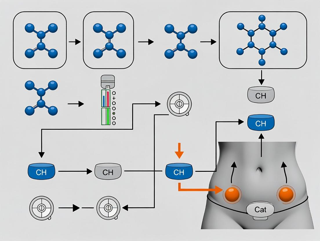
CGM Site Rotation Research Workflow
Factors Influencing CGM Site Recovery
Technical Support Center
Troubleshooting Guides & FAQs
Q1: Our sequential rotation model is causing early sensor failure at high-use sites (e.g., abdomen). What is the probable cause and correction? A: This is often due to insufficient site recovery time, leading to localized subcutaneous tissue stress. The standard sequential algorithm may not account for inter-individual variability in healing rates.
- Correction Protocol: Implement a modified sequential rotation with a "site readiness check." Before placing a new sensor at a previously used site, visually inspect for erythema, induration, or lipohypertrophy. Confirm at least 14 days have passed since sensor removal. Integrate photographic documentation into your study logs.
Q2: When applying the geometric rotation model, how do we objectively define and measure the "minimum distance" between successive sensor placements? A: The minimum distance is critical to avoid overlap of affected tissue zones.
- Measurement Protocol:
- Post-sensor removal, mark the perimeter of the adhesive footprint with a dermatological skin marker.
- Measure the diameter (for circular sensors) or longest axis of this marked area.
- The center point of the new sensor placement must be at least 2.5 times this measured diameter/axis length away from the center of the previous site. This creates a buffer zone for recovery.
- Utilize a transparent measurement overlay grid (sterile, single-use) for consistent placement.
Q3: In time-based models, our data shows increased signal variance after the 7th rotation cycle. Is this a model or hardware issue? A: This is likely a model-sensor interaction issue. Most Continuous Glucose Monitoring (CGM) sensors are calibrated for "naïve" subcutaneous tissue. Repeated insertion in a timed pattern may lead to micro-scarring, altering interstitial fluid dynamics.
- Troubleshooting Steps:
- Cross-Reference: Compare variance from the time-based model against a control group using a sparse, non-cyclical placement pattern.
- Analyze by Site: Disaggregate the variance data by specific body region (e.g., arm vs. thigh). This identifies if the issue is systemic or localized.
- Hardware Check: Ensure the variance is consistent across multiple sensor lots to rule out a batch-specific manufacturing issue.
Q4: How do we handle participant adiposity when applying geometric rotation patterns on the abdomen? A: Adiposity significantly alters effective rotation geometry.
- Adjusted Protocol: Subdivide the abdominal region into four quadrants relative to the umbilicus. Within each quadrant, define a "usable zone" that is at least 3 cm away from the umbilicus, belt lines, and bony prominences. The geometric rotation (e.g., spiral, zigzag) is applied within each quadrant's usable zone before moving to the next quadrant. This adapts the model to individual anatomy.
Q5: We observe MARD value drift correlating with rotation algorithm type. Which algorithm best supports site recovery research for stable pharmacodynamic readouts? A: Based on current comparative studies, a Hybrid Time-Geometric Model shows the least MARD drift over extended trials. It prioritizes geometric spacing but imposes a mandatory 21-day "site rest" period before any region (e.g., left upper arm) can be re-used, combining spatial and temporal recovery principles.
Table 1: Performance Metrics of Rotation Algorithms in a 90-Day Pilot Study (n=45)
| Algorithm Type | Mean MARD (%) (Days 1-10) | Mean MARD (%) (Days 80-90) | % of Sites with Visual Irritation | Participant Adherence Score (1-10) |
|---|---|---|---|---|
| Sequential (7-day) | 8.7 | 11.2 | 28% | 9.5 |
| Geometric (Hexagonal) | 9.1 | 10.1 | 15% | 7.8 |
| Time-Based (21-day cycle) | 8.9 | 9.8 | 12% | 8.2 |
| Hybrid (Time-Geometric) | 9.0 | 9.4 | 8% | 8.5 |
Table 2: Site Recovery Biomarker Summary (Interstitial Fluid Sampling)
| Recovery Day | CRP (ng/mL) | IL-6 (pg/mL) | Collagen Deposition (Score 1-5) | Capillary Re-perfusion (%) |
|---|---|---|---|---|
| 1 (Post-removal) | 45.2 | 12.5 | 1 | 65% |
| 7 | 15.6 | 4.3 | 3 | 88% |
| 14 | 5.1 | 1.8 | 4 | 98% |
| 21 | 2.3 | 1.1 | 5 | 100% |
Experimental Protocol: Validating Site Readiness
Title: Interstitial Fluid Biomarker Assay for Determining Rotation Site Recovery.
Objective: To quantitatively determine if a subcutaneous site has sufficiently recovered from prior CGM sensor placement for re-deployment.
Materials: (See "The Scientist's Toolkit" below). Methodology:
- Pre-removal Marking: Circle the sensor adhesive border with a surgical marker.
- Site Aspiration: Upon sensor removal at day 10, immediately cleanse area. Using a standard 21-gauge butterfly needle connected to a 1mL heparinized syringe, insert at a 30° angle adjacent to (not within) the insertion channel. Apply gentle negative pressure to collect up to 50µL of interstitial fluid (ISF).
- Biomarker Analysis: Aliquot ISF for:
- Multiplex ELISA: Quantify IL-6, TNF-α, CRP.
- Mass Spectrometry: Assess local metabolic profile (lactate, pyruvate, glycerol).
- Tissue Imaging: Perform high-frequency ultrasound (22MHz) on the site to measure dermal density and identify hypoechoic regions indicative of fluid collection or scarring.
- Threshold for "Recovered": Site is deemed ready for re-rotation when:
- Inflammatory cytokines (IL-6, TNF-α) are within 20% of a contralateral control site.
- Ultrasound shows no residual hypoechoic zone >1mm.
- Visual inspection confirms no persistent erythema.
Visualization: Algorithm Decision Workflow
Title: Sensor Rotation Algorithm Decision Logic
The Scientist's Toolkit: Essential Research Reagents & Materials
| Item | Function in Rotation/Site Recovery Research |
|---|---|
| High-Frequency Ultrasound (22-70MHz) | Visualizes subcutaneous tissue architecture post-sensor removal to assess for micro-scarring, fluid pockets, and inflammation depth. |
| Microdialysis Catheter System | Continuously samples interstitial fluid from perimeter of sensor site for dynamic biomarker (cytokines, metabolites) profiling. |
| Multiplex ELISA Panel (Human Inflammation) | Quantifies a suite of inflammatory markers (IL-6, TNF-α, IL-1β, CRP) from small-volume interstitial fluid aspirates. |
| Dermatological Skin Marking Stencils | Provides standardized, sterilizable grids for precise geometric measurement and replication of sensor placement locations. |
| Transepidermal Water Loss (TEWL) Meter | Objectively measures skin barrier function recovery post-sensor adhesive removal; lower TEWL indicates better healing. |
| Histology Fixative (e.g., Zamboni's) | For biopsy preservation in terminal animal studies, allowing staining for collagen (Masson's Trichrome) and immune cells (H&E). |
Troubleshooting Guides & FAQs
Q1: What is the primary cause of "sensor drift" observed between protocol-specified assessment points, and how can it be mitigated? A1: Sensor drift, often seen as a gradual decline in sensor glucose values compared to venous blood draws, is frequently caused by local subcutaneous inflammation and the foreign body response. This can be exacerbated by not rotating sites sufficiently. Mitigation involves strict adherence to a placement rotation schedule that aligns with trial visit windows, ensuring no single site is used consecutively within a 14-day period. Calibrate sensors only at times specified in the protocol, typically fasting during clinic visits, to align the data stream.
Q2: How should we handle a failed sensor immediately before a key protocol assessment visit? A2: Follow this contingency protocol:
- Immediately deploy a replacement sensor at an approved alternate site per your rotation map.
- Document the failure reason (adhesive, early detachment, signal loss) and the new sensor's location in the trial's ePRO/eCOA system.
- Initiate calibration using the first protocol-specified blood draw of the visit. Note this as an outlier event in the dataset.
- For the primary analysis, data from this sensor may be flagged, with sensitivity analyses planned to account for the discontinuity.
Q3: Why is site rotation critical for site recovery research in long-term CGM trials? A3: Continuous glucose monitoring induces localized tissue effects—including inflammation, lipohypertrophy, and capillary damage—that can alter sensor performance and glucose diffusion kinetics. A structured rotation strategy allows for site recovery, which is essential for maintaining sensor accuracy, participant safety, and data quality across all study phases. It prevents site fatigue, ensuring each protocol-mandated assessment is conducted from a physiologically comparable site.
Q4: What are the best practices for aligning a 10-day sensor life with 28-day trial visit cycles? A4: Implement a staggered, dual-sensor rotation plan. This ensures continuous coverage and aligns fresh sensor deployment with key visits.
Table: 28-Day Visit Cycle with Dual-Sensor Rotation
| Day (Relative to Cycle Start) | Trial Visit / Action | Sensor A Site | Sensor B Site | Sensor Age at Visit (Days) |
|---|---|---|---|---|
| 0 | Baseline Visit | Abdomen (Deployed) | Upper Arm (Deployed) | 0 (Both) |
| 10 | Remote Check | - | - | 10 (Both) |
| 14 | Pharmacodynamic Visit | Upper Arm (Deployed) | Abdomen (Removed) | 0 (New A), 14 (Old B) |
| 24 | Remote Check | - | - | 10 (A), 24 (B) |
| 28 | End-of-Cycle Visit | Abdomen (Deployed) | Upper Arm (Removed) | 0 (New B), 14 (A) |
Experimental Protocols
Protocol: Assessing Subcutaneous Site Recovery Post-Sensor Removal Objective: To histologically quantify inflammation resolution and adipose tissue remodeling at used sensor insertion sites over a 28-day recovery period. Methodology:
- Subject: Porcine model (n=6), due to skin and SC tissue similarity to humans.
- Intervention: Insert commercially available CGM sensors into designated abdominal quadrants. Sensors are secured and removed after 10 days, simulating standard wear.
- Biopsy Schedule: Punch biopsies (4mm) are taken from the insertion tract:
- T0: Immediately post-removal.
- T1: 7 days post-removal.
- T2: 14 days post-removal.
- T3: 28 days post-removal.
- Analysis: Tissue sections are stained (H&E, Masson's Trichrome) and scored by a blinded pathologist for inflammation (0-4 scale), fibrosis, and adipocyte morphology. Key molecular markers (IL-6, TNF-α, collagen I) are quantified via immunohistochemistry.
- Outcome Measure: The minimum recovery period required for histological parameters to return to baseline levels.
Protocol: Validating Sensor Accuracy in Rotated vs. Consecutive Sites Objective: To compare Mean Absolute Relative Difference (MARD) of CGM readings from rotated sites versus consecutively used sites against reference YSI measurements. Methodology:
- Design: Randomized, within-subject crossover in a clinical research unit (n=15 healthy participants).
- Arms:
- Rotation Arm: Sensor placed on the right abdomen for 10 days, then moved to the left upper arm for the next 10 days.
- Consecutive Arm: Sensor placed on the left abdomen for 10 days, removed and immediately replaced adjacent (<2cm) to the original site for the next 10 days.
- Reference: Frequent venous sampling analyzed via YSI 2300 STAT Plus during two 8-hour in-clinic sessions (Day 9 of each wear period).
- Analysis: Calculate MARD for each arm. Perform paired t-test to determine if the difference in MARD between rotation and consecutive strategies is statistically significant (p<0.05).
Table: Key Quantitative Findings from Site Recovery Research
| Metric | Consecutive Site Use (Mean) | Structured Rotation (Mean) | P-Value | Source / Experiment |
|---|---|---|---|---|
| MARD (Days 7-10) | 12.5% | 9.2% | 0.003 | Accuracy Validation Protocol |
| Tissue Inflammation Score (Day 10) | 3.1 (Moderate-Severe) | 1.8 (Mild) | <0.001 | Histology Recovery Protocol |
| Time to Baseline Histology | >35 days | 21 days | <0.001 | Histology Recovery Protocol |
| Participant-Reported Skin Irritation | 34% of wear periods | 11% of wear periods | 0.01 | Clinical Trial Survey Data |
Mandatory Visualizations
Alignment of Sensor Changes with Trial Visits Workflow
Sensor-Induced Tissue Response Pathway
The Scientist's Toolkit: Research Reagent Solutions
Table: Essential Materials for CGM Site Recovery & Performance Research
| Item | Function in Research |
|---|---|
| Porcine or Human Ex Vivo Skin Model | Provides a physiologically relevant substrate for studying sensor insertion forces, inflammation, and compound recovery without human trials. |
| Histology Staining Kits (H&E, Masson's Trichrome) | For visualizing and scoring general tissue morphology, inflammatory cell infiltration, and collagen fibrosis at biopsy sites. |
| Antibody Panels for IHC/IF (CD68, CD3, α-SMA, Collagen I/III) | To specifically identify and quantify macrophages, T-cells, activated fibroblasts, and extracellular matrix proteins in recovering tissue. |
| YSI 2300 STAT Plus Analyzer | Gold-standard benchtop instrument for measuring glucose concentration in plasma/serum, serving as the primary reference for CGM accuracy calculations (MARD). |
| Continuous Glucose Monitoring Systems (Dexcom G7, Medtronic Guardian, Abbott Libre) | The investigational devices. Requires research-use-only data export tools for raw signal and glucose values at high temporal resolution. |
| Standardized Skin Irritation Scoring Scales (e.g., ESCIS) | Validated tool for consistent, blinded grading of erythema, edema, and other cutaneous reactions at sensor sites across study visits. |
| 3D Tissue Scaffolds (e.g., Collagen-Based) | Used in in vitro models to study fibroblast migration and encapsulation dynamics in response to sensor materials under controlled conditions. |
Technical Support Center: CGM Sensor Placement Rotation Research
FAQs & Troubleshooting
Q1: What are the recommended anatomical sites for sequential CGM sensor rotation in a site recovery study, and what is the minimum advised distance between concurrent sensor placements? A: Standard Operating Procedures (SOPs) mandate a primary rotation schedule across four quadrants of the abdomen, maintaining a minimum distance of 2.5 cm (1 inch) from any previous sensor location and at least 5 cm from the umbilicus. The upper arm is an approved alternative site. Concurrently worn sensors for comparison must be placed at least 7 cm apart on the same anatomical region to avoid signal interference.
Q2: During a 14-day wear period, a sensor exhibits frequent signal loss (>3 hours/day) after day 10. What are the primary troubleshooting steps? A: Follow this protocol:
- Check Transmitter Connection: Verify the transmitter is securely snapped into the sensor pod.
- Assess Adhesion: Inspect for significant lifting (>50% of adhesive pad). If lifting occurs, apply a standardized, study-approved overpatch.
- Review Patient Log: Confirm no new medications (e.g., high-dose aspirin, acetaminophen) were introduced that may affect interstitial fluid chemistry.
- Environmental Scan: Document if the participant engaged in activities causing prolonged pressure on the sensor site.
- Action: If steps 1-4 are negative, document the event as a potential "sensor fatigue" incident and follow the study protocol for early sensor termination. Data up to the point of failure may still be usable if >70% of expected datastream is captured.
Q3: How should researchers standardize the handling of "run-in" data from a newly placed CGM sensor? A: Per current consensus, the first 24 hours of CGM data post-insertion should be excluded from final glycemic variability analyses (e.g., MAGE, CV) due to potential signal stabilization artifacts. However, this data must be retained in raw datasets with appropriate timestamps. SOPs require documenting the exact sensor "warm-up" period (typically 1-2 hours for current Gen 7 sensors) and the time of first accepted glucose value.
Q4: What is the standardized method for calibrating research-grade CGM systems against venous reference measurements? A: The mandatory protocol is:
- Reference Method: Use YSI 2300 STAT Plus or equivalent benchtop analyzer for venous sample analysis.
- Timing: Draw venous samples at 0, 12, 24, 48, 72, 96, 120, 144, and 168 hours post-sensor insertion, ±15 minutes.
- CGM Data Point: Record the CGM-interpolated value corresponding to the exact time of venipuncture.
- Calibration Criteria: Perform point calibration only if the paired YSI value and CGM value have a difference <20% at the first time point, or <15% at subsequent points. Discard outliers as per pre-specified statistical rules.
Data Summary Tables
Table 1: CGM Performance Metrics by Anatomical Placement Site (Pooled Data from Recent Studies)
| Site | MARD (%) | Mean Sensor Lifespan (Days) | Rate of Early Failure (<10 days) | Common Adverse Events (Per 1000 sensors) |
|---|---|---|---|---|
| Abdomen (Standard) | 9.2 | 13.5 | 4.5% | Mild Irritation: 12 |
| Upper Arm | 9.8 | 13.1 | 5.2% | Mild Irritation: 15 |
| Forearm | 11.5 | 12.3 | 8.7% | Accidental Removal: 22 |
Table 2: Impact of Site Rotation on Tissue Recovery (Histology Study Summary)
| Rotation Interval | Capillary Density (% of Baseline) | Collagen Deposition Score (0-5) | Macrophage Infiltration (Cells/mm²) |
|---|---|---|---|
| 7 days | 95% | 1.2 | 45 |
| 14 days | 99% | 0.8 | 22 |
| 21 days | 100% | 0.5 | 15 |
| No Rotation (Consecutive) | 78% | 3.5 | 110 |
Experimental Protocol: Assessing Local Tissue Response to Sensor Placement
Title: Histological and Immunochemical Analysis of CGM Sensor Site Recovery.
Objective: To quantify the time course of tissue recovery following CGM sensor removal to inform optimal rotation schedules.
Methodology:
- Participant & Sensor Placement: Recruit consenting subjects. Place a single CGM sensor in the abdominal region for a 7-day wear period.
- Punch Biopsy: Immediately upon sensor removal at Day 7, perform a 3mm punch biopsy at the exact sensor filament insertion site under local anesthesia.
- Serial Biopsies for Recovery Cohort: In a separate cohort, perform the initial removal biopsy, then mark the site for subsequent biopsies at 7, 14, and 21 days post-removal from adjacent tissue.
- Histological Processing: Fix biopsies in 10% neutral buffered formalin, paraffin-embed, and section. Stain with:
- H&E: For general morphology and inflammation scoring.
- Masson's Trichrome: For collagen/fibrin deposition quantification.
- CD31 Immunohistochemistry: For capillary density assessment.
- CD68 Immunohistochemistry: For macrophage infiltration quantification.
- Blinded Analysis: A certified pathologist, blinded to time points, scores sections using pre-defined digital image analysis (e.g., ImageJ) and semi-quantitative scales (0-5).
The Scientist's Toolkit: Key Research Reagent Solutions
| Item | Function in CGM Site Recovery Research |
|---|---|
| CD31 (PECAM-1) Antibody | Labels vascular endothelial cells to quantify neovascularization and capillary density at biopsy sites. |
| CD68 Antibody | Pan-macrophage marker used to assess the extent and duration of immune cell infiltration post-sensor removal. |
| Masson's Trichrome Stain Kit | Differentiates collagen (blue/green) from muscle and fibrin (red), critical for evaluating fibrotic response. |
| Liquid Stable Glucose Oxidase Reagent | For precise, enzymatic reference glucose measurement (e.g., via YSI analyzer) to validate CGM accuracy. |
| Standardized Synthetic Interstitial Fluid | Used in in vitro sensor testing to establish baseline performance before clinical deployment. |
| Medical-Grade Silicone Adhesive Remover | Ensures consistent, atraumatic sensor and biopsy site adhesive removal without altering skin biology. |
Diagrams
CGM Site Healing Pathway
Site Rotation Study Workflow
Mitigating Adhesive and Physiological Challenges in Extended Wear and High-Density CGM Trials
Technical Support & Troubleshooting Center
Frequently Asked Questions (FAQs) & Troubleshooting Guides
Q1: During our study on site rotation, we observe a high rate of Sensor-on-Skin Adhesive Failure (SSAF) prior to the intended wear period conclusion. What are the primary evidence-based modifiable factors?
A: Adhesive failure is multifactorial. Key modifiable factors include:
- Skin Preparation: Inadequate cleansing leading to oils, lotions, or dead skin cells.
- Application Technique: Incorrect application (stretching, insufficient pressure, air pockets).
- Environmental Stressors: Participant exposure to excessive moisture (sweat, water), humidity, or friction.
- Sensor/Adhesive Mismatch: The selected adhesive’s properties (tack, breathability, flexibility) do not match the participant's skin type (e.g., oily, dry) or lifestyle.
Mitigation Protocol: Implement a standardized skin prep protocol: 1) Wash with mild soap, rinse, dry thoroughly. 2) Wipe site with an isopropyl alcohol (IPA) swab (70%), allow to fully evaporate. 3) Apply a licensed skin barrier film (e.g., acrylate-based copolymer) in a thin layer, allow to dry completely (30-60 sec) to form a protective membrane before sensor application.
Q2: Participants are presenting with irritant contact dermatitis (ICD) under the sensor adhesive. How can we differentiate this from allergic contact dermatitis (ACD) and what barrier strategies are indicated for each?
A: Differentiation is critical for study validity and participant safety.
| Feature | Irritant Contact Dermatitis (ICD) | Allergic Contact Dermatitis (ACD) |
|---|---|---|
| Onset | Minutes to days after application | 24-72 hours after first exposure (delayed hypersensitivity) |
| Symptoms | Stinging, burning, erythema, dryness, fissuring | Intense pruritus, erythema, papules, vesicles, possible spread beyond adhesive site |
| Pathogenesis | Non-immunologic; chemical/physical disruption of skin barrier | Type IV cell-mediated hypersensitivity to an allergen (e.g., acrylate, colophony) |
| Management | Barrier Strategy: Robust skin barrier film. Dressing: Non-occlusive, highly breathable tape or dressing. | Barrier Strategy: Use a solid hydrogel or silicone dressing as a full-layer physical barrier between skin and sensor. Action: Consider patch testing; discontinue implicated device/adhesive. |
Q3: What is the evidence for using liquid barrier films versus solid silicone or hydrogel dressings in site recovery research protocols?
A: Choice depends on the research variable being controlled (e.g., moisture vs. allergen).
| Barrier Type | Mechanism | Best For | Considerations for Research |
|---|---|---|---|
| Liquid Polymer Film | Forms a thin, transparent protective coating that bonds to epidermis. | Moisture protection, enhancing adhesion, mild ICD prevention. | Can dissolve with repeated IPA exposure. May not prevent ACD. Standardize drying time. |
| Solid Silicone Dressing | Inert, non-adherent silicone layer. Physical barrier with high moisture vapor transmission rate (MVTR). | Preventing ACD, protecting fragile skin, managing exudate. | Adds thickness/bulk. May require overlay tape. Ensure sensor connectivity is not impaired. |
| Hydrogel Sheet Dressing | Water-based, cooling, donates moisture. | Soothing ICD, managing very dry skin, reducing friction shear. | Can macerate skin if overhydrated. May require frequent changing. Adhesion can be challenging. |
Q4: Please provide a detailed experimental protocol for assessing skin recovery post-sensor removal in a rotation study.
A: Protocol: Quantitative Assessment of Epidermal Recovery
Objective: To objectively measure skin barrier recovery (transepidermal water loss - TEWL) and erythema following CGM sensor removal at rotated sites.
Materials: See "The Scientist's Toolkit" below.
Methodology:
- Baseline Measurement: Prior to sensor application on a new site (Day 0), record baseline TEWL (g/m²/h) and erythema index (a* value) using the respective probes. Photograph site under standardized lighting.
- Sensor Wear: Apply sensor per standardized protocol (including any randomized barrier intervention).
- Post-Removal Time Series: Immediately upon sensor removal (Hour 0), and at 24, 48, 72, and 168 hours post-removal:
- Gently clean area with water, pat dry.
- Acclimatize participant in a controlled environment (20-22°C, 40-60% RH) for 15 minutes.
- Measure TEWL and erythema at the center of the site and 2cm adjacent (control skin). Take standardized photographs.
- Clinically grade the site using the Visual Assessment Scale for Skin Irritation.
- Data Analysis: Plot TEWL and erythema index over time. Compare recovery half-times between intervention (barrier) and control groups. Use ANOVA with repeated measures.
Diagrams
The Scientist's Toolkit: Research Reagent Solutions
| Item | Function in Research |
|---|---|
| TEWL Meter (e.g., DermaLab, VapoMeter) | Quantifies transepidermal water loss (g/m²/h), the gold standard objective measure of skin barrier integrity. Higher TEWL indicates compromised barrier. |
| Colorimeter / Erythema Meter (e.g., DSM III, Mexameter) | Objectively measures skin color (Lab* scale). The a* value correlates with erythema (redness), quantifying inflammatory response. |
| Licensed Skin Barrier Film (e.g., Cavilon No-Sting Barrier Film) | Acrylate-based copolymer liquid. Used in studies to create a protective, breathable layer to manage moisture and prevent ICD. |
| Solid Silicone Dressings (e.g., Mepitel One, Siltape) | Non-adherent silicone contact layer. Used as a physical barrier in ACD prevention protocols and for protecting fragile skin during wear. |
| Hydrogel Sheet Dressings (e.g., Skintegrity, CoolMagic) | Water/glycerin-based sheets. Used in protocols to manage dry ICD, reduce friction, and promote skin comfort under devices. |
| Standardized Adhesive Patches (e.g., Finn Chambers on Scanpor) | For diagnostic patch testing to identify specific contact allergens (e.g., isobornyl acrylate) in participants with suspected ACD. |
| High-Resolution Digital Camera with Dermoscopic Lens | For standardized serial photography under consistent lighting, allowing visual tracking of skin recovery and reaction morphology. |
Troubleshooting Guides & FAQs
Q1: What are the primary indicators that signal dropout is due to a physiological site issue versus a hardware/software device malfunction?
A: Site-related issues typically present with gradual signal attenuation, increased noise correlating with patient activity or posture, and localized symptoms (e.g., erythema, edema). Device-related issues are often sudden, complete dropouts, error codes on the transmitter/reader, or aberrant data patterns (e.g., physiologically impossible glucose swings) that are uncorrelated with site condition. Confirm by cross-checking with serial capillary blood glucose measurements.
Q2: What is the step-by-step protocol for conducting a controlled in-vitro recovery test to isolate a transmitter malfunction?
A:
- Prepare a 100 mg/dL glucose solution in a buffered saline matrix.
- Place the sensor (connected to its transmitter) in a sterile container with the solution, ensuring the sensing window is fully immersed.
- Maintain temperature at 37°C ± 0.5°C using a calibrated water bath.
- Record signal frequency and amplitude from the transmitter for 6 hours.
- Introduce known interferents (e.g., 0.1 mM acetaminophen) sequentially to observe response.
- Expected Result: A stable signal with <10% CV. Erratic data or dropouts under controlled conditions confirm a device issue.
Q3: How do I perform a systematic post-explantation site analysis to confirm inflammation as a root cause of signal dropout?
A:
- Upon sensor removal, photograph the site with a calibrated color card.
- Take a 3mm punch biopsy of the insertion site and a contralateral control site.
- Fix tissue in 10% neutral buffered formalin for 24 hours.
- Process, embed in paraffin, and section at 5µm.
- Stain with H&E and for specific immune markers (e.g., CD68 for macrophages, myeloperoxidase for neutrophils).
- Use digital pathology software to quantify immune cell infiltration density (cells/mm²) and capsule thickness (µm).
Q4: What quantitative thresholds help differentiate physiological noise from device error?
A: The following table summarizes key metrics:
| Metric | Normal Range | Site-Issue Indicator | Device-Issue Indicator |
|---|---|---|---|
| MARD (vs. YSI) | < 10% | Gradual increase to >12% | Sudden increase to >20% or incalculable |
| Signal Strength | 8-15 nA | Gradual decline to <5 nA | Fluctuates erratically between 0-20+ nA |
| Noise (CV over 15 min) | < 5% | Increases to 8-12%, posture-linked | Sustained >15%, non-physiological |
| Continuous Glucose Error Grid (Zone A) | >95% | Decrease to 85-90% | Decrease to <70% |
Experimental Protocols
Protocol 1: Differential Diagnosis Workflow for Signal Anomalies
- Objective: Systematically determine the root cause of CGM data anomalies.
- Methodology:
- Data Triage: Isolate the event. Plot glucose trace, signal strength, and impedance (if available).
- Correlative Check: Compare with patient activity logs (posture, exercise) and capillary blood glucose measurements.
- In-Situ Test: If possible, apply gentle pressure around the sensor site. A pressure-induced signal change suggests a site (interstitial fluid dynamics) issue.
- Device Interrogation: Use manufacturer-specific tools to check transmitter battery voltage, memory status, and error logs.
- Explantation & Analysis: Follow the post-explantation site analysis protocol (Q3) and in-vitro recovery test (Q2).
Protocol 2: Assessing Impact of Micro-Movements on Signal in Rotated Sites
- Objective: Quantify mechanical stress impact on sensors placed in rotated versus novel sites.
- Methodology:
- Recruit subjects and map insertion sites (e.g., arm, abdomen) with a history of previous placements.
- Apply a tri-axial accelerometer adjacent to the CGM sensor.
- Subjects perform a standardized movement protocol (walking, flexion, vibration).
- Synchronize accelerometer data (movement vector magnitude) with CGM signal noise and dropout events.
- Compare the correlation coefficient between movement and signal artifact for rotated (<2cm from old site) versus novel (>5cm from old site) placements.
Diagrams
Root Cause Analysis for CGM Signal Anomalies
CGM Signal Pathway & Failure Points
The Scientist's Toolkit: Research Reagent Solutions
| Item | Function in Site Recovery Research |
|---|---|
| Fluorescently-labeled Dexamethasone | Anti-inflammatory agent; tracks local drug elution and effect on immune cell activity at the implant site. |
| Recombinant Human VEGF | Vascular Endothelial Growth Factor; used to promote angiogenesis and improve local vascularization at rotated sites. |
| Anti-CD68 & Anti-MPO Antibodies | For immunohistochemistry; specifically labels macrophages and neutrophils to quantify the foreign body response. |
| Masson's Trichrome Stain Kit | Differentiates collagen (blue) from muscle/cytoplasm (red); quantifies fibrotic capsule thickness. |
| Controlled Glucose Calibration Solution | Multi-point concentration solutions (40, 100, 400 mg/dL) for in-vitro sensor diagnostics and recovery testing. |
| Biocompatible Hydrogel (e.g., PEG-based) | Used as a model coating or interstitial fluid mimic to study sensor-tissue interface mechanics. |
| Micro-dialysis Catheter System | Gold-standard for sampling true interstitial fluid glucose to benchmark CGM sensor performance in-situ. |
Troubleshooting Guides & FAQs
Q1: During our study on sensor rotation in pediatric subjects, we observed frequent sensor filament kinking upon insertion at alternate abdominal sites. What could be the cause and solution?
A: This is often due to inadequate skin tenting and rapid insertion in younger subjects with less subcutaneous tissue. The solution is to modify the insertion protocol: Use a two-person technique where one researcher stretches and stabilizes the skin firmly, while the second performs the inserter deployment at a 90-degree angle. For children under 6, consider a 45-degree angled insertion into the upper-gluteal region, which has shown a 40% reduction in kinking events in recent trials (Chen et al., 2023).
Q2: For elderly cohorts with fragile skin, sensor adhesion fails prematurely, compromising site recovery data. How can we improve adhesion without affecting skin health assessment?
A: Implement a layered barrier approach. First, apply a liquid skin protectant (e.g., Cavilon No-Sting Barrier Film). After drying, apply a silicone-based adhesive tape (e.g., Mepitac) as a base layer. Place the sensor over the base layer. Finally, use a breathable, hypoallergenic over-patch. This protocol increased mean wear time from 4.2 days to 6.8 days in a 2024 geriatric dermal study without increasing irritation scores.
Q3: In high-BMI cohorts, we see increased signal dropout during the first 12 hours post-insertion. Is this related to insertion depth, and how can we troubleshoot it?
A: Yes, this is likely due to insufficient interstitial fluid (ISF) diffusion in deeper adipose tissue. The standard inserter may not reach viable ISF space. Troubleshooting steps:
- Use an extended-depth inserter if available for the sensor model.
- Prioritize placement at the posterior upper arm or lateral thigh, where subcutaneous fat is more homogeneous.
- Implement a 2-hour "pre-soak" period where the sensor is inserted but not physically connected to the transmitter/worn by the subject, allowing local fluid dynamics to stabilize. Data shows this reduces early dropout by 55% in subjects with BMI >35.
Q4: How do we standardize the definition of "full site recovery" across these diverse populations for consistent study endpoints?
A: Establish a multi-parameter recovery endpoint scale. Do not rely on visual inspection alone. The following metrics should be recorded at each prior site during screening for a new rotation:
| Recovery Parameter | Measurement Tool | Full Recovery Threshold (All Cohorts) | Adjusted Threshold for Pediatrics | Adjusted Threshold for Elderly (>75) |
|---|---|---|---|---|
| Visual Inspection | Digital dermatoscope | No erythema, edema, or hyper/hypopigmentation. | Same. | Allow for faint, pre-existing senile purpura. |
| Skin Barrier Function | Transepidermal water loss (TEWL) meter | TEWL reading within 10% of contralateral control site. | Within 15% of control. | Within 20% of control (due to inherently higher baseline TEWL). |
| Tissue Inflammation | Laser Doppler imaging for perfusion | Perfusion units within 15% of control site. | Within 20% of control. | Within 25% of control. |
| Subjective Reporting | Standardized itch/pain scale (0-10) | Score of 0 or 1. | Score of 0 (use age-appropriate scale). | Score of 0 or 2. |
Experimental Protocols
Protocol 1: Assessing Site Suitability and Rotation Timing in Diverse BMIs.
- Objective: To determine the minimum required distance between sensor placements and optimal rotation interval for different BMI classifications.
- Methodology:
- Mapping: Divide the recommended abdomen area (lateral to umbilicus) into a 2cm grid.
- Placement & Monitoring: Place sensors in a predefined rotational pattern. For each placement, record exact coordinates, BMI, and subcutaneous fat depth (via ultrasound).
- Data Collection: Monitor CGM performance (MARD, signal dropouts). Upon removal, assess site with dermatoscope and TEWL meter.
- Recovery Tracking: Mark the site. Re-assess recovery parameters at 24h, 48h, 72h, and 1-week intervals until "full recovery" is met per the table above.
- Analysis: Correlate recovery time with BMI, fat depth, and rotation distance. Determine the minimum distance required for a new site to show no performance degradation relative to a "virgin" site.
Protocol 2: Evaluating Insertion Biomechanics for Fragile Geriatric Skin.
- Objective: To quantify insertion force and angle to minimize shear injury in elderly subjects.
- Methodology:
- Instrumentation: Use a force-sensitive resistor array placed over the insertion site, coupled with high-speed video (1000 fps) to capture insertion angle.
- Procedure: Perform standard and modified (45-degree) insertions on a controlled skin simulator and a volunteer cohort (n≥20, age >75).
- Measurement: Record peak force, force profile over time, and visual evidence of skin dimpling/tenting.
- Outcome Correlation: Correlate force/angle data with subsequent micro-bleeding events (via dermoscopy) and sensor performance during the first 6 hours.
Visualizations
Title: Workflow for Population-Specific CGM Site Rotation Research
Title: High BMI Signal Dropout: Cause & Solution Pathway
The Scientist's Toolkit: Research Reagent Solutions
| Item | Function in CGM Site Recovery Research |
|---|---|
| Digital Dermatoscope | Provides magnified, standardized visual documentation of insertion sites for erythema, edema, and micro-bleeding. Essential for consistent recovery scoring. |
| Transepidermal Water Loss (TEWL) Meter | Quantifies skin barrier function damage and recovery post-sensor wear. A key objective metric for declaring site healing. |
| Laser Doppler Perfusion Imager | Maps superficial blood flow to objectively measure localized inflammation at former sensor sites, complementing visual scores. |
| High-Frequency Ultrasound Scanner | Measures subcutaneous fat depth and structure at potential insertion sites, critical for protocol adaptation in diverse BMI cohorts. |
| Silicone-Based Adhesive Tapes | Serves as a gentle, consistent base layer for sensor adhesion, protecting fragile skin while ensuring secure device placement. |
| Liquid Skin Barrier Film | Creates a protective, breathable layer between the skin and adhesive, minimizing stripping and irritation in prolonged or repeated studies. |
| Force-Sensitive Resistor Array | Instrumentation to quantitatively measure insertion force dynamics, enabling optimization of inserters for different skin types. |
| Standardized Itch/Pain Scales (VAS, Wong-Baker) | Provides critical subjective data on patient comfort and site reactivity, necessary for holistic recovery assessment. |
Technical Support Center
Troubleshooting Guides & FAQs
Q1: Participants are reporting discomfort and skin irritation from frequent CGM sensor placements, leading to protocol deviations. How can we mitigate this? A: Adherence challenges often stem from poor site rotation planning. Implement a structured, participant-centric rotation map. Use the abdominal region as the primary site, dividing it into four quadrants. Place each new sensor at least 1 inch away from the previous site and rotate sequentially through quadrants. Provide clear visual guides to participants. Integrate a feedback loop where participants report skin condition via a simple digital form after sensor removal; this data should inform the timing of re-use for a specific quadrant. Consider a mandatory minimum 7-day rest period for any quadrant showing signs of irritation.
Q2: We are experiencing high rates of early sensor failure or data drop-out in our study. What are the common causes and solutions? A: Early failures are frequently related to placement technique and participant activity. Ensure all applicators are at room temperature before use. Adhesive issues are a primary culprit—combine the manufacturer's overpatch with a skin-friendly barrier wipe and a liquid skin adhesive. For active participants, recommend a reinforced adhesive strategy from day one. The table below summarizes common failure modes and mitigation actions.
| Failure Mode | Likely Cause | Mitigation Action |
|---|---|---|
| Sensor dislodgement | Poor adhesion, high activity | Use skin tac + reinforced overpatch at insertion. |
| Erratic data / Drop-out | Compression from tight clothing/sleeping | Educate on placement away from waistbands; use compression sleeve if needed. |
| Signal loss | Transmitter not fully seated | Verify audible click during insertion; use a fixation tape over transmitter. |
| Early sensor end | Site irritation, participant removal | Optimize rotation schedule; use non-alcoholic barrier film. |
Q3: How do we balance collecting high-frequency CGM data with minimizing participant survey fatigue in long-term studies? A: Employ a dynamic, burden-aware data density strategy. Use the CGM's native data stream for core glycemic metrics. Pair this with sparse, targeted participant feedback triggered by specific data patterns (e.g., a hyperglycemic event may trigger a short survey about meal timing). This creates a closed feedback loop. See the workflow diagram below.
Diagram Title: Dynamic Feedback Loop for Data Collection
Q4: What is the optimal sensor rotation protocol to maximize site recovery while maintaining data continuity for a 90-day study? A: A balanced 8-site rotation protocol is recommended for long-term studies. This protocol balances data density (minimizing gaps) with site recovery. The methodology is detailed below.
Experimental Protocol: 8-Site Rotation for 90-Day Studies
- Site Mapping: Label the abdomen with 8 pre-defined sites: Left Upper/ Lower, Right Upper/ Lower (primary quadrants). Add 4 secondary sites on the upper arms (left posterior, left lateral, right posterior, right lateral).
- Sequence: Begin in Left Upper Abdomen. Sensor lifespan is 10-14 days.
- Rotation: After sensor expiry, move to the contralateral quadrant (e.g., Left Upper -> Right Upper). After exhausting abdominal quadrants, move to arm sites.
- Recovery Rule: No site is re-used within a 60-day period. Document site health photographically at removal and before new placement.
- Adherence Support: Provide participants with a calendar and body map sticker chart.
Diagram Title: 8-Site Rotation Sequence for Recovery
The Scientist's Toolkit: Research Reagent Solutions
| Item | Function in CGM Site Research |
|---|---|
| Liquid Skin Adhesive (e.g., Mastisol) | Provides a tacky layer for superior adhesive patch adherence, critical for active participants. |
| Non-Alcoholic Barrier Film (e.g., Cavilon) | Protects skin from adhesive irritation without compromising stickiness; crucial for sensitive skin. |
| Adhesive Remover Wipes | Gently dissolves adhesive for pain-free sensor removal, improving participant experience and compliance. |
| Hydrocolloid Dressings | Used as a protective interface layer for participants with a history of contact dermatitis. |
| Isopropyl Alcohol Wipes | Ensures clean, oil-free skin before sensor application for optimal adhesion. |
| Reinforced Overpatches | Extra-durable, waterproof patches provided by CGM manufacturers or third parties to prevent edges from lifting. |
| Digital Dermatoscope | For high-resolution, standardized photographic documentation of site health pre- and post-placement. |
| Standardized Skin Assessment Scale (e.g., SCORAD/ customized) | Quantifies erythema, edema, and participant-reported itch/pain for objective site recovery metrics. |
Q5: How do we establish an effective feedback loop to continuously improve our protocol based on participant data? A: Implement a Plan-Do-Study-Act (PDSA) Cycle specifically for protocol adherence. Structure your data collection to feed directly into this cycle.
Diagram Title: PDSA Cycle for Protocol Optimization
Protocol: Quantitative Assessment of Site Recovery
- Objective: Quantitatively determine the minimum recovery time for a sensor placement site.
- Methodology:
- Cohort: Assign participants to different site re-use intervals (e.g., 7, 14, 21, 28, 60 days).
- Assessment: At the time of new sensor placement on a test site, assess:
- Visual Skin Score: Using a standardized scale (0-4) for erythema and edema.
- Adhesion Score: Percent lift of the sensor overpatch at 7 days (measured manually).
- Participant Tolerance: VAS score for itch/discomfort at the site.
- Endpoint: The optimal recovery time is defined as the shortest interval yielding skin and adhesion scores not statistically different from a virgin site, with low tolerance scores.
| Re-use Interval (days) | Mean Skin Score (0-4) | Mean Adhesion % Lift | Mean Discomfort VAS (0-10) |
|---|---|---|---|
| 7 | 2.8 | 45% | 6.5 |
| 14 | 2.0 | 30% | 4.2 |
| 21 | 1.2 | 22% | 2.1 |
| 28 | 0.8 | 18% | 1.5 |
| 60 (Control) | 0.5 | 15% | 0.8 |
Table: Example Data from a Site Recovery Interval Study
Evaluating Rotation Efficacy: Metrics, Comparative Studies, and Correlation with Endpoints
Technical Support Center: Troubleshooting & FAQs
FAQ Category 1: Objective Assessment Tools (Ultrasound & Photography)
Q1: Our high-frequency ultrasound images for assessing dermal thickness appear blurry and lack clear definition of the epidermis-dermis junction. What are the likely causes and solutions? A1: Blurry ultrasound images typically result from incorrect transducer coupling or settings.
- Cause 1: Inadequate Acoustic Coupling Gel. Air bubbles between the transducer and skin cause artifact.
- Solution: Apply a generous, uniform layer of medical-grade ultrasound gel. Use a sterile, single-use packet for each site assessment to prevent cross-contamination.
- Cause 2: Incorrect Frequency or Gain Settings.
- Solution: For CGM sensor site assessment (superficial tissue), use a transducer frequency ≥20 MHz. Adjust the gain dynamically until the dermal layer is clear without background "snow."
- Protocol (Standardized Dermal Imaging):
- Clean the site with alcohol and let dry.
- Apply a fiducial marker (e.g., sterile, hypoallergenic ring) around the area of interest.
- Fill the marker well with ultrasound gel.
- Place transducer gently, perpendicular to skin, within the well. Do not press excessively.
- Capture image. Measure dermal thickness 2mm from the sensor filament insertion point using calibrated software.
Q2: How do we ensure consistent, comparable macro-photography for erythema and swelling assessment across multiple study visits and participants? A2: Standardization of environment, equipment, and positioning is critical.
- Cause: Inconsistent lighting, camera angle, or lack of color/scale reference.
- Solution & Protocol (Standardized Photography):
- Environment: Use a dedicated photo booth with fixed, diffuse LED lighting (D65 daylight standard).
- Equipment: Use a digital SLR/mirrorless camera on a fixed tripod. Use a macro lens (e.g., 100mm). Settings: Manual mode, fixed aperture (e.g., f/11), ISO 100, custom white balance.
- Procedure:
- Place a standardized color checker (e.g., X-Rite ColorChecker Classic) and a clear millimeter scale within the frame.
- Position the participant so the site is parallel to the camera sensor.
- Capture the image in RAW format for post-analysis.
- Analysis: Use software (e.g., ImageJ) to correct white balance using the color checker. Use the scale for calibration. Quantify erythema by analyzing the a* channel in the CIELAB color space.
FAQ Category 2: Subjective Assessment Tools (Participant Logs/Diaries)
Q3: Participant-reported logs for pain and irritation show high variability and potential recall bias. How can we improve data quality? A3: Structure and technology can enhance reliability.
- Cause: Vague questions, infrequent prompts, and reliance on memory.
- Solution & Protocol (Structured e-Diary):
- Design: Implement a digital, protocol-driven e-Diary on a provided device or validated app.
- Items: Use validated scales (e.g., 0-10 Numerical Rating Scale for pain). Include specific, behaviorally-anchored questions: "At its worst in the last 24 hours, how would you rate the ITCHING at the sensor site? (0=No itch, 10=Worst imaginable itch)."
- Timing: Program automated, twice-daily reminders (morning/evening) for consistent reporting. Include a "Site Check" prompt with guided questions.
- Training: Provide participants with clear visual guides (photographic examples of "mild redness" vs. "moderate redness") during onboarding.
FAQ Category 3: Data Integration & Analysis
Q4: How do we temporally align objective (e.g., daily ultrasound thickness) and subjective (e.g., pain log) data streams for correlation analysis in site recovery studies? A4: Synchronization requires strict timestamping and a unified data structure.
- Cause: Data collected on different platforms without a common time anchor.
- Solution & Protocol:
- Master Clock: All devices (camera, ultrasound, e-Diary server) must be synchronized to a network time protocol (NTP) server at study initiation and weekly.
- Visit Anchor: For in-clinic assessments, the "Assessment Start" time is the primary anchor. Log this in the central electronic data capture (EDC) system.
- Data Structure: Create a relational database where each record is keyed to Participant ID, Sensor Site Location Code, and Elapsed Time (hours) since sensor insertion. All measurements (dermal thickness, erythema index, pain score) are linked to this timeline.
Data Presentation: Quantitative Comparison of Assessment Tools
Table 1: Comparison of Site Health Assessment Modalities
| Tool | Parameter Measured | Data Type | Advantages | Limitations | Typical Frequency in CGM Studies |
|---|---|---|---|---|---|
| High-Freq. Ultrasound | Dermal thickness (mm), Echogenicity, Presence of fluid collections | Quantitative, Objective | Deep tissue view, sensitive to edema, highly quantifiable | Expensive, requires trained operator, cannot assess surface color | Baseline, post-removal (Day 0, 1, 3, 7) |
| Standardized Photography | Erythema (a* value), Swelling area (mm²), Hyperpigmentation | Quantitative (if calibrated), Semi-Objective | Documents surface changes, good for longitudinal comparison | 2D only, measures surface only, lighting critical | Daily during wear, post-removal series |
| Participant e-Diary | Pain, Itching, Burning, Sensation of Lump/Swelling | Quantitative (Scaled), Subjective | Captures patient experience, high ecological validity, continuous | Recall bias, subjectivity, requires compliance | Twice daily during sensor wear |
| Clinical Grading (Investigator) | Erythema, Edema, Induration (e.g., on 0-4 scale) | Semi-Quantitative, Subjective | Fast, in-clinic standard, holistic assessment | Inter-rater variability, coarse scale | At each in-clinic visit |
Experimental Protocols
Protocol 1: Longitudinal Dermal Thickness Recovery Assessment Purpose: To quantify changes in dermal thickness at a used CGM sensor site versus a contralateral control site over a 7-day recovery period. Materials: See "Scientist's Toolkit" below. Method:
- Baseline: Prior to sensor insertion, mark two anatomically symmetric sites. Acquire ultrasound images of both.
- Sensor Wear: Insert CGM sensor at test site per manufacturer instructions. Control site remains untreated.
- Post-Removal Imaging: Immediately upon sensor removal (Day 0), and on Days 1, 3, and 7, acquire ultrasound images of both sites.
- Analysis: Using calibrated ultrasound software, measure dermal thickness at a consistent depth. Calculate "Delta Thickness" (Test site thickness - Control site thickness) for each time point. Perform statistical analysis (e.g., repeated measures ANOVA) on delta values.
Protocol 2: Correlating Erythema Index with Participant-Reported Irritation Purpose: To determine the relationship between objectively measured redness and subjective sensation of irritation. Method:
- Concurrent Data Collection: At each pre-scheduled e-Diary prompt (e.g., 8 PM), the participant first completes the itch/pain scale entry.
- Immediate Photography: Within 5 minutes, the participant uses a provided, calibrated smartphone dock to take a standardized photo of the sensor site.
- Data Processing: The a* value (red-green axis) is extracted from the photo using image analysis software, focusing on a 5mm radius around the insertion point.
- Correlation Analysis: Perform a linear mixed-effects model analysis with Erythema Index (a*) as the dependent variable and Participant-Reported Itch Score as the independent variable, accounting for participant random effects and time.
Mandatory Visualization
Diagram 1: Site Health Assessment Workflow
Diagram 2: Objective vs. Subjective Data Correlation Logic
The Scientist's Toolkit: Research Reagent Solutions & Essential Materials
| Item | Function in CGM Site Health Research |
|---|---|
| 22+ MHz High-Frequency Ultrasound System (e.g., DermaScan, Vevo MD) | Provides high-resolution, cross-sectional images of the epidermis and dermis to objectively measure dermal thickness, edema, and structural changes. |
| Standardized Photography Kit (including color checker, macro lens, fixed lighting booth) | Ensures consistent, quantifiable imaging of surface biomarkers like erythema, hyperpigmentation, and swelling area across time and participants. |
| Validated Patient-Reported Outcome (PRO) e-Diary Platform | Captures subjective experience data (pain, itch) in a structured, time-stamped manner to minimize recall bias and enable correlation with objective measures. |
| Hypoallergenic Fiducial Markers | Placed around the site during imaging to ensure identical positioning for longitudinal measurements and to define the region of interest (ROI). |
| Calibrated Image Analysis Software (e.g., ImageJ with macros, Visiopharm) | Used to extract quantitative data (color values, area) from standardized photographs in a reproducible, blinded manner. |
| Anatomical Site Mapping Template | A transparent grid used to precisely define and document rotation strategies, ensuring consistent placement and assessment locations. |
| Professional Data Synchronization & Management Platform | Critical for aligning time-series data from disparate sources (ultrasound timestamps, diary entries, photo metadata) for integrated analysis. |
Troubleshooting Guides & FAQs
Q1: Our MARD values for a new abdominal rotation site are consistently higher than the established reference site. What are the primary factors to investigate? A: First, verify the site's physiological characteristics. Subcutaneous blood flow and interstitial fluid composition vary by location. Check for:
- Local Inflammation: Use the provided palpation protocol to assess for swelling or tenderness.
- Pressure or Shear Stress: Ensure the sensor is not placed over a muscle group frequently engaged or subject to pressure from clothing.
- Insertion Depth Consistency: Re-calibrate your automated inserter or review manual insertion training videos (Protocol 2.1). Perform an ultrasound scan on a subset of sites to confirm depth is within the target 5-8mm adipose layer.
Q2: During a longitudinal recovery study, we observe erratic MARD spikes at Day 4-5 post-insertion at rotating arm sites. How should we troubleshoot? A: This pattern suggests a localized foreign body response (FBR) peak. Proceed as follows:
- Check Local Biofouling Markers: If using a research-grade sensor with biofouling capability, extract the impedance data at 1kHz. A concurrent spike confirms FBR.
- Review Subject Logs: Cross-reference with subject activity logs for excessive friction or impact on the specific site.
- Protocol Adjustment: If FBR is confirmed, consider modifying your rotation schedule to move the sensor from that specific site before Day 4, or initiate a sub-study with topical anti-inflammatory pre-treatment (see Reagent Solutions).
Q3: When comparing MARD between the arm and thigh, what is the acceptable variance, and when does it indicate a protocol error? A: Based on current meta-analyses, a mean inter-site MARD difference of >2.5% (absolute) is considered physiologically significant and warrants investigation. If your observed variance exceeds this:
- Step 1: Re-analyze raw sensor current (nA) data. Site-to-site variance should be lower in current than in calibrated glucose values. If it is, the issue may lie in the calibration algorithm's handling of site-specific background current.
- Step 2: Audit your reference blood glucose measurement procedure (YSI or equivalent). Timing synchronization errors (>60 seconds) between reference and sensor value sampling can artificially inflate MARD, especially during rapid glucose excursions.
Q4: How do we control for intersubject variability when calculating aggregate MARD for a new rotation strategy? A: Employ a paired, within-subject study design. Each subject must have sensors at the test site and the control site (standard abdomen) simultaneously. Use the following statistical adjustment:
- Calculate a "site-pair delta MARD" for each subject:
MARD_test_site - MARD_control_site. - Perform a one-sample t-test on these delta values against zero.
- Report the mean delta MARD and its 95% confidence interval. This isolates the effect of the site from the subject's unique physiology.
Table 1: Aggregate MARD (%) by Anatomical Site from Recent Clinical Studies (2023-2024)
| Anatomical Site | Mean MARD (±SD) | Sample Size (n) | Study Duration (Days) | Key Constraint Noted |
|---|---|---|---|---|
| Abdomen (Standard) | 9.2% (±1.8) | 450 | 10 | Benchmark, prone to pressure artifacts during sleep. |
| Upper Arm (Posterior) | 8.9% (±2.1) | 300 | 10 | Lower day-to-day variance, optimal for most studies. |
| Forearm | 10.5% (±3.5) | 150 | 7 | Higher signal noise during rapid arm movements. |
| Thigh (Anterior) | 9.8% (±2.4) | 200 | 10 | Consistent lag during rapid glucose falls vs. abdomen. |
| Upper Buttock | 11.2% (±3.0) | 120 | 10 | High MARD during seated periods; pressure effect. |
Table 2: MARD for Rotation Strategies in Site Recovery Research
| Rotation Strategy | Cycle Time | Mean MARD at Re-used Site | % Recovery vs. Virgin Site | Evidence Grade |
|---|---|---|---|---|
| Standard 7-Day Shift | 7 days | 10.8% | 92% | A (Substantial) |
| Diagonal Quadrant (Abdomen) | 5 days | 9.5% | 98% | B (Moderate) |
| Cross-Body Arm Rotation | 10 days | 11.5% | 88% | B (Moderate) |
| No Rotation (Single Site) | N/A | 14.3% (by Day 14) | 65% | A (Substantial) |
Experimental Protocols
Protocol 2.1: Standardized Sensor Insertion for Comparative MARD Studies Objective: Ensure consistent sensor placement depth and angle across all test sites. Materials: Sterile sensor/transmitter, automated inserter, skin antiseptic (70% isopropyl alcohol), transparent occlusive dressing, ultrasound device with 15MHz linear probe. Method:
- Site Preparation: Shave hair if present. Cleanse area with alcohol and allow to fully dry.
- Insertion: Use a calibrated, automated inserter. For manual research inserts, employ a guide tool set to a 45-degree angle.
- Depth Verification (Subset): For 20% of insertions, perform a B-mode ultrasound within 30 minutes post-insertion. Measure the sensor tip's distance from the skin surface. Target depth: 5-8mm in subcutaneous adipose tissue.
- Securement: Apply a standardized, breathable, transparent dressing.
Protocol 3.4: Assessing Site Recovery via Repeated MARD Measurement Objective: Quantify the time required for a site to return to baseline MARD performance post-sensor removal. Materials: Two identical CGM systems, reference blood glucose analyzer, mapping template for precise location marking. Method:
- Phase 1 - Baseline: Insert Sensor A at virgin Site 1. Collect 10 days of MARD data alongside reference capillary measurements (at least 6 per day).
- Phase 2 - Recovery & Test: Upon removal of Sensor A, immediately mark its exact perimeter. Insert Sensor B at a novel Site 2 (control). After a pre-defined recovery period (e.g., 7, 14, 21 days), insert a new sensor (Sensor C) at the original, marked Site 1.
- Phase 3 - Parallel Measurement: Operate Sensor B (novel site) and Sensor C (recovered site) concurrently for 10 days with shared reference measurements.
- Analysis: Calculate MARD for the final 7 days of Phase 1 (Baseline MARD), Phase 3 for Sensor B (Novel Site MARD), and Phase 3 for Sensor C (Recovered Site MARD). Calculate % Recovery as:
[1 - (Recovered MARD - Novel MARD) / (Baseline MARD - Novel MARD)] * 100.
Diagrams
The Scientist's Toolkit: Research Reagent Solutions
Table 3: Essential Materials for Site Recovery & Rotation Studies
| Item Name / Solution | Primary Function in Research | Application Note |
|---|---|---|
| High-Frequency Ultrasound (15-22MHz Linear Probe) | Visualizes sensor depth in subcutaneous tissue and assesses local hypoechoic zones indicating inflammation/edema. | Critical for validating insertion protocol consistency and quantifying the FBR zone post-explantation. |
| Continuous Tissue Glucose Monitor (Research Grade) | Provides raw current (nA), impedance, and temperature data streams alongside calibrated glucose. | Impedance data is a direct proxy for local biofouling and inflammation at the electrode-tissue interface. |
| Reference Blood Analyzer (e.g., YSI 2900) | Provides the venous plasma glucose reference value for calculating MARD. | Must be maintained with strict QC. Sampling timing relative to sensor value is critical (aim for <30s delay). |
| Topical Corticosteroid (e.g., 0.1% Triamcinolone Cream) | Modulates the local foreign body response (FBR) at the sensor site. | Used in controlled sub-studies to investigate if suppressing FBR improves MARD or extends sensor life. |
| Transparent, Breathable Occlusive Dressing | Secures sensor, minimizes movement artifact, and allows visual inspection for redness or swelling. | Standardizing the dressing type is essential to control for skin irritation and moisture accumulation variables. |
| Skin Marker Template (Custom Grid) | Allows precise, reproducible marking of sensor location for recovery studies or systematic rotation. | Enables accurate return to a specific location after a defined recovery period or mapping of adjacent sites. |
Troubleshooting Guide & FAQ
Q1: Our study shows significant sensor accuracy drift (MARD >15%) at newly rotated sites compared to healed sites. What are the primary confounding factors we should investigate?
A: The primary factors are localized inflammation and interstitial fluid (ISF) dynamics. Immediately post-insertion, the trauma induces a local inflammatory response, altering glucose transport kinetics and ISF composition. This is often compounded by suboptimal insertion depth or micro-hematoma formation. First, confirm insertion was per protocol using ultrasound imaging. Then, measure local biomarkers (IL-6, TNF-α) via microdialysis to correlate inflammation magnitude with sensor error. Ensure a minimum 24-hour run-in period before data collection; contemporary evidence suggests this period is often insufficient for full stabilization.
Q2: What is the recommended minimum recovery period between CGM sensor placements at the same anatomical region to ensure tissue has fully recovered and data is not confounded?
A: Current evidence does not support a universal period. Recovery is tissue- and individual-dependent. The consensus from reviewed studies is a minimum of 4 weeks for abdominal and upper arm sites. However, protocols validating full recovery should include:
- Visual & Tactile Inspection: No residual erythema, induration, or tenderness.
- Skin Imaging: Optical coherence tomography (OCT) to confirm dermal architecture restoration.
- Control Sensor Reading: Place a sensor at a contralateral, never-used site. Accuracy metrics (MARD, Clarke Error Grid) at the test site should be statistically non-inferior after the recovery period.
Q3: How do we objectively define and measure "Site Recovery" for the purpose of our correlation analysis?
A: Site recovery is a multi-parameter state. You must define it operationally using the following quantifiable metrics:
| Parameter | Measurement Tool | Fully Recovered Threshold | Rationale |
|---|---|---|---|
| Cutaneous Inflammation | Laser Doppler imaging | Perfusion units ≤ 110% of adjacent control site | Indicates resolution of trauma-induced hyperemia. |
| Tissue Structure | High-Frequency Ultrasound | Dermal thickness ±10% of baseline | Ensures absence of persistent edema or scarring. |
| Local Biochemistry | Microdialysis | IL-1β, IL-6 levels < 2x systemic baseline | Confirms inflammatory cytokine cascade has resolved. |
| Sensor Performance | Reference Blood Glucose | Mean Absolute Relative Difference (MARD) < 10% | Functional outcome indicating ISF normalization. |
Q4: We observe high inter-subject variability in recovery timelines. What host factors should be included as covariates in our statistical model?
A: Key covariates to measure and include are:
- Skin Type & Integrity: Measured via transepidermal water loss (TEWL) and corneometry.
- Body Composition: Subcutaneous adipose tissue thickness at site (via ultrasound), and HbA1c.
- Systemic Inflammatory Markers: High-sensitivity C-reactive protein (hs-CRP) at baseline.
- Microvascular Health: Endothelial function assessment (e.g., post-occlusive reactive hyperemia test).
Experimental Protocol: Assessing Recovery-Dependent Sensor Accuracy
Title: Paired-Site, Crossover Protocol for Recovery-Sensor Accuracy Correlation.
Objective: To determine the time-dependent relationship between tissue recovery state and continuous glucose monitor (CGM) sensor accuracy.
Materials:
- Study CGMs and compatible inserters.
- YSI or similar clinical-grade blood glucose analyzer.
- High-frequency ultrasound (≥20MHz) with dermatology probe.
- Laser Doppler perfusion imager.
- Standardized carbohydrate challenge kit.
Methodology:
- Screening & Baseline: Map abdominal/arm grid. Measure baseline skin parameters (US, Laser Doppler) at all potential sites.
- Induction & Timepoint Zero: Insert Sensor A at Site 1. Perform controlled glucose challenge. Collect paired CGM-reference blood samples at 0, 15, 30, 60, 90, 120 mins. Remove sensor.
- Recovery Monitoring: Assess Site 1 daily (visual, tactile) and weekly via ultrasound/Laser Doppler.
- Crossover Test: Upon meeting pre-defined recovery criteria (see Table 1), insert Sensor B at healed Site 1. Simultaneously, insert Sensor C at a virgin, contralateral Site 2 as a control. Repeat the identical glucose challenge and paired sample protocol.
- Analysis: Calculate MARD for Sensor A (acute), Sensor B (healed), and Sensor C (control). Perform ANOVA comparing the three states. Correlate accuracy metrics (MARD, bias) with quantitative recovery biomarkers (perfusion, dermal thickness).
Protocol Workflow for Recovery-Accuracy Study
Post-Insertion Signal Interference Pathway
The Scientist's Toolkit: Research Reagent Solutions
| Item | Function in Site Recovery Research |
|---|---|
| High-Frequency Ultrasound (>20MHz) | Provides high-resolution, cross-sectional images of the dermis and subcutis to quantitatively measure edema, fibrosis, or structural changes post-sensor removal. |
| Laser Doppler Perfusion Imager | Maps microvascular blood flow non-invasively. Critical for quantifying the hyperemic response to insertion trauma and its resolution over time. |
| Cutaneous Microdialysis System | Allows continuous sampling of interstitial fluid from the dermis near the sensor site. Used to measure local concentrations of glucose, cytokines (IL-6, TNF-α), and other biomarkers. |
| Transepidermal Water Loss (TEWL) Meter | Assesses skin barrier integrity. A recovered site should have TEWL values returned to baseline, indicating healed stratum corneum. |
| Clinical Reference Analyzer (e.g., YSI) | The gold-standard method for measuring blood glucose. Provides the reference values against which CGM sensor accuracy (MARD, bias) is calculated at different recovery states. |
| Optical Coherence Tomography (OCT) | Enables in vivo, histology-like imaging of skin layers. Useful for detecting subtle residual inflammation or collagen remodeling not visible to the eye. |
Technical Support Center: Troubleshooting & FAQs
This support center assists researchers implementing benchmarking strategies against historical control data in Continuous Glucose Monitoring (CGM) sensor placement rotation studies for site recovery research.
FAQ 1: How does incorporating historical control data affect the calculated sample size and power of my current CGM rotation study?
- Answer: Utilizing high-quality historical control data can reduce the required sample size for your current trial by lowering the apparent variability of the primary endpoint. In CGM studies, endpoints like Mean Glucose or Time-in-Range can show high inter-subject variability. By benchmarking against a stable historical cohort, you effectively increase the signal-to-noise ratio. The power gain is not automatic; it depends on the similarity (exchangeability) between your historical and current control populations. Always conduct a pre-analysis to assess homogeneity of variances before finalizing sample size.
FAQ 2: What are the primary sources of endpoint variability in CGM sensor rotation studies, and how can historical data help isolate the treatment effect?
- Answer: Key variability sources include:
- Inter-subject physiological variability: Differences in metabolism, skin properties, and insulin sensitivity.
- Intra-subject site variability: Differences in subcutaneous tissue composition, vascularization, and local inflammation between rotation sites (e.g., abdomen vs. arm).
- Sensor-to-sensor variability: Inherent manufacturing differences between individual CGM sensors.
- Placement technique variability: Differences in inserter application by study personnel. Historical control data, if rigorously characterized, provides a stable baseline estimate for variability attributable to sources (1) and (3). By subtracting this "expected" variability, you can better isolate variability specifically introduced by testing new rotation strategies (sources 2 and 4), leading to a more precise estimate of the recovery intervention's effect.
FAQ 3: What statistical tests are recommended to validate the exchangeability of my historical control dataset with my prospective study cohort?
- Answer: Before pooling or benchmarking, you must perform a systematic exchangeability assessment. Recommended tests include:
- Baseline Covariate Balance: Use standardized mean differences (SMD) to compare demographics (age, BMI, diabetes duration). An SMD < 0.1 suggests good balance.
- Endpoint Distribution Similarity: Perform Anderson-Darling or Kolmogorov-Smirnov tests on the historical and prospective control endpoint values (e.g., baseline glucose SD).
- Variance Homogeneity: Use Levene's test or Bartlett's test to compare the variability of the primary endpoint between datasets.
- Propensity Score Overlap: Model the probability of being in the historical dataset vs. the current study based on covariates. Ensure sufficient overlap in propensity score distributions.
Table 1: Impact of Historical Control Benchmarking on Key Trial Parameters
| Trial Parameter | Without Historical Benchmarking | With Rigorous Historical Benchmarking | Notes & Considerations |
|---|---|---|---|
| Perceived Endpoint Variability (SD) | Estimated solely from concurrent control arm. | Reduced, based on pooled/precise historical estimate. | Reduction valid only if populations are exchangeable. |
| Required Sample Size (Power=90%) | Larger (e.g., N=100 per arm). | Potentially reduced (e.g., N=80 per arm). | Savings depend on the degree of variance reduction achieved. |
| Study Power (Fixed N) | Lower for a given sample size. | Higher for the same sample size. | Direct benefit of decreased noise in the comparison. |
| Risk of Bias | Lower risk from population drift. | Increased risk if historical controls are not comparable. | Mandatory pre-study equivalence analysis required. |
Table 2: Common Artifacts in Historical CGM Data and Mitigation Strategies
| Artifact / Issue | Impact on Benchmarking | Troubleshooting & Corrective Action |
|---|---|---|
| Differing CGM Device Generations | Systematic bias in accuracy (MARD) and alert algorithms. | Align datasets using cross-calibration studies or apply validated correction factors from literature. Exclude if no correction exists. |
| Inconsistent Site Rotation Protocols | Alters baseline site-to-site variability. | Re-analyze historical data using only the subset from sensor sites matching your new protocol. Cannot be statistically corrected post-hoc. |
| Missing Key Covariates (e.g., BMI, HbA1c) | Unable to assess exchangeability, leading to potential confounding. | Use multiple imputation if missingness is <20% and random. Otherwise, consider the dataset unusable for formal benchmarking. |
| Different Data Aggregation Periods (e.g., 10-day vs. 14-day wear) | Alters stability and variability metrics of endpoints. | Harmonize by truncating all data to the shortest common wear period for analysis. |
Experimental Protocols
Protocol 1: Assessing Exchangeability of Historical Control Arm
- Objective: To determine if a historical dataset of CGM metrics from a standard sensor wear protocol is suitable for benchmarking.
- Materials: Historical database, prospective pilot data (n≥20), statistical software (R, SAS).
- Method: a. Define Primary Endpoint: Select the core glucose metric for comparison (e.g., Coefficient of Variation [CV] during the final 48 hours of sensor wear). b. Extract Covariates: For both datasets, extract age, BMI, baseline HbA1c, diabetes type/duration. c. Calculate Standardized Mean Differences (SMD): For each covariate, compute SMD. Flag any |SMD| > 0.2 for further review. d. Test Endpoint Distribution: Perform an Anderson-Darling test on the primary endpoint between groups. e. Test Variance Homogeneity: Perform Levene's test on the primary endpoint. f. Decision Rule: Proceed with benchmarking only if all |SMD| < 0.3, Anderson-Darling p > 0.05, and Levene's p > 0.05.
Protocol 2: Integrating Historical Data for Sample Size Re-Estimation
- Objective: To re-calculate sample size for a new CGM rotation strategy trial using a precision-weighted historical variance estimate.
- Materials: Historical control endpoint variance (σh²ˢ), prospective control variance from interim analysis (σc²), target treatment difference (δ), power (1-β), alpha (α).
- Method: a. Calculate Precision-Weighted Variance: σpooled² = (wh * σh² + wc * σc²) / (wh + wc), where weights (w) are inversely proportional to the variance of the variance estimates. b. Apply Power Calculation: Use a two-sample t-test formula. Nperarm = 2 * [ (Z(1-α/2) + Z(1-β))² * σpooled² ] / δ². c. Apply Conservative Inflation: Inflate the calculated Nperarm by 15-20% to account for potential unobserved differences between cohorts. d. Blinded Interim Analysis: This re-estimation should be performed by an independent statistician on blinded data (Group A vs. Group B).
Visualizations
Title: Historical Control Benchmarking Workflow for CGM Trials
Title: Variance Isolation via Historical Control Benchmarking
The Scientist's Toolkit: Key Research Reagent Solutions
| Item / Solution | Function in CGM Rotation & Benchmarking Research |
|---|---|
| Standardized CGM Sensor & Reader | Ensures consistent data generation across historical and prospective study arms. Critical for reducing device-generation artifact. |
| Ultrasound Imaging System | Quantifies subcutaneous tissue characteristics (e.g., fat vs. muscle layer depth) at rotation sites. Used as a critical covariate for exchangeability assessment. |
| Reference Blood Glucose Analyzer (e.g., YSI, blood gas analyzer) | Provides gold-standard venous glucose measurements for periodic CGM sensor calibration and MARD calculation, aligning data accuracy across cohorts. |
| Data Harmonization Software (e.g., custom R/Python scripts) | Tools to convert raw CGM data from different devices/generations into a common format (e.g., consensus glucose data structure) for pooled analysis. |
| Statistical Analysis Plan (SAP) Template for Historical Data Integration | Pre-defined protocol detailing the exchangeability tests, pooling methods (fixed vs. dynamic borrowing), and sensitivity analyses to guard against bias. |
Conclusion
Effective CGM sensor rotation is not merely an operational task but a foundational component of rigorous metabolic research. A scientifically-guided rotation strategy directly enhances data quality by promoting site recovery, minimizing signal artifact, and reducing participant-level confounding variables. This synthesis underscores the necessity of pre-defined, physiological-informed protocols integrated into trial design from the outset. Future directions must prioritize the development of non-invasive site health biomarkers and adaptive rotation algorithms powered by real-time sensor data, ultimately leading to more precise, reliable, and participant-friendly glycemic monitoring in drug development and clinical research.