Mastering the INFOGEST Protocol: A Complete Guide to Simulating Protein Digestion for Biomedical Research
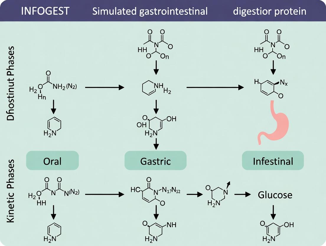
This comprehensive guide details the INFOGEST protocol, a standardized in vitro method for simulating human gastrointestinal protein digestion.
Mastering the INFOGEST Protocol: A Complete Guide to Simulating Protein Digestion for Biomedical Research
Abstract
This comprehensive guide details the INFOGEST protocol, a standardized in vitro method for simulating human gastrointestinal protein digestion. Tailored for researchers, scientists, and drug development professionals, it covers the foundational principles, step-by-step methodology, and practical applications of the protocol. The article provides actionable troubleshooting advice, compares INFOGEST to other digestion models, and outlines validation strategies to ensure experimental reproducibility and physiological relevance in studies of protein digestibility, bioactive peptide release, and nutrient bioavailability.
What is the INFOGEST Protocol? Foundational Principles and Scientific Rationale
Origins and Goals
The INFOGEST network was established by a consortium of international food scientists in 2011 to address the lack of standardized, physiologically relevant in vitro digestion models. Prior methodologies were highly variable, hindering the comparison of data between laboratories studying nutrient bioaccessibility, food structure, and bioactive compound stability. The primary goal was to create a harmonized, consensus static digestion model based on human physiological data, providing a robust and reproducible tool for the scientific community.
The model's development focused on key parameters:
- Physiological Relevance: Simulating oral, gastric, and intestinal phases with enzyme activities, pH, and transit times derived from human in vivo data.
- Simplicity and Accessibility: Ensuring the protocol is implementable in standard laboratories without specialized equipment.
- Reproducibility: Defining exact conditions (e.g., pH, enzyme concentrations, incubation times) to enable direct comparison of results worldwide.
Application Notes and Protocols within Protein Digestion Research
Within the context of protein digestion research, the INFOGEST protocol provides a critical framework for studying protein hydrolysis, peptide release, allergenicity, and the bioavailability of amino acids and bioactive peptides.
Key Experimental Protocol: StaticIn VitroProtein Digestion
Objective: To simulate the human gastrointestinal digestion of a proteinaceous sample and analyze the resulting digest (peptides, free amino acids, undigested protein).
Detailed Methodology:
Phase 1: Simulated Salivary Fluid (SSF) and Oral Phase
- Prepare simulated salivary fluid (SSF) electrolyte stock solution according to the INFOGEST recipe.
- For the oral phase, mix the test protein sample with SSF in a 1:1 ratio (v/v).
- Add α-amylase (75 U/mL final activity in the oral mixture). Human salivary α-amylase is recommended.
- Adjust pH to 7.0 using 1M HCl or NaOH.
- Incubate for 2 minutes at 37°C under gentle agitation.
Phase 2: Simulated Gastric Fluid (SGF) and Gastric Phase
- Prepare simulated gastric fluid (SGF) electrolyte stock solution.
- Combine the entire oral bolus with SGF in a 1:1 ratio (v/v).
- Add porcine pepsin (2000 U/mL final activity in the gastric mixture). Caution: Do not use recombinant pepsin if studying gastric mucolysis.
- Adjust pH to 3.0 using 1M HCl.
- Incubate for 2 hours at 37°C under gentle agitation.
Phase 3: Simulated Intestinal Fluid (SIF) and Intestinal Phase
- Prepare simulated intestinal fluid (SIF) electrolyte stock solution.
- Combine the entire gastric chyme with SIF in a 1:1 ratio (v/v).
- Add a pancreatin enzyme preparation providing final activities of 100 U/mL for trypsin and 25 U/mL for lipase in the intestinal mixture. Bile salts are added at a final concentration of 10 mM.
- Adjust pH to 7.0 using 1M NaOH.
- Incubate for 2 hours at 37°C under gentle agitation.
Termination:
- To stop digestion, place samples on ice and immediately add a protease inhibitor cocktail (e.g., AEBSF) or rapidly lower the pH (<2) for pepsin inactivation or raise it (>9) for pancreatin inactivation, depending on the downstream analysis.
- Centrifuge (e.g., 10,000 g, 20 min, 4°C) to separate the aqueous fraction (containing soluble peptides and amino acids) from the solid pellet (undigested residue).
- Store fractions at -80°C until analysis.
Downstream Analysis for Protein Digestion:
- Degree of Hydrolysis: O-phthaldialdehyde (OPA) method or trinitrobenzenesulfonic acid (TNBS) assay.
- Peptide Profile: SDS-PAGE, Size-Exclusion Chromatography (SEC), or Reversed-Phase HPLC.
- Peptide Identification: Mass Spectrometry (LC-MS/MS).
- Bioactivity Screening: In vitro assays for ACE-inhibition, antioxidant capacity, etc., on the collected aqueous fraction.
Data Presentation: Key INFOGEST Digestion Parameters
Table 1: Standardized Physiological Parameters of the INFOGEST Static Model
| Phase | Duration | pH | Key Enzymes | Concentration / Activity | Bile (Intestinal) |
|---|---|---|---|---|---|
| Oral | 2 min | 7.0 | α-Amylase | 75 U/mL (in mixture) | - |
| Gastric | 2 hours | 3.0 | Pepsin | 2000 U/mL (in mixture) | - |
| Intestinal | 2 hours | 7.0 | Pancreatin (Trypsin, Chymotrypsin, Lipase, etc.) | Trypsin: 100 U/mL; Lipase: 25 U/mL (in mixture) | 10 mM |
Table 2: Research Reagent Solutions for INFOGEST Protein Digestion
| Reagent / Material | Function in Protocol | Critical Specification / Note |
|---|---|---|
| Porcine Pepsin | Gastric protease for protein hydrolysis. | Use ≥2500 U/mg protein. Avoid recombinant forms for certain studies. |
| Pancreatin from Porcine Pancreas | Source of intestinal proteases, amylase, and lipase. | Must be standardized for trypsin activity (e.g., 100 U/mL final). |
| Bile Salts (Porcine) | Emulsifies lipids, solubilizes hydrophobic compounds, affects enzyme activity. | Final concentration of 10 mM in intestinal phase. |
| Simulated Fluids (SSF, SGF, SIF) | Provide physiological ionic background (K+, Na+, Ca2+, Cl-, etc.). | Prepare from concentrated stock solutions per INFOGEST tables. |
| Protease Inhibitor Cocktail (e.g., AEBSF) | Immediately halts enzymatic activity upon sampling. | Essential for accurate endpoint analysis of peptide populations. |
| pH Adjustment Solutions (1M HCl, 1M NaOH) | To achieve exact phase-specific pH values. | Critical for correct enzyme activity and physiological relevance. |
Visualization: Experimental Workflow and Key Pathways
Diagram 1: INFOGEST Static Digestion Workflow for Proteins
Diagram 2: Key Enzymatic Pathways in Protein Digestion
The standardized INFOGEST static simulation protocol provides a foundational framework for studying food digestion, particularly proteins. However, to move from simple simulation to true physiological mimicry, one must engineer systems that replicate the dynamic biophysical and biochemical complexity of the human gastrointestinal (GI) tract. This application note details advanced protocols and considerations for creating more physiologically relevant in vitro environments for protein digestion research, extending the core INFOGEST method.
Quantitative Parameters of Human GI Physiology
Beyond the fixed pH, time, and enzyme concentrations of static protocols, dynamic systems incorporate kinetic and physical parameters.
Table 1: Key Dynamic Parameters for Physiological Mimicry in Protein Digestion
| GI Compartment | Parameter | Physiological Range | Experimental Implementation |
|---|---|---|---|
| Stomach | Gastric Emptying Half-Time | 30 - 90 min (liquid phase) | Use flow rates in dynamic reactors to achieve similar residence times. |
| Gastric Shear Stress | 0.02 - 0.1 Pa | Controlled via impeller speed or peristaltic wall movement in bioreactors. | |
| Pepsin Secretion Pattern | Bolus + continuous | Initial bolus (INFOGEST) followed by continuous infusion of gastric juice. | |
| Duodenum | Bicarbonate Addition | Rapid neutralization to pH ~6.5 | pH-stat titration or controlled infusion of NaHCO₃ solution. |
| Pancreatic Enzyme (Trypsin) Infusion | 10-100 U/mL final, time-dependent | Continuous infusion mimicking postprandial secretion profiles. | |
| Bile Salt Concentration | 3 - 10 mM (fasted-fed) | Fed-state models use ~10 mM bile (e.g., taurocholate). |
Detailed Protocol: Dynamic Gastric Duodenal Sequential Digestion
This protocol extends INFOGEST 2.0 for use in a multi-compartment, computer-controlled dynamic digestion system (e.g., DIDGI, simgi).
Aim: To simulate the temporal kinetics of gastric emptying and duodenal digestion of a protein substrate.
Materials & Pre-Digestion:
- Prepare simulated salivary fluid (SSF), gastric fluid (SGF), and intestinal fluid (SIF) per INFOGEST 2.0.
- Adjust the gastric electrolyte stock to include mucin (1.5 g/L) for rheology.
- Substrate: 5 g of purified protein or protein-rich food homogenate.
Procedure:
- Oral Phase: Mix substrate with SSF (1:1 v/w) for 2 min. Transfer entire bolus to gastric reactor.
- Dynamic Gastric Phase (120 min):
- Maintain temperature at 37°C with jacketed reactor.
- Set initial pH to 5.0, lower to 3.0 over 10 min using 1M HCl.
- Add gastric enzyme cocktail (Pepsin, 2000 U/mL final).
- Initiate gastric emptying: Start peristaltic pump P1 at t=10 min to transfer gastric chyme to duodenal reactor. Use a logarithmic emptying profile (e.g., 50% emptied by 60 min).
- Apply low shear (50 rpm magnetic stirring) mimicking antral contractions.
- Dynamic Duodenal Phase (120 min, starts at t=10 min from gastric start):
- Maintain pH at 6.5 via pH-stat using 0.5M NaHCO₃.
- Upon first arrival of gastric chyme, initiate continuous infusion of SIF containing pancreatin (100 U/mL trypsin activity final) and 10 mM bile salts.
- Maintain stirring at 150 rpm for mixing.
Sampling: Take aliquots from both reactors at defined intervals (e.g., 0, 10, 30, 60, 90, 120 min gastric; matched times in duodenum). Immediately inhibit enzymes (e.g., Pefabloc SC for serine proteases, Pepstatin A for pepsin). Analyze for protein hydrolysis (SDS-PAGE, OPA, MS), peptide release, and aggregation.
Signaling Pathways in GI Secretion Control
Understanding the hormonal control of digestion informs dynamic enzyme and bile delivery.
Hormonal Control of Postprandial Secretions
Experimental Workflow for Advanced Protein Digestibility Studies
Dynamic Protein Digestion Workflow
The Scientist's Toolkit: Key Research Reagent Solutions
Table 2: Essential Materials for Physiological Digestion Models
| Item | Function & Physiological Role | Example/Recommendation |
|---|---|---|
| Porcine Pepsin | Gastric protease, cleaves internal hydrophobic residues. Critical for initial proteolysis. | >2500 U/mg protein, from gastric mucosa. |
| Pancreatin | Provides master mix of pancreatic enzymes (trypsin, chymotrypsin, elastase, amylase, lipase). | Activity standardized per INFOGEST (Trypsin: 100 U/mL). |
| Bile Salts | Emulsify lipids, solubilize hydrophobic peptides, modulate enzyme activity. | Porcine bile extract or synthetic taurocholate/ glycodeoxycholate mix. |
| Gastric Mucin | Provides viscosity, mimics rheology, and can bind proteins/polyphenols altering accessibility. | Porcine gastric mucin Type II. |
| pH-Stat Titrator | Automatically maintains duodenal pH by titrating base (NaHCO₃), simulating neutralization. | Metrohm, Mettler Toledo systems. |
| Computer-Controlled Dynamic Digester | Multi-chamber bioreactor simulating peristalsis, secretion profiles, and emptying kinetics. | DIDGI, simgi, or in-house built systems. |
| Protease Inhibitor Cocktails | Immediately quench digestion at precise timepoints for accurate snapshots of hydrolysis. | Commercial tablets (e.g., cOmplete) or specific inhibitors (Pefabloc). |
| Dialysis Membranes | Simulate passive absorption in small intestine; allow sampling of low molecular weight peptides. | Spectra/Por membranes with 1-10 kDa MWCO. |
Application Notes: The Role of Core Components in the INFOGEST Protein Digestion Protocol
Within the framework of the INFOGEST in vitro simulated gastrointestinal digestion protocol, precise control over enzymatic activities, electrolyte composition, pH, and gastric/intestinal transit times is critical for generating physiologically relevant data on protein hydrolysis, bioactive peptide release, and nutrient/drug bioavailability. Deviations from physiologically mimetic conditions can lead to significant artifacts in digestion kinetics and endpoint analysis.
The following tables consolidate the standard conditions based on the INFOGEST 2.0 consensus and subsequent refinements for protein-focused research.
Table 1: Simulated Digestive Fluids: Electrolytes and pH
| Digestive Phase | Simulated Fluid | Key Electrolytes (Stock Concentration) | Final [Electrolytes] in Reaction | Target pH |
|---|---|---|---|---|
| Oral | Simulated Salivary Fluid (SSF) | KCl, KH₂PO₄, NaHCO₃, MgCl₂, (NH₄)₂CO₃ | Varies by dilution factor (typically 1:1 with sample) | 7.0 ± 0.2 |
| Gastric | Simulated Gastric Fluid (SGF) | KCl, KH₂PO₄, NaHCO₃, NaCl, MgCl₂, (NH₄)₂CO₃ | Varies by dilution factor | 3.0 ± 0.2 (for pepsin activity) |
| Intestinal | Simulated Intestinal Fluid (SIF) | KCl, KH₂PO₄, NaHCO₃, NaCl, MgCl₂ | Varies by dilution factor | 7.0 ± 0.2 (post-pancreatin addition) |
Table 2: Enzymatic Activity & Transit Times
| Digestive Phase | Enzyme(s) | Recommended Activity per mL Final Digest | Typical Transit Time (min) | Key Control Parameter |
|---|---|---|---|---|
| Oral | Human Salivary α-Amylase | 75-150 U (for starch digestion; often omitted in pure protein studies) | 2 | pH, Ca²⁺ presence |
| Gastric | Porcine Pepsin | 2000 U (for total digest volume) | 60-120 (standard: 120) | pH stability at ~3.0 |
| Intestinal | Porcine Pancreatin (Trypsin, Chymotrypsin, etc.) & Bile | Trypsin: 100 U/mL; Chymotrypsin: 25 U/mL; Bile: 10 mM final | 120 (standard) | pH shift to 7.0, bile salts for lipolysis |
Detailed Protocols
Protocol 1: Preparation of Simulated Digestive Fluids (SGF & SIF)
Purpose: To prepare electrolyte stock solutions ensuring reproducible ionic strength and buffering capacity. Materials: KCl, KH₂PO₄, NaHCO₃, NaCl, MgCl₂(H₂O)₆, (NH₄)₂CO₃, HCl (1M, 6M), NaOH (1M), deionized water. Procedure:
- Prepare SGF Electrolyte Stock Solution (5x concentration):
- Dissolve the following in ~800 mL water: KCl (7.0 mL of 0.5M), KH₂PO₄ (1.0 mL of 0.5M), NaHCO₃ (25.0 mL of 1M), NaCl (23.6 mL of 2M), MgCl₂ (2.5 mL of 0.15M), (NH₄)₂CO₃ (2.5 mL of 0.5M).
- Adjust pH to 3.0 using 6M HCl.
- Make up to 1 L with water. Store at 4°C for ≤ 1 week.
- Prepare SIF Electrolyte Stock Solution (5x concentration):
- Dissolve the following in ~800 mL water: KCl (13.5 mL of 0.5M), KH₂PO₄ (4.0 mL of 0.5M), NaHCO₃ (85.0 mL of 1M), NaCl (46.0 mL of 2M), MgCl₂ (0.8 mL of 0.15M).
- Adjust pH to 7.0 using 1M HCl or 1M NaOH.
- Make up to 1 L with water. Store at 4°C for ≤ 1 week.
Protocol 2: Standardized Gastric Digestion Phase for Protein Hydrolysis
Purpose: To simulate gastric protein digestion under controlled pH, enzyme activity, and time. Materials: SGF stock (5x), pepsin from porcine gastric mucosa (≥2500 U/mg), test protein substrate, 1M HCl, 37°C shaking water bath. Procedure:
- Pre-incubation: Warm SGF stock, water, and substrate suspension to 37°C.
- Reaction Mix Assembly: In a digestion vessel, combine:
- 1 part SGF stock (5x)
- 3 parts water
- 1 part protein substrate (pre-dispersed)
- Final volume is calculated for 1x SGF concentration.
- pH Adjustment: Immediately adjust pH to 3.0 ± 0.1 using 1M HCl.
- Enzyme Initiation: Add pre-warmed pepsin solution to achieve a final activity of 2000 U per mL of total gastric digest.
- Incubation: Place vessel in a 37°C shaking water bath (agitation ~150 rpm) for exactly 120 minutes (standard transit time).
- Termination: At t=120 min, immediately raise pH to 7.0 using 1M NaOH to irreversibly inactivate pepsin. Proceed to intestinal phase or sample for analysis.
Protocol 3: Standardized Intestinal Digestion Phase
Purpose: To simulate duodenal digestion following gastric phase. Materials: SIF stock (5x), pancreatin from porcine pancreas, bile salts (e.g., porcine bile extract), 1M NaOH, gastric digest (terminated at pH 7.0). Procedure:
- Preparation: Warm SIF stock, water, and terminated gastric digest to 37°C.
- Reaction Mix Assembly: To the terminated gastric digest, add:
- 1 part SIF stock (5x)
- 1 part water
- Final volume calculated for 1x SIF concentration.
- pH Verification: Confirm pH is 7.0 ± 0.2. Adjust with 1M NaOH if necessary.
- Enzyme/Bile Initiation: Add pancreatin and bile extract solutions to achieve final activities/concentrations of 100 U trypsin/mL, 25 U chymotrypsin/mL, and 10 mM bile salts.
- Incubation: Place vessel in a 37°C shaking water bath (agitation ~150 rpm) for exactly 120 minutes (standard transit time).
- Termination: At t=120 min, immediately place samples on ice or heat-inactivate enzymes (95°C, 5 min) as required by downstream analysis.
Visualizations
Title: INFOGEST Protein Digestion Workflow
Title: pH Regulates Enzyme Activity in INFOGEST
The Scientist's Toolkit: Key Research Reagent Solutions
Table 3: Essential Materials for INFOGEST Protein Digestion Studies
| Item | Function & Specification | Critical Notes |
|---|---|---|
| Porcine Pepsin | Primary gastric protease for protein hydrolysis. ≥2500 U/mg activity. | Activity varies by lot. Must be titrated for accurate dosing (2000 U/mL final digest). |
| Porcine Pancreatin | Source of intestinal proteases (trypsin, chymotrypsin), lipases, and amylases. | Must be standardized by trypsin activity (e.g., 100 U/mL final digest using BAPNA assay). |
| Bile Extract (Porcine) | Emulsifies lipids, facilitates lipolysis, and affects micelle formation for peptide/bioactive absorption. | Typically used at a physiological concentration of 10 mM in final intestinal digest. |
| SSF/SGF/SIF Electrolyte Stocks (5x) | Provide physiological ionic strength, osmolarity, and partial buffering capacity. | Prepare fresh weekly; pH adjustment after warming and dilution is critical. |
| Precision pH Meter & Electrodes | For accurate adjustment to pH 3.0 and 7.0 at 37°C. | Use temperature compensation. Electrodes must be compatible with proteinaceous samples. |
| 37°C Shaking Incubator/Water Bath | Maintains physiological temperature with agitation to simulate peristalsis. | Consistent shaking speed (~150 rpm) is vital for reproducible particle size and mixing. |
| SDS-PAGE & Staining System | For time-resolved monitoring of protein substrate hydrolysis. | Use Tricine gels for small peptides (<10 kDa). |
| HPLC-MS/MS System | For identification and quantification of released peptides and bioactive compounds. | Essential for detailed peptidomics and bioactivity correlation studies. |
The INFOGEST in vitro static simulation of gastrointestinal digestion is a standardized international protocol designed to harmonize research across disciplines. This article frames its application notes within the broader thesis that a unified digestion model is critical for generating comparable, reproducible data on protein fate. This enables direct correlation between food protein functionality (e.g., allergenicity, bioactivity) and pharmaceutical protein/peptide stability, informing both nutraceutical design and oral drug delivery strategies.
Application Notes & Quantitative Data Summaries
Table 1: Key Application Areas & Measurable Endpoints Using INFOGEST
| Application Domain | Primary Objective | Key Quantitative Endpoints | Typical Analytical Methods |
|---|---|---|---|
| Food Science & Allergenicity | Assess protein digestibility & epitope stability. | % Intact protein remaining; IC₅₀ values for IgE binding post-digestion; Bioactive peptide release (µg/mL). | SDS-PAGE, ELISA, HPLC-MS/MS. |
| Nutraceutical Development | Evaluate bioactive peptide release & stability. | ACE-inhibitory activity (IC₅₀); Antioxidant capacity (ORAC, TEAC); Peptide sequence identification. | Spectrophotometry, Fluorometry, LC-MS/MS. |
| Oral Biologic & Drug Delivery | Test stability of therapeutic proteins/peptides. | % Recovery of intact drug; Pharmacological activity retention; Permeability (Papp) in Caco-2 models. | HPLC/UV-MS, Cell-based assays, Ussing chamber. |
| Excipient & Formulation Screening | Determine protective effects of delivery systems. | Encapsulation Efficiency (%); Release kinetics in intestinal phase; Degradation rate constant (k). | Dynamic Light Scattering, In vitro release profiling. |
Table 2: Example INFOGEST Digestion Parameters (Adapted for Protein Focus)
| Phase | Duration | Key Conditions | Enzymes (Activity per mL) | Target Compound |
|---|---|---|---|---|
| Oral | 2 min | pH 7.0, α-amylase (optional for proteins) | - | Initial mixing. |
| Gastric | 2 hours | pH 3.0, 37°C | Pepsin: 2000 U (from porcine) | Protein denaturation & hydrolysis. |
| Duodenal (Intestinal) | 2 hours | pH 7.0, 37°C, Bile salts | Trypsin: 100 U (from porcine), Chymotrypsin: 25 U, Pancreatin: 100 U (lipase), Bile: 10 mM | Peptide & amino acid release. |
Detailed Experimental Protocols
Protocol 1: INFOGEST 2.0 for Allergenicity Assessment of a Novel Food Protein Objective: To simulate the gastrointestinal fate of a purified protein and assess residual immunoreactive fragments. Materials: Purified protein of interest, INFOGEST simulated fluids (SSF, SGF, SIF), enzymes (pepsin, pancreatin, bile salts), HCl/NaOH for pH adjustment, water bath at 37°C. Methodology:
- Oral Phase: Mix 1 mL protein solution (e.g., 2 mg/mL in SSF) with 1 mL SSF (pH 7.0) for 2 minutes.
- Gastric Phase: Lower pH of oral bolus to 3.0 with 1M HCl. Add pepsin solution (final 2000 U/mL). Incubate for 2 hours at 37°C with gentle agitation.
- Intestinal Phase: Raise pH to 7.0 with 1M NaOH. Add SIF and pancreatin/bile extract (final concentrations: trypsin 100 U/mL, bile 10 mM). Incubate for 2 hours at 37°C.
- Reaction Termination: Heat-inactivate enzymes at 95°C for 5 minutes or use protease inhibitors.
- Analysis: Centrifuge. Analyze supernatant via SDS-PAGE for fragment size and competitive ELISA for IgE binding.
Protocol 2: Bioactivity & Stability Testing for an Oral Peptide Drug Candidate Objective: To determine the recovery and activity retention of a synthetic peptide post-digestion. Materials: Synthetic peptide, INFOGEST reagents, Caco-2 cell monolayers (for permeability), activity assay kits (e.g., cAMP assay for a GLP-1 analog). Methodology:
- Digestion: Subject peptide (in relevant formulation) to full INFOGEST protocol (as in Protocol 1).
- Sample Processing: Terminate digestion, centrifuge, and filter (0.22 µm).
- Quantification: Use RP-HPLC to calculate the percentage of intact peptide recovered against a non-digested standard.
- Functional Assay: Apply digested sample to a cell-based receptor activation assay. Compare dose-response to non-digested control to calculate % activity retention.
- Permeability: Apply sample to apical side of Caco-2 monolayers in a Transwell system. Measure peptide appearance in basolateral compartment over 2 hours via LC-MS.
Signaling & Workflow Visualizations
Diagram Title: INFOGEST Workflow for Protein Analysis
Diagram Title: Protein Digestion Pathways & Formulation Impact
The Scientist's Toolkit: Research Reagent Solutions
Table 3: Essential Materials for INFOGEST-Based Protein Studies
| Reagent/Material | Function & Rationale | Example Source/Product |
|---|---|---|
| Pepsin (from porcine gastric mucosa) | Primary gastric protease; cleaves proteins at aromatic amino acids, simulating stomach digestion. | Sigma-Aldrich P7000 (≥2500 U/mg). |
| Pancreatin (from porcine pancreas) | Contains trypsin, chymotrypsin, amylase, lipase; simulates intestinal digestion. | Sigma-Aldrich P7545. |
| Bile salts (porcine) | Emulsifies lipids, solubilizes hydrophobic compounds, and affects enzyme kinetics. | Sigma-Aldrich B8631 (glyco- and tauro-conjugated). |
| Simulated Gastric/Intestinal Fluids (SSF, SGF, SIF) | Provide consistent ionic strength and electrolyte composition mimicking in vivo conditions. | Prepared per INFOGEST 2.0 recipe (KCl, KH₂PO₄, NaHCO₃, NaCl, etc.). |
| pH-Stat Titrator | For dynamic digestion models; automatically maintains pH by adding acid/base, quantifying hydrolysis. | Metrohm 902 Titrando. |
| Caco-2 Human Intestinal Cell Line | Gold-standard in vitro model for predicting intestinal permeability and absorption of peptides/drugs. | ATCC HTB-37. |
| Protease Inhibitor Cocktail (Halts digestion) | To precisely stop enzymatic reaction at defined timepoints for accurate snapshot analysis. | Thermo Scientific 78430. |
Within the field of simulated protein digestion research, the lack of reproducibility and comparability between studies using disparate in vitro models was a significant impediment to progress. The INFOGEST protocol, developed by a international consortium, was established to address this by providing a standardized, physiologically relevant static model. This application note details the advantages of this standardized approach over older, variable models, providing protocols and data to support its adoption in research and drug development.
Comparative Analysis of Key Parameters
The following tables summarize quantitative data comparing key aspects of the INFOGEST standardized model with typical older, non-standardized models.
Table 1: Gastric Phase Parameter Comparison
| Parameter | INFOGEST Standardized Model | Typical Older Models (Range) | Advantage of Standardization |
|---|---|---|---|
| pH | 3.0 (Fixed) | 1.5 - 5.0 | Mimics physiological fed state; prevents pepsin denaturation. |
| Pepsin Activity (U/mL) | 2000 U/mL | 500 - 10,000 U/mL | Ensures consistent proteolytic activity across labs. |
| Incubation Time | 120 min | 10 - 180 min | Accounts for gastric emptying kinetics. |
| Ionic Composition | Simulated Gastric Fluid (SGF) with electrolytes | Often just HCl/NaCl solution | Provides correct ionic strength for enzyme activity & stability. |
Table 2: Intestinal Phase & Outcome Metrics
| Parameter | INFOGEST Standardized Model | Typical Older Models (Range) | Impact on Data Quality |
|---|---|---|---|
| Bile Salt Concentration (mM) | 10 mM (porcine) / 5 mM (bovine) | 0.1 - 50 mM | Standardized micelle formation for lipid digestion & peptide solubility. |
| Pancreatin Activity (Trypsin) | 100 U/mL (trypsin basis) | Highly variable by source/batch | Directly controls major proteolytic rate; enables cross-study comparison. |
| Final Digesta Analysis | Recommended: SDS-PAGE, HPLC, MS | Often only OPA or Bradford | Enables detailed molecular weight & peptide profile comparison. |
| Inter-laboratory CV for Protein Hydrolysis* | < 15% (for well-characterized proteins) | Often > 50% | Drastically improves reproducibility and collaborative potential. |
*Data based on inter-laboratory validation studies for β-lactoglobulin and casein.
Detailed Experimental Protocols
Protocol 3.1: Standardized INFOGEST 2.0 Static Digestion
This is the core protocol for simulating gastrointestinal digestion of proteins.
A. Pre-digestion Preparation:
- Sample Preparation: Weigh protein substrate. For solid foods, homogenize. Adjust to 5% (w/v) in simulated oral fluid (SSF) electrolyte stock.
- Solution Preparation: Prepare fresh Simulated Gastric Fluid (SGF) and Simulated Intestinal Fluid (SIF) electrolyte stocks as per INFOGEST tables. Chill on ice.
- Enzyme/Bile Preparation: Dissolve porcine pepsin (≥2500 U/mg) in SGF to a 25x concentrated stock. Dissolve pancreatin (porcine) and bile salts (porcine) in SIF to 5x concentrated stocks. Keep on ice.
B. Gastric Phase:
- Mix protein sample with SSF electrolyte stock (1:1 v/v) in a suitable tube. Incubate 2 min at 37°C with agitation (e.g., in a shaking water bath).
- Lower pH to 3.0 using 1M HCl.
- Add the pre-prepared pepsin stock solution (in SGF) to achieve a final activity of 2000 U/mL. Adjust final volume with SGF stock.
- Incubate for 120 minutes at 37°C with constant agitation.
C. Intestinal Phase:
- After gastric digestion, raise pH to 7.0 using 1M NaOH.
- Add the pre-prepared pancreatin-bile stock solution (in SIF) to achieve final concentrations of 100 U/mL trypsin activity and 10 mM bile salts.
- Adjust final volume with SIF stock.
- Incubate for 120 minutes at 37°C with constant agitation.
- Enzyme Inactivation: At time = 120 min, immediately place tubes in a 90°C water bath for 5-10 min to halt enzymatic activity. Cool on ice.
- Centrifuge (e.g., 10,000 g, 20 min, 4°C) to separate soluble fraction (digesta) from any pellets. Store at -80°C for analysis.
Protocol 3.2: Critical Control Experiment: Assessing Inter-Laboratory Reproducibility
This protocol validates the standardization using a reference protein.
- Select a well-characterized protein (e.g., β-lactoglobulin, BSA) as a universal substrate.
- Precisely follow Protocol 3.1 in at least three independent replicates.
- Analyze the final intestinal digesta using: a. SDS-PAGE: To visualize the degradation pattern of the parent protein. b. Degree of Hydrolysis (DH): Using the o-phthalaldehyde (OPA) assay or pH-stat. Calculate DH using standard formulas. c. Peptide Profile: Via reverse-phase HPLC or LC-MS/MS.
- Compare the DH values and SDS-PAGE profiles with data from collaborating laboratories using the same INFOGEST protocol. The coefficient of variation (CV) for DH should be minimal (<15%).
Visualizations
Title: Standardization Solves Reproducibility Crisis
Title: INFOGEST 2.0 Standardized Experimental Workflow
The Scientist's Toolkit: Essential Research Reagent Solutions
Table 3: Key Reagents for INFOGEST Protocol Implementation
| Item | Function in Protocol | Critical Specification / Note |
|---|---|---|
| Porcine Pepsin | Primary gastric protease. Cleaves peptide bonds adjacent to aromatic amino acids. | Activity ≥2500 U/mg protein. Must be from gastric mucosa. Store lyophilized at -20°C. |
| Porcine Pancreatin | Source of intestinal proteases (trypsin, chymotrypsin), lipases, and amylases. | Standardize by trypsin activity (≥25 U/mg powder). Batch-to-batch variability necessitates activity checks. |
| Bile Extract (Porcine) | Emulsifies lipids, forms micelles, facilitates lipolysis and solubilizes hydrophobic peptides. | Primary bile salts (e.g., glycocholate, taurocholate). Use consistent concentration (10 mM). |
| Simulated Fluids Electrolyte Stocks | Provides physiologically accurate ionic environment for enzyme function and stability. | Must be prepared precisely per INFOGEST tables (KCl, KH₂PO₄, NaHCO₃, NaCl, MgCl₂, (NH₄)₂CO₃). |
| Protease Inhibitor Cocktail (e.g., AEBSF, Aprotinin) | For immediate quenching of enzymatic reactions for specific downstream analyses (e.g., bioactive peptide assay). | Add instead of heat inactivation if heat may degrade target analytes. |
| Reference Protein (e.g., β-Lactoglobulin, Casein) | Positive control substrate to validate protocol performance and inter-lab reproducibility. | Use a highly pure, well-characterized commercial standard. |
Executing the INFOGEST Protocol: A Step-by-Step Methodological Guide
Within the standardized INFOGEST in vitro static simulation protocol for food digestion (Brodkorb et al., 2019), Phase 1: Oral Digestion serves as the critical initial step. Its primary objectives are the mechanical reduction of solid food matrices and the initiation of starch hydrolysis via salivary α-amylase. For protein digestion research, this phase is essential for standardizing the initial breakdown of protein-containing food or drug formulations, which significantly influences subsequent gastric and intestinal proteolysis kinetics. Simulating mastication establishes a reproducible particle size distribution, while α-amylase activity can modify the food bolus viscosity and structure, potentially affecting protein accessibility. These Application Notes detail the standardized protocol and key considerations for implementing Phase 1 to ensure inter-laboratory reproducibility in protein digestion studies.
Experimental Protocol: Oral Phase Simulation
Principle: The solid or semi-solid test sample is combined with simulated salivary fluid (SSF) and human salivary α-amylase, and subjected to a defined period of mechanical agitation to simulate the chewing process.
Materials & Pre-Experimental Preparation:
- Simulated Salivary Fluid (SSF): Prepare stock electrolyte solution as per INFOGEST 2.0. For 1 L: KCl (0.15 g), KH₂PO₄ (0.08 g), NaHCO₃ (0.84 g), MgCl₂(H₂O)₆ (0.01 g), (NH₄)₂CO₃ (0.005 g). Adjust pH to 7.0 ± 0.2.
- Human Salivary α-Amylase (HSA): Purified enzyme. Activity must be verified.
- CaCl₂(H₂O)₂ solution: 0.3 M stock.
- Ultrapure Water.
- Masticatory Simulator: An incubator equipped with a horizontal shaking platform capable of maintaining 37°C and a frequency of 1 Hz (60 oscillations/min) is standard. For solid foods, a mechanical device that provides standardized shear/crushing may be used.
- pH Meter.
Procedure:
- Sample Preparation: Weigh the appropriate mass of test food/formulation. For solid foods, a pre-cut size of ~4x4x4 mm³ is recommended as a starting material.
- SSF/Enzyme Mixture Preparation: For a final volume of X mL:
- Volume of SSF stock = 0.493 * X mL.
- Volume of α-Amylase solution = Calculated to provide a final activity of 75 U/mL in the final oral bolus.
- Volume of 0.3 M CaCl₂ = 0.002 * X mL (final [Ca²⁺] = 0.6 mM).
- Add Ultrapure Water to a total volume of X mL.
- Warm the mixture to 37°C.
- Oral Digestion:
- Combine the sample with the SSF/Enzyme mixture in a suitable container. The standard food-to-liquid ratio is 1:1 (w/v) for solids or a defined mass for liquids.
- Immediately place the mixture in the shaking incubator at 37°C, 1 Hz for a defined period. The standard incubation time is 2 minutes.
- For solid samples, manual or mechanical shearing may be applied during the first 30 seconds to simulate chewing.
- Termination: After 2 min, the oral bolus is collected and immediately combined with simulated gastric fluid (SGF) to initiate the gastric phase. For time-point analysis, α-amylase activity can be inhibited by lowering pH (<4.0) or adding a specific inhibitor (e.g., acarbose).
Table 1: Standardized Composition of Simulated Salivary Fluid (SSF) for INFOGEST 2.0
| Electrolyte | Final Concentration (mM) | Function in Digestion |
|---|---|---|
| KCl | 15.1 | Maintains ionic strength & osmolality |
| KH₂PO₄ | 3.7 | Buffer capacity |
| NaHCO₃ | 13.6 | Main pH buffer, mimics saliva alkalinity |
| MgCl₂(H₂O)₆ | 0.15 | Cofactor for various enzymes |
| (NH₄)₂CO₃ | 0.06 | Source of NH₄⁺ ions |
| HCl/NaOH | - | pH adjustment to 7.0 ± 0.2 |
Table 2: Key Operational Parameters for Oral Phase Simulation
| Parameter | Standardized Condition | Rationale / Variability Allowed |
|---|---|---|
| Temperature | 37°C | Physiological body temperature |
| Incubation Time | 2 min | Average oral processing time |
| Agitation | 1 Hz (60 osc/min) | Simulates tongue movement |
| Final α-Amylase Activity | 75 U/mL | Average human salivary activity |
| pH | 7.0 ± 0.2 | Typical resting saliva pH |
| Ca²⁺ Concentration | 0.6 mM (final in bolus) | Stabilizes α-amylase structure/activity |
| Food/Saliva Ratio (solid) | 1:1 (w/v) | Standard bolus formation ratio |
Visualization of Experimental Workflow
Oral Digestion Phase Workflow
α-Amylase Catalytic Action & Outcome
The Scientist's Toolkit: Key Research Reagent Solutions
Table 3: Essential Materials for Oral Phase Simulation
| Item / Reagent | Function / Rationale | Key Consideration for Protein Research |
|---|---|---|
| Purified Human Salivary α-Amylase | Catalyzes starch hydrolysis. Essential for realistic bolus formation. | Verify specific activity (U/mg). Bacterial amylases differ in pH/thermal stability. |
| SSF Electrolyte Stock Solution | Provides physiologically relevant ionic environment, pH buffering. | Consistent preparation is critical for inter-lab reproducibility of downstream proteolysis. |
| Calcium Chloride (CaCl₂) Stock | Maintains α-amylase stability and activity as a cofactor. | Final [Ca²⁺] of 0.6 mM in bolus must be ensured for consistent enzyme kinetics. |
| pH Meter & Standard Buffers | To accurately adjust SSF pH to 7.0. | Small pH deviations can significantly affect amylase activity and initial protein structure. |
| Temperature-Controlled Shaker | Provides mechanical agitation (mastication simulation) at 37°C. | 1 Hz frequency is standardized. For very hard solids, a dedicated chewing simulator may be needed. |
| Protease/Amylase Inhibitors | To "freeze" the oral phase at specific time points for analysis. | Use acarbose (amylase inhibitor) if analyzing intermediate starch products without affecting proteins. |
Within the standardized INFOGEST protocol for simulating human gastrointestinal digestion in vitro, Phase 2 (gastric phase) is critical for protein breakdown. This phase models the complex interplay of acidic pH, the proteolytic enzyme pepsin, and mechanical forces. Accurate simulation is essential for research in food science, nutraceutical development, drug delivery (particularly for biologics), and understanding protein allergenicity. This application note provides detailed protocols and current data for implementing a robust gastric digestion simulation.
Quantitative Parameters for Gastric Phase Simulation
The following table summarizes the standard and optimized conditions for Phase 2 digestion as per the INFOGEST 2.0 consensus and subsequent research.
Table 1: Standardized and Variant Conditions for Simulated Gastric Phase
| Parameter | INFOGEST 2.0 Standard | Alternative Conditions (Application-Specific) | Rationale & Notes |
|---|---|---|---|
| pH | 3.0 | 1.5-2.0 (fasted), 4.0-5.0 (fed state), 5.0 (infant) | Low pH denatures proteins, activates pepsinogen to pepsin. Fasted state is more acidic. |
| Pepsin Activity | 2000 U/mL per digesta | 500-3000 U/mL depending on protein substrate | Activity is pH-dependent with optimum ~pH 2. Porcine pepsin is standard model enzyme. |
| Electrolytes | See Table 2 | Adjustable for specific ionic strength studies | Maintains physiological osmolarity and provides ions for enzymatic function. |
| Incubation Time | 120 min | 30-180 min (common for drug release studies) | Duration impacts extent of proteolysis. Can be sampled at intervals for kinetics. |
| Temperature | 37°C | Constant 37°C via water bath or incubator | Maintains physiological reaction rates. |
| Mechanical Stress | Intermittent agitation (e.g., vortex every 30 min) | Continuous shaking (orbital, 100 rpm), or dynamic gastric model systems | Influences particle size reduction, enzyme-substrate mixing, and shear-induced denaturation. |
Table 2: Gastric Electrolyte Stock Solution (SGF) Composition (per Liter)
| Compound | Concentration (mM) | Mass for 1L Stock | Function |
|---|---|---|---|
| KCl | 6.9 | 0.514 g | Maintains ionic strength and membrane potentials. |
| KH₂PO₄ | 0.9 | 0.122 g | Buffer component. |
| NaHCO₃ | 25.0 | 2.100 g | Provides bicarbonate, key for pH adjustment. |
| NaCl | 47.2 | 2.757 g | Main osmotic agent. |
| MgCl₂(H₂O)₆ | 0.1 | 0.020 g | Cofactor for some enzymes. |
| (NH₄)₂CO₃ | 0.5 | 0.048 g | Source of ammonium ions. |
Detailed Experimental Protocol
Protocol 1: Standard INFOGEST Gastric Phase Digestion
Title: Preparation and Execution of In Vitro Gastric Digestion.
Principle: A standardized bolus (from oral phase) is mixed with Simulated Gastric Fluid (SGF) containing pepsin at pH 3.0 and incubated at 37°C for 2 hours with mechanical agitation to simulate gastric peristalsis.
Materials & Reagents:
- Simulated Gastric Fluid Electrolyte Stock (Table 2)
- Porcine Pepsin (e.g., ≥2500 U/mg protein)
- 1M HCl
- 1M NaOH
- pH Meter
- Water Bath or Incubator with shaking capability (37°C)
- Vortex mixer or magnetic stirrer
- Gastric Bolus (from INFOGEST Phase 1 or test substrate in simulated saliva)
Procedure:
- SGF Preparation: For a final gastric digestion volume of X mL, calculate required volumes. Dilute the Gastric Electrolyte Stock (Table 2) with distilled water (typically 1:1 v/v). Adjust temperature to 37°C.
- pH Adjustment: Lower the pH of the SGF to 3.0 using 1M HCl. Note: Pepsin is added after pH adjustment to prevent auto-digestion.
- Pepsin Addition: Add the required amount of porcine pepsin to achieve a final activity of 2000 U/mL in the total gastric digestion volume. Gently mix.
- Initiation of Digestion: Combine the gastric bolus (typically 50% of final volume) with the SGF-pepsin solution (50% of final volume) in a suitable reaction vessel. Start the timer. Record this as t=0.
- Incubation: Incubate the mixture at 37°C for 120 minutes. Apply mechanical stress by:
- Standard Protocol: Vortexing or vigorous manual shaking for 10-15 seconds every 30 minutes.
- Enhanced Protocol: Continuous orbital shaking at 100-150 rpm.
- Sampling & Termination: At desired time points (e.g., 30, 60, 120 min), withdraw aliquots. Immediately increase pH to ≥7.0 using 1M NaOH or a suitable inhibitor cocktail (e.g., Pepstatin A) to stop pepsin activity for downstream analysis.
- Analysis: Centrifuge samples (e.g., 10,000 g, 10 min) to separate soluble fraction (digesta) from undigested particles. Analyze for protein concentration (BCA, Bradford), peptide profile (SDS-PAGE, HPLC), or other target analytes.
Protocol 2: Investigating pH-Dependent Proteolysis Kinetics
Title: Quantifying Pepsin Activity and Protein Degradation Across pH Gradients.
Principle: This protocol characterizes the extent of protein hydrolysis as a function of gastric pH, which is vital for modeling different physiological states (e.g., hypochlorhydria, fed vs. fasted).
Procedure:
- Prepare separate SGF batches and adjust them to target pH values: 1.5, 2.0, 3.0 (standard), 4.0, and 5.0.
- Add identical pepsin activity concentrations (2000 U/mL final) to each.
- Aliquot a standardized protein substrate (e.g., β-lactoglobulin, 2.5 mg/mL final) into each pH-conditioned SGF.
- Incubate at 37°C with continuous shaking.
- Withdraw aliquots at t=0, 5, 15, 30, 60, 120 minutes.
- Immediately terminate reactions by raising pH to 9.0.
- Measure the degree of hydrolysis (DH) using the o-phthalaldehyde (OPA) assay or analyze by trichloroacetic acid (TCA) soluble nitrogen.
- Plot DH vs. time for each pH to determine kinetic parameters.
The Scientist's Toolkit: Key Research Reagent Solutions
Table 3: Essential Materials for Gastric Digestion Studies
| Item | Function & Specification | Example Product/Catalog # |
|---|---|---|
| Porcine Pepsin | Primary gastric protease. Activity ~2500-3500 U/mg. Critical to verify activity via hemoglobin assay. | Sigma-Aldrich P7000 |
| Pepstatin A | Specific, potent inhibitor of pepsin and other aspartic proteases. Used to instantly terminate digestion for analysis. | Sigma-Aldrich P5318 |
| Simulated Gastric Fluid (SGF) Electrolyte Stock | Provides physiologically relevant ionic environment. Can be prepared in-house per Table 2 or purchased as a kit. | BioRelevant FaSSGF/FeSSGF solutions |
| pH-Stable Fluorogenic Protease Substrate | For real-time, continuous kinetic assays of pepsin activity under different conditions (pH, inhibitors). | MCA-peptide substrates (e.g., MCA-AKVKPPRSSSR-lys(Dnp)-NH₂) |
| Dynamic Gastric Model (DGM) Systems | Advanced apparatus simulating shear forces, gradual acidification, and emptying. For high-fidelity mechanical stress simulation. | In vitro Gastric Simulation Equipment (e.g., from Ghent University ESGI consortium designs) |
| o-Phthalaldehyde (OPA) Reagent | For rapid spectrophotometric measurement of primary amines released during proteolysis, indicating Degree of Hydrolysis (DH). | Prepared fresh per published methods (Nielsen et al.) |
Visualizations
Gastric Phase Experimental Workflow
Factors in Gastric Protein Digestion
Within the standardized INFOGEST 2.0 in vitro simulated gastrointestinal digestion protocol, Phase 3 represents the intestinal digestion stage. This phase models the duodenal environment, where pancreatic enzymes and bile salts act on gastric-digested chyme. For protein digestion research, this stage is critical for simulating the generation of final peptides and amino acids prior to absorption, making it essential for nutritional, pharmacological, and toxicological studies.
Key Biochemical Agents: Functions & Specifications
Pancreatic Enzymes
The pancreatic extract is a complex mixture. For standardization, INFOGEST recommends using porcine-derived pancreatin with defined activity.
Table 1: Key Pancreatic Enzyme Activities & Recommended Concentrations (INFOGEST 2.0)
| Enzyme | Primary Substrate | Recommended Activity in Final Digesta | Physiological Role in Protein Digestion |
|---|---|---|---|
| Trypsin | Peptide bonds (C-term of Lys, Arg) | 100 U/mL | Primary endopeptidase; activates other zymogens. |
| Chymotrypsin | Peptide bonds (C-term of aromatic, bulky hydrophobic) | 25 U/mL | Endopeptidase; broad specificity. |
| Elastase | Peptide bonds (C-term of small, neutral amino acids) | >0.5 U/mL | Endopeptidase. |
| Carboxypeptidase A | C-terminal amino acids (aromatic, aliphatic) | - | Exopeptidase. |
| Carboxypeptidase B | C-terminal amino acids (basic: Arg, Lys) | - | Exopeptidase. |
Bile Salts
Bile salts solubilize lipids and facilitate the formation of mixed micelles. Their concentration significantly impacts lipolysis and can affect protein digestibility by interacting with hydrophobic peptides.
Table 2: Bile Salt Composition & Concentrations
| Bile Salt/Extract | Typical Composition | INFOGEST Recommended Concentration (Fast State) | Primary Function |
|---|---|---|---|
| Porcine Bile Extract | Mixture of glycine & taurine conjugates | 10 mM (total bile salts) | Emulsification, micelle formation, lipase activation. |
| Synthetic (e.g., Taurocholate) | Pure sodium taurocholate | 5-10 mM (for standardization) | Defined chemical composition; reduces variability. |
Core INFOGEST Protocol for Phase 3 Intestinal Digestion
Reagent Preparation
- SIF (Simulated Intestinal Fluid): 5 mM KCl, 6 mM KH₂PO₄, 10 mM NaHCO₃, 85 mM NaCl, 0.3 mM MgCl₂(H₂O)₆, 1.6 mM CaCl₂(H₂O)₂. Adjust pH to 7.0 ± 0.1 at 37°C. Pre-warm to 37°C.
- Pancreatin Solution: Dissolve porcine pancreatin in SIF. Centrifuge briefly (e.g., 1500 x g, 10 min, 4°C) to remove insoluble material. The supernatant activity must be verified (see Table 1). Prepare fresh daily.
- Bile Salt Solution: Dissolve porcine bile extract or pure taurocholate in SIF. Filter sterilize (0.22 µm). Stable at 4°C for 1 week.
Step-by-Step Experimental Procedure
Input: pH-adjusted gastric chyme from Phase 2 (Gastric Digestion).
Transition to Intestinal Conditions:
- To the gastric chyme, add a calculated volume of pre-warmed SIF containing CaCl₂ to maintain a final free Ca²⁺ concentration of ~0.6 mM.
- Adjust pH to 7.0 using 1M NaOH.
Addition of Intestinal Secretions:
- Add the pre-warmed bile salt solution to achieve a final concentration of 10 mM.
- Immediately add the prepared pancreatin solution to achieve the final trypsin activity of 100 U/mL.
- Record the final volume.
Digestion Incubation:
- Incubate the mixture at 37°C with constant agitation (e.g., orbital shaking, magnetic stirring) for 120 minutes.
- Maintain pH at 7.0 ± 0.2 using an automated pH-stat titrator (with 0.25M NaOH) or via manual checks and adjustments. pH-stat is preferred for kinetic studies.
Reaction Termination:
- At the end of the incubation, immediately inactivate enzymes. Common methods include:
- Heat Inactivation: 85°C for 10 minutes.
- Protease Inhibitors: Add a cocktail (e.g., AEBSF, Leupeptin, Pepstatin A).
- Rapid Cooling & pH Shift: Place on ice and adjust pH to <4 or >9.
- Centrifuge (e.g., 10,000 x g, 20 min, 4°C) to separate soluble and insoluble fractions.
- Aliquot and store at -80°C for downstream analysis (e.g., SDS-PAGE, LC-MS/MS for peptides, OPA assay for amino acids).
- At the end of the incubation, immediately inactivate enzymes. Common methods include:
Visualization of Phase 3 Workflow & Biochemistry
Title: INFOGEST Phase 3 Intestinal Digestion Protocol Workflow
Title: Pancreatic Enzyme Cascade & Bile Salt Function in Protein Digestion
The Scientist's Toolkit: Key Research Reagent Solutions
Table 3: Essential Materials for INFOGEST Phase 3 Experiments
| Item | Function/Justification | Example & Specification |
|---|---|---|
| Porcine Pancreatin | Source of key digestive enzymes (trypsin, chymotrypsin, etc.). Must be assayed for activity. | Sigma-Aldrich P7545; verify trypsin activity per INFOGEST SOP. |
| Bile Salts (Porcine or Synthetic) | Creates physiological duodenal conditions; emulsifies lipids. | Sigma-Aldrich B8631 (porcine) or T4009 (sodium taurocholate). |
| pH-Stat Titrator | Critical for maintaining constant pH 7.0 during digestion, mimicking pancreatic bicarbonate secretion. | Metrohm 916 Ti-Touch with 807 Dosimat. |
| SIF Electrolyte Stock Solutions | Provides correct ionic strength and calcium concentration for enzyme activity. | Prepare concentrated stocks (e.g., 10x) for reproducibility. |
| Protease Inhibitor Cocktail (Animal-Free) | For immediate, irreversible enzyme termination post-digestion, preserving peptide profiles. | MilliporeSigma 535140 (AEBSF-based). |
| Activity Assay Kits | For standardizing pancreatin activity (Trypsin, Chymotrypsin) pre-experiment. | Thermo Scientific EIATRYP (Trypsin) or colorimetric substrates (BAPNA, BTEE). |
| O-Phthaldialdehyde (OPA) Reagent | For rapid spectrophotometric quantification of primary amine groups (free amino acids). | Prepare fresh with β-mercaptoethanol and SDS. |
Sample Preparation and Substrate Considerations for Proteins
Within the framework of INFOGEST, an internationally standardized protocol for simulating human gastrointestinal digestion, reproducible sample preparation and appropriate substrate characterization are foundational. The behavior of dietary or therapeutic proteins during gastric and intestinal phases is critically dependent on initial physical and chemical states. This document details application notes and protocols for preparing protein substrates prior to in vitro digestion studies, ensuring data alignment with physiological conditions and inter-laboratory reproducibility.
Key Considerations for Protein Substrates
Physicochemical State
The digestibility of a protein is influenced by its native conformation, aggregation state, and matrix interactions. Denatured proteins are typically more susceptible to pepsin and pancreatin hydrolysis than native, globular structures. Furthermore, proteins embedded in complex food matrices or encapsulated for delivery require specialized preparation to mimic consumption.
Buffer and pH Environment
The initial immersion buffer must not pre-activate or denature the protein prematurely. For many studies, a mild buffer like PBS or simulated oral fluid electrolyte solution is used, adjusting the pH to mimic the mouth (typically pH 6.8-7.0) before the gastric phase commences.
Homogenization and Particle Size
Particle size directly impacts enzyme accessibility. A protocol-standardized homogenization step is crucial for solid substrates. The INFOGEST suggests aiming for particles <2 mm for solid foods, though for pure protein isolates, achieving a homogeneous suspension or solution is the goal.
Table 1: Impact of Sample Preparation on Digestibility Metrics
| Preparation Variable | Typical Range | Effect on Hydrolysis Rate (vs. Native) | Recommended for INFOGEST |
|---|---|---|---|
| Heat Denaturation | 70-95°C, 5-15 min | Increase of 20-50% | Recommended only if mimicking processed food. |
| pH Pre-treatment | pH 2-3 or 9-11, 30 min | Increase of 15-60% | Not standard; alters native state. |
| Homogenization Speed | 5,000-15,000 rpm | Increase of 10-30% (for solids) | Required for solids; 10,000 rpm for 2 min suggested. |
| Final Particle Size | < 0.5 mm - 2 mm | Smaller size increases rate by up to 40% | Target < 2 mm for solid matrices. |
| Concentration | 1-10% (w/v) protein | High conc. may reduce % hydrolysis due to enzyme saturation. | 4-5% (w/v) is commonly used. |
Table 2: Common Buffer Systems for Pre-Gastric Phase
| Buffer Name | Key Components | pH Range | Use Case |
|---|---|---|---|
| Simulated Salivary Fluid (SSF) | KCl, KH₂PO₄, NaHCO₃, MgCl₂, (NH₄)₂CO₃ | 6.8-7.0 | Initial suspension for oral phase. |
| Phosphate Buffered Saline (PBS) | NaCl, KCl, Na₂HPO₄, KH₂PO₄ | 7.2-7.4 | General protein suspension where electrolyte match is needed. |
| HEPES Buffer | HEPES, NaCl | 7.0-7.4 | When phosphate interferes with analysis. |
| Water (Ultra-pure) | N/A | Variable (often ~6.5) | Only for highly soluble isolates where ions are confounding. |
Detailed Experimental Protocols
Protocol 1: Standard Preparation of Liquid Protein Solutions/Suspensions for INFOGEST
Objective: To prepare a homogeneous, native (or defined) protein sample for introduction to the simulated oral phase.
Materials:
- Protein isolate (e.g., whey, casein, soy, purified enzyme).
- Simulated Salivary Fluid (SSF) or appropriate buffer (see Table 2).
- pH meter and adjusters (1M HCl/NaOH).
- Magnetic stirrer or vortex mixer.
- Analytical balance.
Methodology:
- Weighing: Precisely weigh the required mass of protein powder to achieve the target concentration (e.g., 5% w/v) in the final volume of digestion experiment.
- Dispersion: Gradually add the powder to the required volume of pre-chilled SSF buffer in a beaker under constant magnetic stirring (or vortex in a tube) to avoid clumping.
- Hydration: Continue stirring for 30-60 minutes at 4°C to ensure complete hydration without promoting denaturation.
- pH Adjustment: Measure the pH of the suspension. Adjust to pH 7.0 ± 0.2 using microliter volumes of 1M HCl or NaOH. Record the final volume and any dilution.
- Equilibration: Allow the sample to equilibrate in a water bath at 37°C for 10 minutes immediately before initiating the INFOGEST oral phase.
- Aliquot: If necessary, aliquot into individual reaction vessels for replicated digestions.
Protocol 2: Preparation of Solid or Complex Protein Matrices
Objective: To standardize the physical form of solid proteinaceous foods (e.g., meat, legumes, powdered supplements) for digestion studies.
Materials:
- Solid protein sample.
- Food processor or laboratory homogenizer (e.g., Ultra-Turrax).
- Sieve or particle size analyzer.
- SSF Buffer.
- Cold chamber or ice bath.
Methodology:
- Size Reduction: For large solids, dice into ~5 mm pieces using a clean blade.
- Homogenization: Add the pieces to a pre-chilled homogenization vessel with a calculated volume of cold SSF buffer. Homogenize at 10,000 rpm for 2 minutes, keeping the vessel on ice to prevent heating.
- Particle Size Verification: Pass a representative aliquot through a 2 mm sieve. If material is retained, repeat homogenization in short bursts until >95% passes.
- pH Adjustment: Measure and adjust the homogenate to pH 7.0 as in Protocol 1. The homogenate is now considered the "bolus."
- Weight/Volume Recording: Record the total mass of the bolus. The effective protein concentration can be calculated from the known composition of the original solid.
The Scientist's Toolkit
Table 3: Essential Research Reagent Solutions & Materials
| Item | Function in Sample Prep |
|---|---|
| Simulated Salivary Fluid (SSF) | Electrolyte solution mimicking mouth fluid; initial hydration medium for proteins. |
| Pepsin from Porcine Gastric Mucosa | Primary gastric protease. Must be activity-verified (e.g., 2500 U/mL per INFOGEST). |
| Pancreatin from Porcine Pancreas | Mixture of intestinal proteases (trypsin, chymotrypsin), lipases, amylases. |
| Bile Extract (Porcine/Ovine) | Emulsifies lipids, influences protease accessibility to hydrophobic protein regions. |
| pH-Stat Titrator | Critical for maintaining exact pH during gastric (pH 3.0) and intestinal (pH 7.0) phases. |
| Ultra-Turrax Homogenizer | Provides standardized, high-shear homogenization for solid matrices. |
| 0.22 μm Syringe Filters | For clarifying digest samples prior to HPLC or MS analysis, removing enzymes/particulates. |
| Protease Inhibitor Cocktails | Added immediately post-digestion time points to irreversibly halt enzymatic activity for analysis. |
Visualization of Workflows
Title: Protein Sample Preparation Workflow for INFOGEST
Title: Sample Prep Role in Digestion Research Cycle
Within the broader thesis on standardizing simulated gastrointestinal digestion using the INFOGEST protocol, this document addresses the critical parameters that directly impact the reproducibility and physiological relevance of in vitro protein digestion studies. Rigorous control of temperature, precise timing, and validated enzyme sourcing are non-negotiable prerequisites for generating reliable, comparable data on protein hydrolysis, bioactive peptide release, and nutrient/drug bioavailability.
Temperature Control: Protocols and Data
Temperature directly influences enzyme kinetics, protein denaturation, and the physiological simulation of gastric and intestinal phases.
Protocol 1.1: Calibration and Monitoring of Incubation Systems
Objective: To ensure the digestion chamber maintains 37±0.5°C throughout the experiment. Materials: Temperature-calibrated water bath or dry incubator, NIST-traceable thermometer, independent temperature logger. Procedure:
- Place the temperature probe of the independent logger in the digestion vessel filled with simulated electrolyte solution.
- Set the incubator to 37°C and allow to equilibrate for 1 hour.
- Record the temperature from the independent logger every minute for 30 minutes.
- Calculate the mean and standard deviation. The system is validated if the mean is 37.0°C ± 0.5°C and the standard deviation is <0.2°C.
- Perform this validation monthly and before critical experiments.
Table 1: Impact of Temperature Variation on Pepsin Activity
| Temperature (°C) | Relative Pepsin Activity (%) | Mean Hydrolysis Degree (%) after 60 min (Casein) |
|---|---|---|
| 34.0 | 78.2 ± 3.1 | 15.4 ± 1.2 |
| 37.0 | 100.0 ± 2.5 | 24.7 ± 0.8 |
| 40.0 | 118.5 ± 4.0 | 28.1 ± 1.5 |
| 43.0 | 95.7 ± 5.2 | 22.3 ± 2.1 |
Timing: Phase Transition and Sampling Protocols
Adherence to defined gastric and intestinal phase durations is critical for comparative studies.
Protocol 2.1: Standardized Gastric-to-Intestinal Phase Transition
Objective: To precisely terminate the gastric phase and initiate the intestinal phase. Materials: pH meter, 1M NaOH, 1M HCl, pre-warmed intestinal electrolytes and enzyme solution. Procedure:
- At t=120 min (gastric phase conclusion), immediately add the pre-warmed intestinal electrolyte solution.
- Rapidly adjust pH to 7.0 ± 0.1 using 1M NaOH.
- Then add the pancreatin+bile extract solution. Critical Step: pH must be adjusted before enzyme addition to prevent pancreatin inactivation.
- Record the exact time of pancreatin addition as t=0 for the intestinal phase.
- Maintain pH at 7.0 via an automated titrator or manual checks every 10 min.
Table 2: Effect of Gastric Phase Duration on Final Hydrolysis Products
| Gastric Phase Duration (min) | % of Parent Protein Intact | Dominant Peptide Size Range (kDa) |
|---|---|---|
| 60 | 42.5 ± 3.2 | 3-10 |
| 120 (INFOGEST Standard) | 18.7 ± 1.8 | 1-5 |
| 180 | 12.1 ± 2.1 | 0.5-3 |
Enzyme Sourcing and Activity Validation
Enzyme preparation is the largest source of inter-laboratory variability. Sourcing and validation are paramount.
Protocol 3.1: Titration of Enzyme Activity for Standardization
Objective: To determine the lipase, protease, and amylase activities of pancreatin batches. Materials: Porcine pancreatin (e.g., Sigma P7545, BioUltra), bile extract (e.g., Sigma B8631), substrates (e.g., BAPNA for trypsin, pH-stat for lipase). Procedure for Trypsin Activity (adapted from INFOGEST):
- Prepare 1 mM BAPNA in DMSO and 50 mM Tris-HCl buffer (pH 8.2).
- Add 50 µL of suitably diluted pancreatin solution to 2.45 mL of buffer in a cuvette.
- Start reaction by adding 500 µL of BAPNA solution.
- Immediately measure the increase in absorbance at 410 nm for 3 minutes at 37°C.
- Calculate activity using ε410=8800 M⁻¹cm⁻¹. Activity should be ≥25 U/mg where 1 U = 1 µmol BAPNA hydrolyzed per minute.
Table 3: Comparison of Commercially Sourced Enzyme Preparations
| Supplier & Product Code | Reported Trypsin Activity (U/mg) | Lot-to-Lot Variability | Cost per 10g (USD) |
|---|---|---|---|
| Sigma-Aldrich P7545 (BioUltra) | 30 ± 3 | Low | 450 |
| BioCatalytics PAN-1 | 28 ± 5 | Moderate | 320 |
| Megazyme P-ANCA5 | 32 ± 2 | Very Low | 520 |
| In-House Porcine Preparation | 15 - 40 | Very High | N/A |
The Scientist's Toolkit: Research Reagent Solutions
Table 4: Essential Materials for INFOGEST Protein Digestion Studies
| Item & Example Source | Function in Protocol |
|---|---|
| Porcine Pepsin (Sigma P6887) | Gastric protease; hydrolyzes proteins at low pH. Must be >2500 U/mg. |
| Porcine Pancreatin (Sigma P7545) | Contains key intestinal enzymes (trypsin, chymotrypsin, lipase, amylase). |
| Porcine Bile Extract (Sigma B8631) | Emulsifies lipids, facilitates lipolysis, and solubilizes hydrophobic compounds. |
| Gastric Electrolyte Stock Solution | Provides physiologically relevant ionic strength and pH for gastric phase simulation. |
| pH-Stat Titrator (e.g., Mettler) | Critical for maintaining constant intestinal pH 7.0, enabling accurate lipase assay. |
| Temperature Logger (e.g., Testo) | Independent verification of incubation stability at 37°C. |
| HPLC-MS System | For definitive analysis of protein hydrolysis products and peptide sequences. |
Visualized Workflows and Pathways
Diagram 1: INFOGEST Protein Digestion Workflow
Diagram 2: Enzyme Activity Validation Pathway
Diagram 3: Temperature Impact on Digestion Kinetics
Application Notes
The INFOGEST static in vitro simulation of gastrointestinal digestion has become a cornerstone for studying the fate of dietary proteins. Downstream analysis following this simulated digestion is critical for elucidating bioactivity, bioavailability, and potential allergenicity of resulting peptides. These Application Notes detail three core downstream strategies: temporal sampling for kinetic profiles, targeted enzyme inhibition to probe mechanism, and advanced peptide characterization.
Sampling Dynamics: Strategic sampling at defined timepoints (e.g., 0, 5, 15, 30, 60, 120 minutes of intestinal phase) allows for the construction of digestion kinetics. This is vital for identifying transient bioactive peptides or assessing the resistance of proteins, such as allergens or drug-delivery vehicles, to proteolytic breakdown.
Enzyme Inhibition Studies: The targeted inhibition of specific digestive enzymes (e.g., pepsin, trypsin, chymotrypsin) during the INFOGEST protocol provides a mechanistic understanding of proteolysis. By comparing peptide profiles from inhibited and non-inhibited digestions, researchers can pinpoint which enzymes are responsible for generating specific peptides or degrading target proteins. This has direct applications in designing enzyme-targeted therapies or functional foods.
Peptide Characterization: The end-goal of many INFOGEST experiments is the identification and quantification of released peptides. Techniques like liquid chromatography-tandem mass spectrometry (LC-MS/MS) coupled with bioinformatics are employed. This characterization links digestive stability to potential bioactivities (e.g., ACE-inhibitory, antioxidant) and informs structure-function relationships crucial for pharmaceutical and nutraceutical development.
Quantitative Data Summary: Key Parameters in Downstream Analysis
Table 1: Common Timepoints for Kinetic Sampling in INFOGEST Intestinal Phase
| Timepoint (min) | Primary Purpose |
|---|---|
| 0 | Baseline, "start of intestinal phase" sample. |
| 5, 15 | Capture early, rapid hydrolysis events and transient peptide populations. |
| 30, 60 | Monitor mid-phase stabilization of peptide profiles. |
| 120 | Endpoint analysis, representing near-complete digestion under standardized conditions. |
Table 2: Commonly Inhibited Enzymes and Their Selective Inhibitors
| Enzyme | Selective Inhibitor | Typical Working Concentration in INFOGEST | Primary Function Probed |
|---|---|---|---|
| Pepsin | Pepstatin A | 1-10 µM | Gastric-phase proteolysis specificity. |
| Trypsin | Trypsin Inhibitor (Soybean) | 0.1-1.0 mg/mL | Cleavage after Lys/Arg residues. |
| Chymotrypsin | Chymostatin | 10-100 µM | Cleavage after hydrophobic residues. |
| Pancreatic Elastase | Elastatinal | 10-50 µM | Cleavage after small neutral residues. |
Table 3: Core LC-MS/MS Parameters for Peptide Characterization
| Parameter | Typical Setting / Method | Purpose |
|---|---|---|
| Chromatography | C18 reversed-phase column, 60-120 min gradient | Peptide separation based on hydrophobicity. |
| MS Mode | Data-Dependent Acquisition (DDA) | Automatically selects top N ions for fragmentation. |
| MS1 Resolution | ≥ 60,000 @ m/z 200 | Accurate mass measurement for peptide identification. |
| MS2 Fragmentation | HCD (Higher-energy C-trap Dissociation) | Generates sequence-informative fragment ions. |
| Database Search | Against specific protein database (e.g., UniProt) | Matches MS2 spectra to theoretical peptide sequences. |
| Quantification | Label-free (peak area intensity) or TMT/iTRAQ | Relative abundance of peptides across samples. |
Experimental Protocols
Protocol 2.1: Time-Course Sampling During the INFOGEST Intestinal Phase
Objective: To collect aliquots at specific timepoints during the intestinal digestion phase for kinetic analysis of protein hydrolysis.
Materials:
- INFOGEST digestion reaction (currently in intestinal phase).
- Pre-heated heating block or water bath (37°C).
- 1.5 mL microcentrifuge tubes (pre-labeled for timepoints).
- Timer.
- Piper and appropriate tips.
- 0.5 M NaHCO₃ (for gastric phase termination, if sampling pre-intestinal phase).
- Pefabloc SC or other serine protease inhibitor (for intestinal phase termination).
Procedure:
- Preparation: Prior to initiating the intestinal phase, label microcentrifuge tubes for each desired timepoint (e.g., T0, T5, T30, T120). Add 10 µL of 100 mM Pefabloc SC stock to each tube (final inhibitor concentration ~1 mM upon sample addition).
- Initiation & T0 Sampling: Start the intestinal phase by adding pancreatin and bile salts to the gastric chyme. Immediately withdraw a 100 µL aliquot and add it to the prepared "T0" tube. Vortex immediately to inhibit enzymes.
- Sequential Sampling: At each predetermined timepoint, withdraw a 100 µL aliquot and transfer it to the corresponding inhibitor-containing tube. Vortex immediately.
- Sample Handling: Place all samples on ice immediately after collection. Centrifuge at 10,000 x g for 10 minutes at 4°C to pellet any insoluble material or precipitated bile salts.
- Storage: Transfer the clear supernatant to a new tube. Store at -80°C until analysis (e.g., by OPA assay, SDS-PAGE, or LC-MS/MS).
Protocol 2.2: Enzyme Inhibition Within the INFOGEST Workflow
Objective: To assess the specific role of a digestive enzyme (e.g., trypsin) in the proteolytic degradation of a target protein or generation of a bioactive peptide.
Materials:
- Standard INFOGEST reagents (pepsin, pancreatin, bile salts, buffers).
- Target protein substrate.
- Selective enzyme inhibitor (e.g., Soybean Trypsin Inhibitor - SBTI).
- Control inhibitor vehicle (e.g., digestion buffer).
Procedure:
- Experimental Design: Set up two parallel digestion reactions: (A) INFOGEST Control, (B) INFOGEST + Inhibitor.
- Inhibitor Pre-incubation: For reaction (B), pre-incubate the pancreatin extract with the selective inhibitor (e.g., 0.5 mg/mL SBTI) in intestinal buffer at 37°C for 10 minutes prior to initiating the intestinal phase. Note: For gastric-phase inhibitors like pepstatin, pre-incubate with pepsin.
- Gastric Phase: Perform the standard 2-hour gastric phase on the target protein for both reactions (A) and (B).
- Intestinal Phase Initiation: For reaction (A), add standard pancreatin+bile mix. For reaction (B), add the inhibitor-pre-treated pancreatin with bile salts.
- Digestion: Continue the intestinal phase for 120 minutes under standard conditions (37°C, agitation).
- Termination & Analysis: At endpoint, inactivate both reactions with protease inhibitors. Centrifuge, collect supernatants, and analyze. Comparative analysis can use SDS-PAGE to visualize differential protein degradation, or LC-MS/MS to identify peptides whose generation is blocked by the inhibitor.
Protocol 2.3: Peptide Characterization by LC-MS/MS and Bioinformatics
Objective: To identify and quantify peptides present in an INFOGEST digest sample.
Materials:
- Desalted, INFOGEST digest supernatant.
- LC-MS/MS system (nanoflow or analytical flow).
- Solvent A: 0.1% Formic acid in water.
- Solvent B: 0.1% Formic acid in acetonitrile.
- Bioinformatics software (e.g., MaxQuant, Proteome Discoverer, Mascot).
Procedure:
- Sample Preparation: Desalt peptides using C18 solid-phase extraction tips or columns. Dry samples in a vacuum concentrator and reconstitute in 3% acetonitrile / 0.1% formic acid.
- LC-MS/MS Setup: Inject sample onto a C18 column. Apply a gradient from 3% to 35% Solvent B over 60-120 minutes at a flow rate of 300 nL/min (nanoflow).
- Mass Spectrometry: Operate the mass spectrometer in positive ion mode with a DDA cycle. Acquire full MS scans (m/z 350-1600) at high resolution. Select the top 15-20 most intense ions for MS/MS fragmentation using HCD.
- Database Searching: Convert raw files to .mgf or .ms2 format. Search against a database containing the sequence(s) of the target protein(s) using search engines (e.g., Andromeda in MaxQuant).
- Search Parameters: Enzyme: "None" (as peptides are from a complex digest); Fixed modifications: Carbamidomethyl (C); Variable modifications: Oxidation (M); Peptide mass tolerance: 10 ppm; MS/MS tolerance: 0.05 Da.
- Data Filtering: Filter results for high-confidence peptides using a False Discovery Rate (FDR) threshold of ≤ 1% at the peptide-spectrum-match level.
- Quantification & Analysis: Use label-free intensity values or spectral counts to compare peptide abundance across different samples (e.g., different timepoints or inhibition conditions).
Diagrams
Diagram 1: Downstream Analysis Workflow after INFOGEST
Diagram 2: Enzyme Inhibition Points in INFOGEST Protocol
The Scientist's Toolkit: Research Reagent Solutions
Table 4: Essential Materials for Downstream Analysis of INFOGEST Digests
| Item / Reagent | Function / Purpose in Downstream Analysis |
|---|---|
| Soybean Trypsin Inhibitor (SBTI) | Selective, irreversible inhibitor of trypsin. Used to probe the specific contribution of trypsin to intestinal proteolysis. |
| Pepstatin A | Potent and specific inhibitor of aspartic proteases (pepsin). Used to selectively block gastric-phase digestion. |
| Pefabloc SC (AEBSF) | Broad-spectrum serine protease inhibitor. Used for rapid, irreversible termination of intestinal-phase digestion in samples. |
| C18 Solid-Phase Extraction (SPE) Tips | For desalting and concentrating peptide mixtures prior to LC-MS/MS analysis, removing buffers and salts. |
| LC-MS Grade Solvents | High-purity water, acetonitrile, and formic acid. Essential for reproducible, high-sensitivity LC-MS/MS peptide analysis. |
| Standard Protein/Peptide Calibration Mix | For instrument calibration and monitoring LC-MS/MS system performance and mass accuracy. |
| Bioinformatics Software Suite (e.g., MaxQuant, PEAKS) | For processing raw MS data, database searching, peptide identification, and quantitative analysis. |
| Kinetic Analysis Software (e.g., GraphPad Prism) | For modeling digestion kinetics from time-course data (e.g., fitting hydrolysis curves from OPA/Ninhydrin assays). |
Troubleshooting the INFOGEST Protocol: Common Pitfalls and Optimization Strategies
Application Notes: Within the INFOGEST Protein Digestion Framework
Low digestibility yields in simulated protein digestion studies, particularly those employing the INFOGEST protocol, present significant challenges for accurately predicting bioaccessibility and allergenicity. The primary factors leading to these low yields are intrinsic enzyme activity variations and substrate-related issues. These must be systematically diagnosed and addressed to ensure experimental validity.
Key Factors Influencing Digestibility Yield:
- Enzyme Activity: Lot-to-lot variability in pepsin, pancreatin, and other digestive enzyme preparations directly impacts proteolytic efficiency. Sub-optimal pH, incorrect ionic strength, or the absence of necessary co-factors (e.g., bile salts for lipase activity) can further reduce functional activity.
- Substrate Issues: Protein denaturation state, aggregation, presence of anti-nutritional factors (e.g., trypsin inhibitors), and food matrix effects (e.g., lipid encapsulation, dietary fiber) can severely limit enzyme accessibility.
Table 1: Common Causes and Diagnostic Indicators of Low Digestibility Yields
| Category | Specific Issue | Observable Diagnostic Indicator |
|---|---|---|
| Enzyme Activity | Low pepsin activity (pH >2.5, old stock) | Low hydrolysis in gastric phase; intact proteins post-gastric. |
| Low trypsin/chymotrypsin activity | Accumulation of large peptides post-intestinal phase. | |
| Insufficient bile salt concentration | Reduced solubilization of lipolytic products; lower amino acid release. | |
| Substrate Properties | Highly aggregated/insoluble protein | High pellet mass after centrifugation; low soluble protein/peptide. |
| Presence of protease inhibitors | Sudden plateau in degree of hydrolysis over time. | |
| Dense food matrix (high fiber/fat) | Low bioaccessibility despite high enzyme activity in supernatant. |
Experimental Protocols for Diagnosis and Resolution
Protocol 1: Verification of Enzyme Activity
Objective: To confirm the proteolytic activity of enzyme stocks against standard protein substrates. Materials: Pepsin from porcine gastric mucosa (≥2500 U/mg), Porcine pancreatin (trypsin activity ≥25 U/mg), Bile salts (porcine), Bovine Serum Albumin (BSA) or β-lactoglobulin, Trichloroacetic Acid (TCA, 20% w/v), Lowry or BCA assay reagents.
- Prepare simulated gastric fluid (SGF) without enzyme (pH 3.0) and simulated intestinal fluid (SIF) without enzyme (pH 7.0) per INFOGEST 2.0.
- Gastric Activity Assay: Dissolve BSA (2 mg/mL) in SGF. Add pepsin to 2000 U/mL final activity. Incubate at 37°C with shaking (300 rpm).
- Intestinal Activity Assay: Pre-hydrolyze BSA with pepsin (15 min). Neutralize pH to 7.0. Add pancreatin/bile mix (final trypsin activity 100 U/mL, bile 10 mM).
- At intervals (0, 5, 15, 30, 60 min), withdraw aliquots. For gastric, immediately raise pH to 7.0 to stop reaction. Add equal volume of 20% TCA to precipitate intact proteins (incubate 4°C, 15 min).
- Centrifuge (10,000 x g, 15 min, 4°C). Analyze TCA-soluble fraction (peptides) for amino groups using the O-phthaldialdehyde (OPA) assay or measure total protein in supernatant (TCA-soluble) via Lowry/BCA assay.
- Calculation: Calculate Degree of Hydrolysis (DH) or plot TCA-soluble protein vs. time. Compare activity to historical lab controls or literature values.
Protocol 2: Assessment of Substrate Limitations
Objective: To determine if low yield is due to enzyme inhibition or physical inaccessibility. Materials: Same as Protocol 1, plus model inhibitor (e.g., soybean trypsin inhibitor). Part A: Inhibition Test
- Conduct standard intestinal digestion (Protocol 1, Part 2) with pre-hydrolyzed substrate.
- In parallel, include a condition with a known, pure trypsin inhibitor added at a molar excess to trypsin.
- Compare hydrolysis curves. If both your sample and the inhibitor control show similarly depressed hydrolysis, endogenous inhibitors may be present. Part B: Accessibility Test
- Perform a standard INFOGEST digestion on the test protein/food.
- At the end of the intestinal phase, centrifuge (10,000 x g, 30 min, 4°C).
- Separately analyze the supernatant (accessible fraction) and the pellet (inaccessible fraction) for protein content (Kjeldahl or Dumas). Resuspend pellet in fresh SIF with fresh enzymes and digest for an additional 2 hours.
- A significant protein mass in the pellet after the first digestion, which is then hydrolyzed in the second digestion, indicates a physical accessibility barrier (e.g., aggregation, encapsulation).
Table 2: Research Reagent Solutions Toolkit
| Reagent/Material | Function in INFOGEST Digestion | Critical Consideration |
|---|---|---|
| Pepsin (Porcine) | Primary gastric protease; cleaves at aromatic/leucine residues. | Verify activity (U/mg) upon receipt. Aliquot and store at -80°C. Avoid repeated freeze-thaw. |
| Pancreatin (Porcine) | Provides intestinal proteases (trypsin, chymotrypsin), lipases, amylases. | High variability between batches. Pre-test each lot for trypsin activity. |
| Bile Salts Extract | Emulsifies lipids, solubilizes lipolytic products, activates lipase. | Concentration critical (10 mM final in SIF). Affects micelle formation & proteolysis. |
| Calcium Chloride (CaCl₂) | Cofactor for lipase and protease activity; simulates physiological ionic conditions. | Required stepwise addition (gastric: 0.15 mM, intestinal: 0.6 mM final). |
| Protease Inhibitor Cocktails | For immediate quenching of enzymatic reactions at sampling timepoints. | Must be added immediately upon sampling. Choice depends on phase (e.g., pepstatin for gastric). |
| OPA Reagent | Spectrophotometric quantification of primary amines (hydrolysis products). | Must be prepared fresh. Sensitive to light. Correlates with Degree of Hydrolysis. |
| TCA (Trichloroacetic Acid) | Precipitates intact proteins and large peptides for separation from small peptides. | Standard quenching method for gastric phase. Typical final concentration 10% (w/v). |
Diagnostic and Experimental Workflow Diagrams
Low Digestibility Yield Diagnosis Workflow
INFOGEST Protocol with Integrated Quality Control
pH Stability Problems During Gastric and Intestinal Phases
Within the standardized INFOGEST protocol for simulating gastrointestinal digestion, maintaining precise pH stability during the gastric and intestinal phases is a critical, yet challenging, aspect for reproducible protein digestion research. The protocol mandates specific pH setpoints: pH 3.0 for the gastric phase and pH 7.0 for the intestinal phase. Deviations from these targets, caused by the buffering capacity of food matrices or reagents, can significantly alter enzyme activities (e.g., pepsin, pancreatin), compromise proteolysis kinetics, and lead to irreproducible results in assessing protein digestibility, allergenicity, or bioactive peptide release.
The primary challenges and their quantitative effects on digestion parameters are summarized below.
Table 1: Impact of pH Deviations on Key Digestion Parameters
| Phase | Target pH | Common Deviation | Impact on Pepsin Activity (%) | Impact on Trypsin/Chymotrypsin Activity (%) | Key Consequence |
|---|---|---|---|---|---|
| Gastric | 3.0 | Increase to pH 4.0-5.0 | Reduction by 70-90% | N/A | Incomplete proteolysis, overestimation of protein stability. |
| Gastric | 3.0 | Decrease to pH <2.5 | Potential denaturation; variable impact. | N/A | Potential enzyme inactivation, non-physiological conditions. |
| Intestinal | 7.0 | Decrease to pH 6.0-6.5 | N/A | Reduction by 50-80% (Trypsin) | Incomplete peptide release, altered bioaccessibility data. |
| Intestinal | 7.0 | Increase to pH >7.5 | N/A | Reduction by 30-60% (Chymotrypsin) | Non-physiological conditions, altered enzyme specificity. |
Data synthesized from recent studies on INFOGEST method optimization (2020-2023).
Detailed Experimental Protocols
Protocol 3.1: Pre-Digestion Buffering Capacity Assessment
Objective: To predict and compensate for the pH-stabilizing effect of the test sample.
- Prepare 5 mL of your protein sample (e.g., 5% w/v protein isolate) in simulated gastric fluid (SGF, without enzymes).
- Titrate the sample incrementally (e.g., 10 µL steps) with 1M HCl under stirring, monitoring pH with a calibrated micro-electrode.
- Record the volume of acid required to reach pH 3.0. Compare to the volume needed for SGF alone (blank).
- Calculate the additional acid (or base, for intestinal phase) needed for the full digestion volume. Incorporate this adjusted volume into the main digestion protocol.
Protocol 3.2: Real-Time pH Monitoring and Adjustment During Gastric Phase
Objective: To maintain pH at 3.0 ± 0.1 throughout the gastric digestion.
- Set up the digestion vessel with SGF, pre-warmed to 37°C, in a temperature-controlled incubator with stirring.
- Initiate digestion by adding pepsin. Immediately insert a calibrated, sterilized pH electrode.
- Monitor pH continuously via a connected data logger. Set alarms for deviations >0.1 pH units.
- Adjust using micro-additions (e.g., 1-5 µL) of 1M HCl or 1M NaOH as needed. Record all additions.
- Sample carefully to avoid introducing air or affecting the liquid volume significantly.
Protocol 3.3: Standardized pH Adjustment for Intestinal Phase Transition
Objective: To achieve a rapid and precise shift from gastric pH 3.0 to intestinal pH 7.0.
- Pre-warm simulated intestinal fluid (SIF) and 1M NaOH to 37°C.
- At the end of the gastric phase, place the vessel on a stirrer.
- Add the SIF concentrate (with bile salts and pancreatin) according to the INFOGEST volume ratios.
- Immediately titrate the mixture to pH 7.0 using the pre-warmed 1M NaOH. Perform this adjustment within 1 minute to minimize enzyme exposure to suboptimal pH.
- Verify stability at pH 7.0 for 2 minutes before proceeding with the timed intestinal digestion.
The Scientist's Toolkit: Key Reagent Solutions
Table 2: Essential Materials for pH Management in INFOGEST Protocols
| Item | Function & Rationale |
|---|---|
| Calibrated Micro-pH Electrode | Enables precise, real-time measurement in small volumes; essential for monitoring. |
| 1M HCl & 1M NaOH (Sterile) | High-concentration titrants allow for minute volume adjustments, minimizing dilution effects. |
| pH Data Logger/Controller | Allows for continuous recording and can be linked to automated titrators for stability. |
| Simulated Gastric Fluid (SGF, electrolyte stock) | Provides consistent ionic background. Must be prepared without acid to allow controlled acidification. |
| Simulated Intestinal Fluid (SIF, electrolyte stock) | Provides consistent ionic background. Must be prepared without bicarbonate to allow controlled basification. |
| Thermostated Stirring Digestion Vessel | Ensures uniform temperature and pH throughout the sample, preventing localized gradients. |
Visualization of Workflow and Problem-Solving
Diagram 1: pH Control Workflow in INFOGEST Digestion
Diagram 2: Causes & Solutions for pH Instability
Context: The INFOGEST in vitro static simulation of gastrointestinal digestion has become a foundational tool for food and pharmaceutical science. This protocol extends its application to assess the digestibility, stability, and potential allergenicity of novel protein entities (NPEs), including plant/insect-based novel proteins, recombinant allergens, and engineered biologics (e.g., peptide drugs, enzyme therapies). The core thesis is that modulating INFOGEST parameters (pH, enzyme ratios, transit times) can predict the fate of these complex proteins and inform their safety and efficacy profiles.
Table 1: Standard vs. Optimized INFOGEST Phases for NPEs
| Phase | Standard INFOGEST Condition | Optimized Condition for Novel Proteins | Optimized Condition for Engineered Biologics | Rationale |
|---|---|---|---|---|
| Oral | pH 7.0, 75 U/mL α-amylase | pH 6.5-7.5, 75 U/mL α-amylase | Omit or pH 7.4, no α-amylase | Mimics variable saliva pH; biologics often bypass oral phase. |
| Gastric | pH 3.0, 2000 U/mL pepsin, 60 min. | pH 2.5-5.0, 500-4000 U/mL pepsin, 30-120 min. | pH 1.5-3.0, 2000-6000 U/mL pepsin, 5-30 min. | Adjust for gastric resistance (allergens) or extreme sensitivity (some biologics). |
| Intestinal | pH 7.0, 100 U/mL pancreatin, 10 mM bile, 120 min. | pH 7.0, 200 U/mL pancreatin, 10 mM bile, 180 min. | pH 7.0, 50 U/mL pancreatin, 2-10 mM bile, 5-60 min. | Extended time for complex breakdown; reduced enzymes/bile for fragile biologics. |
| Sampling | End-point aliquots. | Time-series (0, 5, 15, 30, 60, 120, 180 min). | Frequent time-series (0, 2, 5, 10, 20, 30 min). | Captures kinetic digestion profiles and transient peptide formation. |
Table 2: Key Analytical Readouts and Target Values
| Analytical Method | Target for Allergenicity Assessment | Target for Biologic Stability | Key Metrics |
|---|---|---|---|
| SDS-PAGE & Immunoblot | Persistence of >~20 kDa IgE-reactive bands. | ≥70% intact primary post-gastric phase. | Band intensity, molecular weight shift. |
| ELISA (IgE-binding) | ≥50% reduction in immunoreactivity post-digestion. | Not Primary. | IC50 values, % inhibition. |
| LC-MS/MS Peptidomics | Identification of stable, immunogenic peptides (>9 aa). | Identification of cleavage hot-spots. | Peptide sequence, abundance, half-life. |
| Cell-Based Assay (e.g., basophil activation) | ≥30% reduction in degranulation vs. native protein. | Not Primary. | %CD63+ basophils, histamine release. |
Detailed Experimental Protocols
Protocol 1: Kinetic INFOGEST Digestion of a Novel Protein/Allergen Objective: To monitor the time-dependent degradation of a novel protein and its IgE-binding epitopes.
- Sample Preparation: Prepare 1 mg/mL protein solution in simulated salivary fluid (SSF) electrolyte stock.
- Oral Phase (Optional): Adjust to pH 6.8 with 1M HCl/NaOH. Add α-amylase (75 U/mL final). Incubate 2 min, 37°C, with agitation.
- Gastric Phase: Lower pH to 3.0 (or test value). Add porcine pepsin (2000 U/mL final). Immediately take a t=0 sample, inactivate with 20mM Na₂CO₃. Continue incubation at 37°C. Withdraw aliquots at t=5, 15, 30, 60, 120 min, and inactivate.
- Intestinal Phase: Adjust gastric digest pH to 7.0. Add pancreatin (200 U/mL trypsin activity) and bile salts (10 mM final). Take t=0 sample, inactivate by heating (95°C, 5 min). Incubate at 37°C, sampling at t=30, 60, 120, 180 min, inactivating each.
- Analysis: Clarify samples by centrifugation. Analyze supernatants by SDS-PAGE, IgE-ELISA using patient sera pools, and LC-MS/MS.
Protocol 2: Forced Degradation Stability Testing for an Engineered Biologic Objective: To identify vulnerable sites in a therapeutic enzyme or peptide under stressed digestive conditions.
- Stress Conditions Setup: Prepare four 0.5 mL aliquots of biologic (1 mg/mL in SSF).
- A: Control (pH 7.0, no enzymes).
- B: Low Stress Gastric (pH 3.0, 500 U/mL pepsin, 10 min).
- C: High Stress Gastric (pH 2.0, 4000 U/mL pepsin, 30 min).
- D: Intestinal Stress (pH 7.0, 100 U/mL pancreatin, 5mM bile, 30 min).
- Digestion & Quenching: Incubate all (except A) at 37°C. Quench B, C with pepstatin A (final 10 µM). Quench D with 4x protease inhibitor cocktail + heating.
- Activity/Binding Assay: Perform functional assay (e.g., enzymatic activity, receptor-binding ELISA) on all quenched samples. Express as % residual activity/binding vs. Control A.
- Structural Analysis: Run quenched samples on native-PAGE and size-exclusion chromatography (SEC-HPLC) to assess aggregation and fragmentation.
Visualization
Title: Decision Workflow for Digestibility Assessment of Novel Biologics
Title: Pathway Linking Digestion-Resistant Proteins to Allergic Sensitization
The Scientist's Toolkit: Key Research Reagent Solutions
Table 3: Essential Materials for INFOGEST-based NPE Studies
| Item | Function & Rationale | Example/Specification |
|---|---|---|
| Porcine Pepsin | Primary gastric protease. High purity ensures reproducible activity. | ≥2500 U/mg protein, lyophilized powder. |
| Pancreatin from Porcine Pancreas | Source of intestinal proteases (trypsin, chymotrypsin), lipases, amylases. | Activity standardized per INFOGEST (trypsin: 100 U/mL final). |
| Bile Salts (Porcine) | Emulsifies lipids, affects protease accessibility to protein. | Mixture of taurocholate, glycocholate, etc. |
| Simulated Fluids (SSF, SGF, SIF) | Electrolyte solutions (K+, Na+, Ca2+, etc.) providing physiologically relevant ionic strength. | Prepared per INFOGEST 2.0 monograph. |
| Protease Inhibitor Cocktails | For precise quenching of digestion at time points. | E.g., Pepstatin A for pepsin, AEBSF for pancreatin. |
| Human IgE Sera Pools | For immunoreactivity assessment of potential allergens. | Sera from multiple patients allergic to the source material. |
| Standard Allergen Controls | Positive controls for digestion stability and immunoassays. | E.g., Ara h 2 (peanut), β-lactoglobulin (milk). |
| LC-MS/MS Grade Solvents | For peptidomic analysis to identify stable peptide sequences. | Acetonitrile, water, formic acid of ultra-high purity. |
Adapting the Static Model for Dynamic Elements (e.g., Secretion Rates).
Application Notes
Within the INFOGEST in vitro simulated protein digestion research framework, the static gastric model provides a standardized, reproducible baseline. However, physiological digestion is dynamic, characterized by feedback-regulated secretion of gastric acid, pepsin, and other components. Adapting the static model to incorporate dynamic secretion rates, particularly of HCl and pepsinogen/pepsin, is critical for predicting the proteolysis of complex substrates, such as therapeutic proteins or novel food matrices, with higher physiological relevance. This adaptation bridges the gap between standardized screening and mechanistic understanding of digestion kinetics.
Key dynamic parameters include:
- Titratable Acid Secretion Rate: Mimicking the cephalic and gastric phases of acid response to a meal.
- Pepsin Secretion/Activation Kinetics: Coupling pepsin activity to pH drop, reflecting the in vivo zymogen activation cascade.
- Gastric Emptying: While INFOGEST defines a fixed gastric phase duration, a dynamic model can integrate gradual emptying, affecting substrate concentration and enzyme kinetics.
The following table summarizes core dynamic parameters adapted from recent literature for integration into the INFOGEST gastric phase:
Table 1: Key Dynamic Parameters for Adapting the INFOGEST Gastric Phase.
| Parameter | Typical Physiological Range/Profile | Adapted In Vitro Implementation | Primary Impact on Proteolysis |
|---|---|---|---|
| Acid Secretion Rate | 10-40 mmol H+/h postprandial (variable) | Continuous or pulsed titration via pH-stat to maintain a prescribed pH curve (e.g., from pH 6.5 to 2.5 over 60 min). | Controls pH-dependent substrate denaturation and pepsin activation kinetics. |
| Pepsin Activity Rate | Secreted as pepsinogen, activated below pH 5.0. Max activity ~pH 2.0-3.5. | Initial low pepsin activity, with incremental addition of active pepsin or pepsinogen aligned with pH drop. | Determines temporal proteolytic profile; avoids non-physiological early hydrolysis at high pH. |
| Gastric Phase Duration | 2-4 h (variable by meal). INFOGEST: 120 min standard. | Can be modeled as a variable parameter, with sampling to track hydrolysis kinetics over time. | Affects extent, not just endpoint, of digestion. Enables kinetic modeling. |
Experimental Protocols
Protocol 1: Dynamic Gastric Acidification Using a pH-Stat System
This protocol details the adaptation of the standard INFOGEST gastric phase (37°C, 120 min, 0.15 M NaCl, initial pH 3.0) to incorporate a physiologically dynamic acidification profile.
I. Materials & Pre-Experimentation Setup
- Reaction Vessel: Jacketed beaker connected to a 37°C circulating water bath.
- pH-Stat Titrator: Automated titrator (e.g., Metrohm, Mettler Toledo) equipped with a combined pH electrode and a motorized burette.
- Gastric Electrolyte Solution: Prepare per INFOGEST (pH 7.0 ± 0.2): 6.9 mM KCl, 0.9 mM KH₂PO₄, 25 mM NaHCO₃, 47.2 mM NaCl, 0.1 mM MgCl₂(H₂O)₆, 0.15 mM (NH₄)₂CO₃. Pre-warm to 37°C.
- Titrant: 1.0 M HCl (for acid secretion simulation). Calibrate concentration precisely.
- Substrate: Protein of interest (e.g., 5-10 mg/mL final concentration in gastric electrolyte).
- Enzyme Stock: Porcine pepsin stock solution (25,000 U/mL in gastric electrolyte). Keep on ice.
II. Method
- Place the gastric electrolyte solution (containing the substrate) in the jacketed beaker under constant magnetic stirring.
- Immerse the pH electrode. Ensure the titrator burette is filled with 1.0 M HCl.
- Initialization: Manually adjust the pH of the mixture to 6.5 ± 0.2 using small aliquots of 1.0 M HCl. This simulates the initial meal bolus pH.
- Program the Dynamic Profile: Set the pH-Stat to "pH-stat" mode. Instead of holding a constant pH, program a dynamic setpoint curve. Example profile:
- t=0-10 min: Linear decrease from pH 6.5 to 5.0.
- t=10-60 min: Linear decrease from pH 5.0 to 2.5.
- t=60-120 min: Maintain pH at 2.5. The titrator will automatically add 1.0 M HCl to follow this curve. Record the volume of titrant added every 30 seconds.
- Pepsin Addition: At t=0, add a minimal dose of pepsin (e.g., 50 U/mL). At the moment the pH reaches 5.0, add the remainder of the target INFOGEST activity (e.g., 2000 U/mL final). This simulates pH-dependent pepsinogen activation.
- Sampling: Take aliquots at defined time points (e.g., 0, 5, 15, 30, 60, 90, 120 min). Immediately increase pH to ≥7.0 using 1 M NaHCO₃ to halt pepsin activity. Store samples at -80°C for subsequent analysis (e.g., SDS-PAGE, LC-MS/MS, OPA assay).
Protocol 2: Kinetic Sampling for Proteolysis Modeling
This protocol complements Protocol 1 by providing a framework for data collection to model hydrolysis kinetics under dynamic conditions.
I. Method
- Perform the dynamic digestion experiment as per Protocol 1.
- At each sampling time point, remove a precise aliquot (e.g., 500 µL).
- Immediately transfer the aliquot to a pre-chilled microtube containing 100 µL of 1 M NaHCO₃ (for pH inactivation) and/or a specific protease inhibitor cocktail (e.g., containing pepstatin A).
- Centrifuge aliquots if necessary to remove insoluble material (e.g., 10,000 x g, 4°C, 10 min). Collect the supernatant.
- Analyze samples for:
- Degree of Hydrolysis (DH): Using O-phthaldialdehyde (OPA) assay or trinitrobenzenesulfonic acid (TNBSA) assay at each time point.
- Peptide Profile: Via RP-HPLC or capillary electrophoresis.
- Intact Protein Depletion: Via SDS-PAGE densitometry.
- Data Modeling: Fit the DH vs. time data to kinetic models (e.g., first-order, Michaelis-Menten-based population models) using software like R or Python. Compare rate constants between static (constant pH 3.0) and dynamic pH profiles.
Diagrams
Diagram 1: Workflow for dynamic gastric digestion.
Diagram 2: Static vs dynamic digestion model logic.
The Scientist's Toolkit
Table 2: Essential Research Reagent Solutions for Dynamic INFOGEST Adaptation.
| Item | Function in Dynamic Adaptation | Key Consideration |
|---|---|---|
| Automated pH-Stat System | Enables precise, programmable acid addition to simulate dynamic gastric secretion in real-time. | Ensure system has fast response time and software capable of following a pH-vs-time profile, not just holding a setpoint. |
| High-Purity Pepsin (Porcine) | The primary gastric protease. Allows for staged addition to model zymogen activation kinetics. | Verify activity (U/mg) via hemoglobin assay. Aliquot to avoid freeze-thaw cycles. Consider human-recombinant pepsin for specific applications. |
| Gastric Electrolyte Stock Solutions | Provides physiologically relevant ionic background. Critical for enzyme activity and substrate stability. | Prepare fresh NaHCO₃ solution. Filter-sterilize (0.22 µm) to prevent microbial growth during long experiments. |
| Specific Protease Inhibitors (e.g., Pepstatin A) | Immediate quenching of pepsin activity at sampling timepoints for accurate kinetic snapshots. | Use at recommended concentration. Prepare stock in suitable solvent (e.g., methanol). Include in quenching solution. |
| Substrate (Therapeutic Protein/Peptide) | The target of digestion. Kinetics of its breakdown is the primary readout. | Characterize thoroughly (purity, aggregation state) beforehand. Use a physiologically relevant concentration. |
| Quantitative Assay Kits (OPA, TNBSA, BCA) | For measuring the degree of hydrolysis (DH) at multiple time points to build kinetic curves. | Validate kit compatibility with gastric electrolyte components. Use same standard curve matrix for all samples. |
Guidelines for Modifying the Protocol for Specific Research Questions
Within the broader thesis on adapting the INFOGEST in vitro simulated gastrointestinal digestion protocol for protein-focused research, this document provides structured guidelines for protocol modification to address specific research questions. The INFOGEST protocol offers a standardized framework, but deliberate alterations are often required to model specific physiological conditions or answer targeted mechanistic questions.
Rationale for Protocol Modification
The static INFOGEST protocol (version 3.0) provides a robust baseline. Modifications may be necessary to:
- Model specific populations (e.g., infants, elderly, individuals with digestive impairments).
- Investigate the impact of specific food matrices or pharmaceutical formulations.
- Isolate the effect of individual digestive parameters (e.g., pH, enzyme activity, transit time) on protein hydrolysis, bioaccessibility, and allergenicity.
- Interface digestion with subsequent absorption models (e.g., Caco-2 cell monolayers).
Key Modifiable Parameters and Experimental Considerations
The following table summarizes core parameters within the INFOGEST protocol that can be tailored, alongside their typical baseline values and rationales for modification.
Table 1: Modifiable Parameters in the INFOGEST Protocol for Protein Digestion Studies
| Parameter | Baseline (INFOGEST 3.0) | Rationale for Modification | Example Research Question |
|---|---|---|---|
| Gastric pH | 3.0 (Standard) | To model hypo-/achlorhydria (elderly, drug-induced) or infant conditions. | How does elevated gastric pH affect the pepsinolysis of milk proteins and subsequent peptide profiles? |
| Gastric Digestion Time | 120 min (Standard) | To model accelerated or delayed gastric emptying. | What is the impact of reduced gastric time on the release of bioactive peptides from plant proteins? |
| Enzyme Activities | Pepsin: 2000 U/mL; Trypsin: 100 U/mL; Chymotrypsin: 25 U/mL; etc. | To model enzyme deficiencies or to deconvolute the contribution of specific proteases. | Which proteolytic enzyme (trypsin vs. chymotrypsin) is primarily responsible for cleaving a specific allergenic epitope? |
| Bile Salt Concentration | 10 mM (Duodenal) | To model variations in bile secretion (fasting vs. fed state, liver health). | How does sub-physiological bile concentration affect the aggregation and solubilization of hydrolyzed whey protein? |
| Redox Environment | Addition of antioxidants (e.g., TCEP) | To preserve labile amino acids (e.g., methionine) or study disulfide bond reduction. | Does maintaining a reducing environment prevent the oxidation of essential amino acids during digestion? |
| Food/Test Matrix | Standardized meal / pure protein | To study the effect of macronutrient composition (lipids, fiber) on protein digestibility. | Does the presence of dietary fiber in a matrix protect a therapeutic protein from gastric degradation? |
Detailed Methodologies for Modified Experiments
Protocol 3.1: Modeling Infant Gastric Conditions for Protein Digestion
Aim: To assess the digestion of infant formula proteins under physiologically relevant pH and enzyme conditions. Modifications from Standard INFOGEST:
- Gastric Phase:
- Adjust simulated gastric fluid (SGF) to pH 5.0 using 1M HCl.
- Reduce pepsin activity to 500 U/mL final concentration in the gastric chyme.
- Maintain digestion time at 120 min.
- Intestinal Phase: Proceed as per standard protocol (pH 7.0, standard enzyme activities). Analysis: Sample at gastric and intestinal endpoints for SDS-PAGE, degree of hydrolysis (OPA assay), and LC-MS/MS peptide profiling.
Protocol 3.2: Deconvoluting Protease-Specific Protein Hydrolysis
Aim: To identify the primary protease responsible for cleaving a target protein region. Modifications from Standard INFOGEST:
- Gastric Phase: Perform standard gastric digestion.
- Intestinal Phase (Modified):
- Prepare separate digestion vessels, each containing only one primary pancreatic protease (e.g., trypsin-only, chymotrypsin-only, elastase-only) at standard activity.
- Omit other enzymes and pancreatin.
- Maintain standard bile salt concentration and pH. Analysis: Use targeted mass spectrometry (MRM) to quantify the release of specific peptides from the region of interest in each single-enzyme digest compared to the full pancreatic digest.
Visualization of Experimental Workflow
Diagram Title: Protocol Modification Decision Workflow
Diagram Title: INFOGEST Flow with Modification Points
The Scientist's Toolkit
Table 2: Essential Research Reagent Solutions for Modified Protein Digestion Studies
| Reagent / Material | Function & Rationale |
|---|---|
| Pepsin (from porcine gastric mucosa) | Primary gastric protease. Activity (U/mL) must be verified and adjusted for modeling different physiological states. |
| Pancreatin & Individual Proteases (Trypsin, Chymotrypsin, Elastase) | Pancreatin provides a physiological enzyme mix. Pure enzymes are critical for deconvolution experiments to assign specific proteolytic effects. |
| Bile Salts (e.g., Porcine Bile Extract) | Emulsifies lipids, affects protein solubilization and enzyme activity. Concentration is key for modeling fed/fasted states. |
| Antioxidant Cocktail (e.g., TCEP/GSH) | Preserves redox-sensitive protein structures and prevents amino acid oxidation, crucial for accurate nutritional or structural analysis. |
| pH-adjusted Simulated Fluids (SGF, SIF) | The ionic composition and buffering capacity of SGF and SIF are critical. pH must be precisely tuned for the model system (e.g., infant vs. adult). |
| Protease Inhibitor Cocktails (e.g., AEBSF, Pepstatin A) | Required to immediately and irreversibly halt digestion at precise time points for accurate kinetic analysis. |
| Standardized Protein/Meal Substrate | A well-characterized control protein (e.g., β-lactoglobulin, casein) is essential for benchmarking modified protocols against the standard. |
Validating INFOGEST Data: Comparison to In Vivo Models and Other In Vitro Methods
The standardized INFOGEST static in vitro digestion protocol represents a critical advancement in simulating gastrointestinal conditions for food and pharmaceutical research. Within the broader thesis on INFOGEST for simulated protein digestion, this application note specifically addresses the protocol's validation cornerstone: its correlation with human digestibility data. Establishing the predictive power of this in vitro tool is essential for its adoption in replacing or reducing human trials, optimizing bioactive peptide release, and assessing nutrient bioavailability and drug formulation stability.
The correlation between INFOGEST outcomes and human data varies by nutrient/substrate type, endpoint measured, and specific digestive phase. The following table summarizes quantitative findings from recent studies.
Table 1: Correlation of INFOGEST Outcomes with Human Digestibility Data
| Nutrient/Substrate | Correlation Endpoint | INFOGEST Metric | Human Metric | Correlation Coefficient/Strength | Key Study Reference |
|---|---|---|---|---|---|
| Dietary Proteins (Milk, Soya, Meat) | Nitrogen Digestibility | % Peptide Release / Soluble Nitrogen | Ileal Digestibility (from ileostomy patients) | R² = 0.92 (Strong) | (Egger et al., 2017) |
| Lipids (Oily Emulsions) | Fatty Acid Release | % Free Fatty Acids Released | Plasma Triglyceride Response | Qualitative agreement in kinetics & extent | (Bohn et al., 2018) |
| Starch (Various Sources) | Glucose Availability | % Maltose/Glucose Released | Glycemic Index (Human) | Moderate to Strong (Rank order preserved) | (Woolnough et al., 2008)* |
| Beta-Carotene (from vegetables) | Bioaccessibility | % Micellarized in Digesta | Serum Response in humans | Relative trends matched; absolute values differ | (Reboul et al., 2006)* |
| Drug Formulations (Immediate Release) | Disintegration & Dissolution | % API Released in Gastric + Intestinal | Human Pharmacokinetics (Tmax, Cmax) | Good predictive power for disintegration-limited release | (Minekus et al., 2014) |
| Plant-Based Meat Analogs | Protein Digestibility | Degree of Hydrolysis (DH) | Predicted in vivo digestibility (PDCAAS) | Significant correlation (p<0.05) | (Sousa et al., 2023) |
Note: Studies marked * used pre-INFOGEST harmonized methods, now validated under the INFOGEST framework.
Detailed Experimental Protocol: CorrelatingIn VitroProtein Digestibility with Human Ileal Digestibility
This protocol details the specific adaptation of the INFOGEST 2.0 static method for validating protein digestibility against gold-standard human ileal digestibility data.
Research Reagent Solutions Toolkit
Table 2: Essential Reagents and Materials for INFOGEST Protein Digestibility Correlation Studies
| Item | Function / Specification | Example Supplier / Catalog |
|---|---|---|
| Simulated Gastric Fluid (SGF) Electrolyte Stock | Provides ionic composition of gastric phase. Must be prepared per INFOGEST [1]. | Prepare in-lab: KCl, KH₂PO₄, NaHCO₃, NaCl, MgCl₂, (NH₄)₂CO₃ |
| Simulated Intestinal Fluid (SIF) Electrolyte Stock | Provides ionic composition of intestinal phase. Must be prepared per INFOGEST [1]. | Prepare in-lab: KCl, KH₂PO₄, NaHCO₃, NaCl, MgCl₂ |
| Porcine Pepsin (≥2500 U/mg) | Gastric protease. Activity must be verified. | Sigma, P6887 |
| Porcine Pancreatin (e.g., 4x USP) | Source of intestinal proteases (trypsin, chymotrypsin), lipases, amylases. | Sigma, P7545 or BioConcept, 22028 |
| Bile Salts (Porcine) | Critical for emulsion formation and lipase activity. Defined mixture recommended. | Sigma, B8631 or Glycodeoxycholate/Taurocholate mix |
| pH-Stat Titrator (e.g., Metrohm, Titrando) | For precise real-time monitoring and control of intestinal phase pH. | Metrohm, 905 Titrando |
| Centrifugal Filter Units (10 kDa MWCO) | To separate soluble (digested) peptides from undigested protein and enzymes post-incubation. | Amicon Ultra, UFC901024 |
| OPA Reagent or TNBS Reagent | For quantifying primary amines (degree of hydrolysis) or soluble protein/peptides. | Thermo Fisher, PI28360 (for OPA) |
| Ileal Digesta Samples (Human) | Reference material from ileostomy patients for direct in vitro-in vivo comparison. | Collaborator clinical study collections |
Step-by-Step Protocol
Aim: To determine the in vitro degree of protein hydrolysis (DH) using INFOGEST and correlate it with human ileal digestibility values.
Procedure:
Sample Preparation: Weigh test protein substrates (e.g., casein, soy isolate, cooked meat) to provide ~0.5 g of protein. Include a blank (no enzyme) and a standard reference protein (e.g., casein).
Oral Phase (Optional): For solid foods, perform a brief (2 min) oral simulation with simulated salivary fluid (SSF) and α-amylase (75 U/mL final) at pH 7.
Gastric Phase:
- Add SGF electrolyte stock, water, and 0.3 M CaCl₂ to the sample.
- Adjust pH to 3.0 using 1M HCl.
- Add porcine pepsin to a final activity of 2000 U/mL relative to the gastric volume.
- Incubate for 2 hours at 37°C with continuous agitation (e.g., in a shaking incubator).
Intestinal Phase Setup:
- Transfer the entire gastric chyme to the vessel of a pH-stat titrator maintained at 37°C.
- Add SIF electrolyte stock, water, and 0.3 M CaCl₂.
- Immediately adjust pH to 7.0 using 1M NaOH.
Intestinal Phase Digestion:
- Add porcine pancreatin to a final trypsin activity of 100 U/mL relative to the final intestinal volume.
- Add bile salts to a final concentration of 10 mM.
- Initiate pH-stat titration. The titrator will automatically add 0.1M NaOH to maintain pH 7.0 for 2 hours.
- Record the volume of NaOH consumed every minute. This is directly proportional to the release of free fatty acids (from lipids) and, to a lesser extent, amino acids from proteins.
Termination and Sampling:
- After 2 hours, immediately withdraw aliquots for analysis.
- For Protein Digestibility: Transfer 1 mL of digesta to a tube containing a protease inhibitor (e.g., Pefabloc SC, 1 mM final) and place on ice. Heat at 85°C for 5 minutes to fully inactivate enzymes.
- Centrifuge (10,000 x g, 20°C, 15 min) or use centrifugal filters (10 kDa MWCO) to obtain the soluble fraction (<10 kDa peptides/amino acids).
Analysis - Degree of Hydrolysis (DH):
- Use the o-phthaldialdehyde (OPA) method as the primary assay.
- Prepare OPA reagent fresh daily (OPA, β-mercaptoethanol, borate buffer).
- Mix 10-50 µL of the soluble fraction (or appropriate standard/dilution) with 1 mL OPA reagent.
- Incubate for exactly 2 min at room temperature.
- Measure absorbance at 340 nm.
- Calculate DH using the formula: DH% = (h / htot) * 100, where:
- h = mmol serine NH₂ equivalents per g protein from sample (from OPA standard curve).
- htot = total number of peptide bonds per g protein (theoretical for the specific protein).
Data Correlation:
- Plot the INFOGEST-derived DH% (or % soluble nitrogen) for a set of diverse proteins against their published human ileal digestibility (%) values obtained from ileostomy studies.
- Perform linear regression analysis to determine the coefficient of determination (R²). A value close to 1.0 indicates high predictive power.
Visualizations of Workflows and Mechanisms
Validation Workflow for INFOGEST vs. Human Data
INFOGEST Protein Digestibility Measurement Protocol
Benchmarking Against Other Static Models (e.g., Boisen, Minekus et al. 2014 vs. 2019)
1.0 Application Notes Within the standardizing framework of the INFOGEST protocol for simulated protein digestion, benchmarking against preceding static digestion models is critical for contextualizing methodological evolution and validating improvements. Two seminal models are the Boisen (1991/1994) model, as adapted by Minekus et al. in the 2014 INFOGEST consensus, and its refined successor presented by Minekus et al. in 2019. The 2014 INFOGEST model established a harmonized international baseline, while the 2019 update integrated key physiological adjustments based on subsequent research, particularly regarding gastric phase dynamics and enzyme activity.
2.0 Comparative Data Summary
Table 1: Benchmarking Key Parameters of Static Digestion Models
| Parameter | Boisen Model (as in INFOGEST 2014) | Minekus et al. 2019 (Updated INFOGEST) |
|---|---|---|
| Gastric Phase Duration | 120 min | 120 min (maintained) |
| Gastric pH | 3.0 (constant) | Dynamic: 5.0 → 2.5 (over 30 min), then 2.5 constant |
| Gastric Enzyme (Pepsin) Activity | 2000 U/mL | 2000 U/mL (corrected from erroneous 2014 listing) |
| Intestinal Phase pH | 7.0 | 7.0 (maintained) |
| Bile Concentration | 10 mM | Varied levels (e.g., 2, 5, 10 mM) for dose-response |
| Pancreatin Activity (Trypsin) | 100 U/mL (based on BAPNA) | 100 U/mL (maintained, based on BAPNA) |
| Primary Innovation | Harmonization of variables (pH, time, enzymes) | Introduction of dynamic gastric acidification and clarified enzyme activities. |
3.0 Experimental Protocols for Benchmarking
3.1 Protocol: Comparative Protein Hydrolysis Assessment Objective: To compare the degree of protein hydrolysis (DH%) achieved using the 2014 vs. 2019 INFOGEST gastric conditions on a standard protein substrate (e.g., β-lactoglobulin). Materials: Protein substrate, pepsin (≥2500 U/mg), simulated gastric fluid (SGF) electrolytes, HCl/NaOH, pH-stat apparatus, OPA reagent. Procedure:
- Sample Preparation: Prepare identical 2.5% (w/v) protein solutions in SGF electrolytes.
- Gastric Digestion (Parallel Runs):
- Arm A (2014 Protocol): Adjust pH to 3.0. Add pepsin to 2000 U/mL. Incubate at 37°C with agitation for 120 min. Maintain pH at 3.0.
- Arm B (2019 Protocol): Start at pH 5.0. Add pepsin to 2000 U/mL. Immediately initiate a linear pH reduction to 2.5 over 30 min using a pH-stat. Hold at pH 2.5 for the remaining 90 min (total 120 min).
- Reaction Termination: At time points (0, 30, 60, 90, 120 min), withdraw aliquots and increase pH to >7.0 with 1M NaOH to inactivate pepsin.
- Analysis: Determine DH% using the o-phthalaldehyde (OPA) spectrophotometric assay. Calculate mean and standard deviation (n=3).
- Data Comparison: Plot DH% over time for both arms. Statistical analysis (e.g., t-test) at endpoint (120 min) to identify significant differences.
3.2 Protocol: Bile Dose-Response on Peptide Release Objective: To evaluate the impact of the 2019 model's variable bile recommendation on intestinal peptide profile. Materials: Gastric digesta (from 3.1), pancreatin, bile porcine, simulated intestinal fluid (SIF) electrolytes, dialysis tubing (10 kDa MWCO), HPLC-MS. Procedure:
- Intestinal Phase Initiation: Adjust gastric digesta pH to 7.0. Divide into equal volumes.
- Bile Addition: Add pancreatin (constant 100 U/mL trypsin) and bile to final concentrations of 2 mM, 5 mM, and 10 mM (2014 standard) in separate vessels.
- Incubation: Incubate at 37°C for 120 min.
- Sampling: Collect permeate from a dialysis system (simulating absorption) at 60 and 120 min.
- Analysis: Analyze low-MW peptide profiles (<10 kDa) via RP-HPLC-MS. Quantify total peak area of peptides in the 500-3000 Da range.
- Data Comparison: Correlate bile concentration with total absorbable peptide yield.
4.0 Visualization
Diagram 1: Protocol Evolution Workflow (76 chars)
Diagram 2: Dynamic vs. Constant Gastric pH (73 chars)
5.0 The Scientist's Toolkit: Key Research Reagent Solutions
Table 2: Essential Materials for INFOGEST Benchmarking Studies
| Item | Function in Benchmarking | Key Specification/Note |
|---|---|---|
| Porcine Pepsin | Primary gastric protease for protein breakdown. | Activity ≥2500 U/mg. Critical: Use same source/vendor when comparing 2014 vs. 2019 models. |
| Porcine Pancreatin | Provides intestinal enzyme cocktail (trypsin, chymotrypsin, lipase, amylase). | Standardize by trypsin activity (using BAPNA assay) to 100 U/mL in final SIF. |
| Porcine Bile Extract | Emulsifies lipids and influences peptide solubilization & proteolysis. | Use for dose-response studies (2-10 mM) as per 2019 model refinement. |
| pH-Stat System | Precisely controls and records pH dynamics. | Essential for implementing the 2019 dynamic gastric acidification protocol. |
| SGF & SIF Electrolytes | Provide physiologically relevant ionic environment (K+, Na+, Ca2+, Cl-, etc.). | Prepare stock solutions per INFOGEST tables to ensure reproducibility. |
| OPA Reagent | For rapid spectrophotometric determination of Degree of Hydrolysis (DH%). | Must contain fresh β-mercaptoethanol or DTT for consistent reactivity. |
| Dialysis Tubing (10 kDa) | Simulates passive absorption in the intestinal phase. | Enables fractionation of "absorbable" low-MW peptides for downstream analysis. |
Comparison to Dynamic In Vitro Systems (TIM, SHIME, DIDGI)
Application Notes: Static vs. Dynamic Digestion Models in Protein Research
Within the framework of INFOGEST protocol research on simulated protein digestion, understanding the complementary role of dynamic, multi-compartmental in vitro systems is crucial. The standardized INFOGEST static model provides a robust, reproducible, and accessible method for studying discrete digestive phases. In contrast, dynamic systems like TIM (TNO Gastro-Intestinal Model), SHIME (Simulator of the Human Intestinal Microbial Ecosystem), and DIDGI (Dynamic Digestion System) introduce continuous flow, gradual pH changes, physiological peristalsis, and, in some cases, integrated microbial compartments. This comparison is essential for researchers selecting a model that best aligns with their research objectives, whether it's screening protein digestibility and bioaccessibility (favoring INFOGEST) or investigating complex, time-dependent interactions, transit kinetics, and colonic fermentation (favoring dynamic models).
Key Comparative Advantages:
- INFOGEST: High throughput, cost-effective, excellent for mechanistic studies and comparative screening under standardized conditions.
- TIM/SHIME/DIDGI: High physiological relevance, ability to simulate absorption, continuous sampling from different compartments, and integration of microbiota (SHIME). These systems are particularly valuable for studying sustained-release formulations, probiotic survival, and the generation of microbial metabolites from undigested protein.
Quantitative Comparison of Key Parameters
Table 1: Comparison of INFOGEST and Dynamic Digestion Systems
| Parameter | INFOGEST (Static) | TIM (TNO) | SHIME (UC/Ghent) | DIDGI (INRAE) |
|---|---|---|---|---|
| System Type | Batch, static | Continuous, multi-compartment | Continuous, multi-compartment | Continuous, multi-compartment |
| Flow Dynamics | None (sequential incubation) | Peristaltic mixing, controlled gastric/intestinal emptying | Peristaltic mixing, sequential transfer between vessels | Computer-controlled peristalsis and emptying |
| pH Control | Adjusted manually at phase start | Gradually adjusted via computer, mimics in vivo curves | Pre-programmed gradual change | Real-time programmed gradients |
| Digestive Secretions | Added as bolus at phase start | Continuously infused via pumps at physiological rates | Continuously infused via pumps | Continuously infused via syringe pumps |
| Absorption Simulation | Dialysis or centrifugation only | Semi-permeable membranes for water & nutrient removal | Dialysis membranes; optional absorption columns | Integrated filtration modules |
| Microbiota Integration | Not included | Optional (TIM-2 for colon) | Central feature (proximal/distal colon reactors) | Not standard, can be coupled |
| Throughput | High (multiple samples per run) | Low (typically 1-2 samples/run) | Low (typically 1-2 samples/run) | Medium (modular design) |
| Relative Cost & Complexity | Low | Very High | High | High |
| Primary Application in Protein Research | Protein digestibility kinetics, peptide release, bioaccessibility | Nutrient availability, drug/probiotic formulation performance, allergenicity | Protein fermentation, microbial metabolite production (SCFAs), gut health impact | Protein digestion kinetics under dynamic conditions, structure-function analysis |
Experimental Protocols
Protocol 1: TIM System for Simulated Protein Digestion & Bioaccessibility
- Objective: To measure the bioaccessibility of amino acids from a novel protein ingredient under physiologically dynamic gastric and small intestinal conditions.
- Materials: TIM-1 system, test protein sample, simulated gastric and intestinal juices, dialysis membranes, NaOH and HCl for pH control, peristaltic pump fluids.
- Procedure:
- System Priming: Initialize TIM-1 (stomach, duodenum, jejunum, ileum compartments) with fasted-state secretions. Set temperature to 37°C.
- Parameter Programming: Load gastric emptying curve (e.g., half-life of 60-90 min), duodenal pH curve (from ~6.5 to 7.5), and secretion rates for enzymes, bile, and bicarbonate.
- Sample Introduction: Introduce a standardized meal containing the test protein into the gastric compartment.
- Dynamic Digestion: Start the system. Chyme moves via peristalsis; pH is automatically titrated; secretions are infused continuously.
- Sampling: Collect ileal efflux (bioaccessible fraction) continuously from the ileal compartment over time. Collect non-absorbed fraction via dialysis membranes.
- Analysis: Pool time-dependent ileal effluents. Analyze for total nitrogen, free and total amino acids (HPLC), and peptide profiles (MS).
Protocol 2: SHIME System for Colonic Fermentation of Undigested Protein
- Objective: To assess the impact of protein digestates on gut microbial composition and metabolic activity.
- Materials: SHIME reactor setup (stomach, small intestine, ascending/descending colon vessels), protein digestate (from INFOGEST or TIM), colon nutrient medium, human fecal inoculum, anaerobic workstation.
- Procedure:
- Inoculation & Stabilization: Inoculate the ascending colon vessel with a characterized fecal microbiota under anaerobic conditions. Operate the system for 2-3 weeks with standard feed to stabilize a representative community.
- Digestate Introduction: Replace the standard feed in the nutritional medium with the protein digestate (ileal effluent from a digestion model). Maintain for a defined treatment period (e.g., 1-2 weeks).
- Continuous Operation: Operate the multi-vessel system with controlled pH, retention time, and anaerobic atmosphere. Daily feed the stomach/small intestinal compartment.
- Monitoring: Daily monitor pH and SCFA production (GC-FID) in colon vessels.
- Endpoint Analysis: Sample microbial biomass from colon vessels for 16S rRNA gene sequencing (microbiota composition) and targeted metabolomics.
System Visualization: Workflow and Decision Pathway
Diagram 1: Model Selection Pathway for Protein Digestion Studies
Diagram 2: Core Functional Components of Dynamic Systems
The Scientist's Toolkit: Key Research Reagent Solutions
Table 2: Essential Materials for Dynamic In Vitro Digestion Studies
| Item | Function in Dynamic Systems | Example/Note |
|---|---|---|
| Computer-Controlled Pump Systems | Precisely infuse simulated salivary, gastric, intestinal, and biliary secretions at physiological rates over time. | Multi-channel peristaltic or syringe pumps, often integrated into system software (e.g., TIM, DIDGI). |
| pH Electrodes & Titration Units | Monitor and automatically adjust pH in each compartment using HCl and NaOH to follow in vivo curves. | In-line electrodes with feedback loops to dosing pumps. Critical for enzyme activity simulation. |
| Semi-Permeable Membrane Modules | Simulate absorption of water, digested nutrients (e.g., amino acids, peptides), and electrolytes from the small intestinal lumen. | Hollow-fiber or flat-membrane filters with selective molecular weight cut-offs. |
| Peristaltic Mixing/Emptying Pumps | Mimic gastric and intestinal motility to control chyme mixing, grinding, and transit kinetics. | Pumps with alternating pressure sequences to simulate antral contraction waves. |
| Complex Simulated Digestive Juices | More elaborate than static models, often including mucins, lipoproteins, and specific electrolytes for realistic rheology. | Prepared fresh daily; compositions are system-specific (e.g., TIM vs. SHIME recipes). |
| Anaerobic Workstation & Gassing | For colon reactor modules (SHIME, TIM-2), maintaining an oxygen-free environment is essential for microbiota viability. | Anaerobic chambers or continuous gassing with N₂/CO₂ mixes to control redox potential. |
| Pre-characterized Fecal Inoculum | Source of human gut microbiota for colon fermentation studies. Requires ethical approval and standardized processing. | Often pooled from multiple donors to increase representativeness, used to inoculate colon vessels. |
The standardized INFOGEST in vitro simulated gastrointestinal digestion protocol provides a robust framework for studying protein fate. However, its scientific value hinges on the application of precise validation metrics. Within a broader thesis, this document details the critical application notes and protocols for measuring three core validation endpoints: the Degree of Hydrolysis (DH) to quantify proteolysis extent, Peptide Profiles to characterize proteolysis products, and Bioaccessibility to estimate the fraction available for intestinal absorption. Together, these metrics transform the INFOGEST protocol from a mere simulation into a predictive and analytical tool for food science, nutraceutical, and pharmaceutical development.
Application Notes & Protocols
Metric 1: Degree of Hydrolysis (DH)
Purpose: To quantitatively assess the extent of protein breakdown during digestion. Principle: DH measures the percentage of peptide bonds cleaved. The o-phthaldialdehyde (OPA) method is favored for its speed and compatibility with complex INFOGEST matrices.
Protocol: OPA Assay for DH Determination
- Reagent Preparation:
- OPA Reagent: Dissolve 40 mg OPA in 1 mL ethanol (96%), add 25 mL 100 mM sodium tetraborate buffer (pH 9.7), 2.5 mL 20% SDS, and 100 µL β-mercaptoethanol. Adjust final volume to 50 mL with borate buffer. Prepare daily.
- Serine Standard: Prepare a standard curve using L-serine (0.1 to 1.0 mM) in water.
- Sample Processing: Withdraw aliquots (e.g., 100 µL) from the gastric and intestinal phases of the INFOGEST digestion at defined time points. Immediately dilute 1:10 in 0.75 M Tris-HCl buffer (pH 8.5) to stop enzyme activity. Clarify by centrifugation (10,000 × g, 10°C, 10 min).
- Assay Procedure: In a 96-well plate, mix 10 µL of sample/standard with 200 µL of OPA reagent. Incubate at room temperature for exactly 2 min.
- Measurement: Read absorbance at 340 nm using a microplate reader.
- Calculation:
- Determine serine equivalent concentration from standard curve.
- Calculate DH using the formula:
DH (%) = (hsample - h0) / (htotal - h0) × 100wherehsampleis serine amino groups in the sample (mmol/g protein),h0is the initial free amino groups in undigested protein (mmol/g), andhtotalis the total amino groups from amino acid analysis (typically 7.5-8.0 mmol/g for many proteins).
Table 1: Typical DH Values for Major Protein Types Using INFOGEST
| Protein Source | Initial DH (%) Gastric Phase (End) | Final DH (%) Intestinal Phase (End) | Key Notes |
|---|---|---|---|
| Whey Protein Isolate | 5-8% | 20-25% | Rapid hydrolysis due to soluble nature. |
| Casein (Micellar) | 3-6% | 15-20% | Slow initial gastric phase, accelerates intestinal. |
| Soy Protein Isolate | 4-7% | 18-22% | Plant proteins may show lower final DH. |
| Pea Protein | 3-5% | 16-20% | Structure and antinutritionals can limit DH. |
| Collagen (Gelatin) | 8-12% | 25-35% | High proline/hydroxyproline content influences rate. |
Metric 2: Peptide Profiles
Purpose: To characterize the molecular weight distribution and sequence of peptides generated during digestion. Principle: Size-exclusion (SEC) or reversed-phase (RP) chromatography coupled with mass spectrometry (MS) provides a comprehensive profile.
Protocol: UPLC-SEC-UV/MS for Peptide Profiling
- Sample Preparation: Withdraw intestinal phase digesta. Immediately add protease inhibitor cocktail (1:100 v/v) and centrifuge (15,000 × g, 20 min, 4°C). Filter supernatant through 0.22 µm PVDF membrane.
- Chromatography (SEC):
- Column: BioSep-SEC-s2000 or equivalent (Phenomenex).
- Mobile Phase: 50 mM Sodium phosphate, 150 mM NaCl, pH 7.0.
- Flow Rate: 0.5 mL/min.
- Detection: UV at 214 nm.
- Calibration: Use protein/peptide MW standards (e.g., cytochrome C, aprotinin, vitamin B12).
- Mass Spectrometry Analysis:
- Ionization: NanoESI or ESI source.
- Mass Analyzer: Q-TOF or Orbitrap.
- Data Acquisition: Full scan (m/z 300-2000) followed by data-dependent MS/MS for top ions.
- Data Analysis: Use software (e.g., Mascot, MaxQuant) to identify peptides by searching against a protein database. Generate profiles based on peptide count/intensity vs. molecular weight or hydrophobicity.
Diagram Title: Peptide Profiling Workflow from INFOGEST Digesta
Metric 3: Bioaccessibility
Purpose: To estimate the fraction of protein/peptides released from the food matrix and available for intestinal absorption. Principle: Bioaccessibility is operationally defined as the proportion of nitrogen or specific amino acids in the soluble fraction of the intestinal digesta after centrifugation.
Protocol: Centrifugation-Based Bioaccessibility
- Digestion Completion: Conduct the full INFOGEST intestinal phase.
- Fractionation: Transfer the total intestinal chyme to a pre-weighed centrifuge tube. Centrifuge at 10,000 × g for 60 min at 4°C (simulating physiological conditions).
- Separation: Carefully separate the supernatant (bioaccessible fraction) from the pellet (non-bioaccessible residue). Weigh both fractions.
- Nitrogen/Protein Quantification: Analyze total nitrogen in the initial digest, supernatant, and pellet using the Dumas combustion method (preferred over Kjeldahl for speed and lack of chemicals).
- Instrument: Dumas/N/protein analyzer.
- Calibration: Ethylenediaminetetraacetic acid (EDTA).
- Calculation:
Bioaccessibility (%) = (Ns / Nt) × 100whereNsis nitrogen content in the supernatant andNtis total nitrogen in the intestinal chyme.
Table 2: Comparative Bioaccessibility of Protein Forms
| Protein Formulation | Mean Bioaccessibility (%) ± SD | Primary Factor Influencing Value |
|---|---|---|
| Native Soluble Protein (Whey) | 92 ± 3 | High solubility in intestinal conditions. |
| Aggregated Protein | 65 ± 8 | Insoluble aggregate formation. |
| Protein in Lipid Emulsion | 78 ± 5 | Partitioning and interface protection. |
| Plant Protein in Fibrous Matrix | 55 ± 10 | Entrapment in cell wall structures. |
The Scientist's Toolkit: Research Reagent Solutions
Table 3: Essential Materials for Validation of INFOGEST Digestion
| Item | Function in Validation | Example Product/Catalog |
|---|---|---|
| OPA (o-Phthaldialdehyde) | Derivatization agent for primary amines in the DH assay. | Sigma-Aldrich, P0657 |
| Sodium Tetraborate Decahydrate | Provides alkaline buffer (pH 9.7) for OPA reaction. | Sigma-Aldrich, S9640 |
| β-Mercaptoethanol | Reducing agent in OPA reagent, stabilizes isoindole product. | Thermo Fisher, 21985023 |
| Protease Inhibitor Cocktail (EDTA-free) | Halts digestion instantly during sampling for peptide profiling. | Roche, cOmplete 4693132001 |
| SEC UPLC Columns (e.g., BioSep) | Separate peptides by hydrodynamic size for profile analysis. | Phenomenex, 00H-2145-K0 |
| Molecular Weight Standards (Proteins/Peptides) | Calibrate SEC columns for accurate MW distribution. | Sigma-Aldrich, MWGF200-1KT |
| L-Serine | Primary standard for constructing the DH calibration curve. | Merck, 84959 |
| PVDF Syringe Filters (0.22 µm) | Clarify samples for UPLC-MS by removing particulate matter. | Millipore, SLGV033RS |
| Ethylenediaminetetraacetic Acid (EDTA) | Calibration standard for Dumas nitrogen analysis. | Thermo Fisher, 178920010 |
Diagram Title: Interrelationship of INFOGEST Validation Metrics
Inter-laboratory Validation and the Importance of SOP Adherence
1. Introduction and Context within INFOGEST Protein Digestion Research
Within the framework of a broader thesis on the INFOGEST in vitro simulated gastrointestinal digestion protocol for protein research, the principles of inter-laboratory validation and strict Standard Operating Procedure (SOP) adherence are paramount. The INFOSTEST protocol, developed to standardize food digestion studies, is highly sensitive to methodological variations. Inter-laboratory studies are essential to confirm its robustness for analyzing protein hydrolysis, bioactive peptide release, and allergenicity assessment across different research settings. This application note details protocols and findings from recent validation efforts, emphasizing the critical steps where SOP deviation compromises data comparability.
2. Key Validation Data from Recent Inter-laboratory Studies
Recent validation studies have quantified the impact of protocol adherence on key protein digestion outcomes. The following tables summarize quantitative data on critical parameters.
Table 1: Impact of Gastric Phase pH Deviation on Casein Hydrolysis (Degree of Hydrolysis, DH%)
| Laboratory Code | Adherent pH 3.0 | Deviated pH 2.5 | Deviated pH 4.0 |
|---|---|---|---|
| Lab A | 12.3% ± 0.5 | 15.1% ± 0.7 | 8.2% ± 0.4 |
| Lab B | 11.8% ± 0.6 | 14.6% ± 0.8 | 7.9% ± 0.5 |
| Lab C | 12.5% ± 0.4 | 16.0% ± 0.6 | 8.5% ± 0.3 |
| Mean ± SD | 12.2% ± 0.4 | 15.2% ± 0.7 | 8.2% ± 0.3 |
Table 2: Inter-laboratory Variability in Peptide Release (µg/mL) under Strict SOP vs. Common Deviations
| Condition (Whey Protein) | Strict SOP Adherence | Common Deviation: Enzyme Activity Not Verified | Common Deviation: Incorrect Stirring Speed |
|---|---|---|---|
| Mean Peptide Conc. | 450 ± 35 | 385 ± 102 | 510 ± 88 |
| Coefficient of Variation (CV) across 8 labs | 7.8% | 26.5% | 17.3% |
3. Detailed Experimental Protocols
Protocol 3.1: INFOGEST Simulated Protein Digestion (Gastric Phase) for Inter-laboratory Calibration Objective: To standardize the gastric digestion phase for protein hydrolysis studies. Materials: See "Scientist's Toolkit" (Section 6). Procedure:
- Prepare Simulated Gastric Fluid (SGF) according to INFOGEST SOP: Add pepsin to SGF electrolyte stock to achieve 2000 U/mL in the final digestion mix. Pre-warm to 37°C.
- Adjust pH of protein substrate solution (e.g., 5 mg/mL β-lactoglobulin in water) to 3.0 using 1M HCl. Equilibrate to 37°C.
- Initiate digestion by mixing equal volumes of SGF-pepsin solution and protein substrate. Maintain at 37°C with constant magnetic stirring (ensuring vortex formation without aeration).
- At t=0, 5, 30, 60, and 120 minutes, withdraw aliquots.
- Immediately inhibit pepsin by raising pH to 7.0-7.5 with 1M NaHCO₃.
- Analyze aliquots for Degree of Hydrolysis (by OPA method) and peptide profile (by RP-HPLC or MS).
Protocol 3.2: Protocol for Verification of Enzyme Activity Prior to Digestion Objective: To ensure inter-lab consistency in enzyme activity, a major source of variability. Procedure:
- Pepsin Activity Assay: Prepare 0.5% hemoglobin substrate in 0.03M HCl, pre-warm to 37°C.
- Mix 500 µL substrate with 500 µL of diluted pepsin solution. Incubate exactly 10 min at 37°C.
- Stop reaction with 1 mL of 5% (w/v) trichloroacetic acid (TCA). Centrifuge.
- Measure A₂₈₀ of the TCA-soluble supernatant against a blank. One unit (U) is defined as a ΔA₂₈₀ of 0.001 per minute at pH 3.0 and 37°C.
- Adjust enzyme stock concentration used in Protocol 3.1 based on measured activity, not nominal weight.
4. Visualizing Workflows and Relationships
Title: Impact of SOP Adherence on Inter-laboratory Data Comparability
Title: Key Steps in a Protein Digestion Inter-laboratory Study
5. Critical Control Points and Common Deviations
| Critical Control Point | Common Deviation | Consequence for Protein Digestion |
|---|---|---|
| Gastric pH (3.0) | Using incorrect pH due to uncalibrated meter or different adjustment acid. | Alters pepsin activity/ specificity, changing hydrolysis rates & peptide profiles (see Table 1). |
| Enzyme Activity | Using nominal concentration without verifying activity units. | Major source of inter-lab CV; under/overestimation of proteolysis extent. |
| Incubation Timing | Inconsistent aliquot withdrawal or inhibition times. | Prevents accurate kinetic modeling of protein hydrolysis. |
| Stirring Efficiency | Inadequate or excessive mixing leading to mass transfer issues. | Creates concentration gradients, causing inconsistent enzyme-substrate contact. |
| Inhibition Step | Delayed or incomplete enzyme inactivation (pH adjustment/TCA). | Continued digestion post-sampling skews endpoint analysis. |
6. The Scientist's Toolkit: Key Research Reagent Solutions
| Item | Function in INFOGEST Protein Digestion | Critical Specification for Inter-lab Consistency |
|---|---|---|
| Simulated Gastric/Intestinal Fluids (SGF/SIF) Electrolyte Stocks | Provides physiologically relevant ionic strength and pH buffer capacity. | Must be prepared from high-purity salts per SOP; batch preparation recommended. |
| Purified Porcine Pepsin | Primary protease for gastric phase hydrolysis of proteins. | Activity (U/mg) must be verified spectrophotometrically (see Prot. 3.2); not weight-based. |
| Purified Porcine Pancreatin | Enzyme mixture for intestinal phase (trypsin, chymotrypsin, etc.). | Requires lipase, protease, amylase activity checks; batch pre-screening is essential. |
| Bile Salts (e.g., Porcine) | Emulsifies lipids, affects enzyme accessibility to protein aggregates. | Specific mixture (e.g., taurocholate) and concentration must be standardized. |
| pH Adjustment Solutions (HCl, NaOH, NaHCO₃) | For precise simulation of gastric and intestinal pH transitions. | Must be prepared at correct molarity; use of calibrated pH meter is non-negotiable. |
| Protein/Peptide Degradation Inhibitors (e.g., Pefabloc, AEBSF) | Immediate quenching of serine proteases in intestinal phase samples. | More specific than TCA for peptidomics; must be added at precise concentration. |
Conclusion
The INFOGEST protocol has emerged as an indispensable, standardized tool for simulating human protein digestion in vitro, bridging the gap between simplistic models and complex in vivo studies. By providing a physiologically relevant framework, it enables reproducible research into protein quality, allergenicity, bioactive peptide generation, and drug delivery systems. Successful application requires strict adherence to its methodological details, mindful troubleshooting, and appropriate validation against relevant comparators. Future directions will likely involve further refinements to incorporate individual digestive variability, gut microbiota interactions, and integration with advanced cell-culture models of intestinal absorption. For biomedical and clinical research, mastering INFOGEST paves the way for more reliable, cost-effective, and ethically sound pre-clinical screening of nutritional and therapeutic proteins.