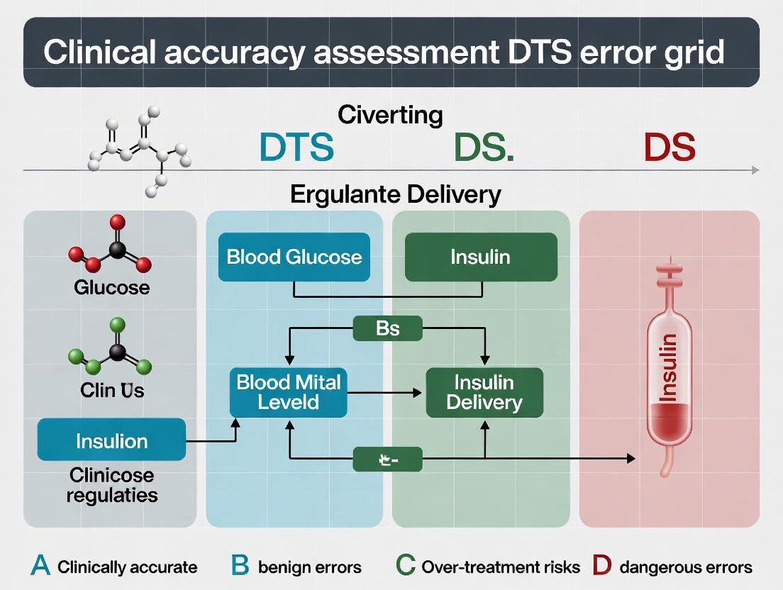
DTS Error Grid Analysis: The Complete Guide to Clinical Accuracy Assessment for Biomarkers and Wearables
This comprehensive guide explores DTS (Diabetes Technology Society) Error Grid Analysis, a critical methodology for evaluating the clinical accuracy of continuous glucose monitors (CGMs) and other vital sign monitoring technologies.
DTS Error Grid Analysis: The Complete Guide to Clinical Accuracy Assessment for Biomarkers and Wearables
Abstract
This comprehensive guide explores DTS (Diabetes Technology Society) Error Grid Analysis, a critical methodology for evaluating the clinical accuracy of continuous glucose monitors (CGMs) and other vital sign monitoring technologies. Aimed at researchers, scientists, and drug development professionals, the article provides a foundational understanding of the DTS grid's origins and purpose, details its methodological application for new device validation, offers strategies for troubleshooting and optimizing studies that employ it, and compares its performance and acceptance against legacy tools like the Clarke Error Grid. The synthesis provides actionable insights for robust clinical accuracy assessment in biomedical research and regulatory submissions.
What is DTS Error Grid Analysis? Origins, Purpose, and Clinical Significance
1. Introduction and Thesis Context
This application note is framed within a broader thesis investigating the clinical accuracy assessment of Digital Therapeutics (DTS). DTS are evidence-based, software-driven interventions for preventing, managing, or treating medical disorders. Traditional error grid analyses, such as the Clarke Error Grid Analysis (EGA) for blood glucose monitoring and the Parkes (Consensus) EGA, were designed for single-parameter, physiological metrics. DTS, however, often involve multi-parameter inputs, behavioral outcomes, and composite risk scores. This necessitates a novel analytical framework—DTS Error Grid Analysis (DTS-EGA)—to evaluate the clinical concordance and risk of DTS-generated recommendations or outputs against a clinical reference standard, moving beyond the limitations of Clarke and Parkes.
2. DTS-EGA Conceptual Framework
DTS-EGA is a multi-axis, risk-stratified plot that maps DTS-generated outputs (e.g., recommended therapy adjustment, risk score, behavioral prompt) against a clinician-panel-derived gold standard. The grid zones are defined by the potential for clinical harm, incorporating dimensions such as therapeutic efficacy, safety, and adherence impact.
Table 1: Proposed DTS Error Grid Zones and Clinical Implications
| Zone | Name | Clinical Risk Definition | Consequence for DTS Efficacy |
|---|---|---|---|
| A | Optimal Action | DTS output is clinically concordant with expert consensus. No risk. | Maximally beneficial. |
| B | Suboptimal but Safe | Deviation from consensus, but low probability of adverse outcomes or missed benefit. | Potentially reduced efficacy; requires design refinement. |
| C | Mild Risk | Action may lead to unnecessary user burden, mild side effects, or moderate delay in optimal care. | Questionable benefit-risk profile. |
| D | Significant Risk | Action carries high probability of moderate harm, significant care delay, or safety issue. | Unacceptable for clinical use. |
| E | Critical Risk | Action has high probability of severe, direct harm (e.g., toxic dose recommendation, critical warning omission). | DTS is dangerous and clinically invalid. |
Diagram Title: DTS-EGA Analysis Workflow
3. Experimental Protocol: Establishing the DTS-EGA Reference Standard
- Objective: To generate a gold-standard dataset of "clinically appropriate actions" for a given set of simulated or de-identified patient cases.
- Materials: See "The Scientist's Toolkit" (Section 6).
- Methodology:
- Case Development: Develop a comprehensive set of N (e.g., 500-1000) virtual patient cases. Each case includes a full data profile a DTS would process (e.g., historical data, real-time biosensor stream, patient log entries).
- Panel Selection & Blinding: Convene an independent, multidisciplinary expert panel (e.g., 5-7 clinicians, pharmacologists, behavioral scientists). Panelists are blinded to the DTS output and to each other's initial assessments.
- Independent Rating: Each panelist reviews each case independently and provides:
- The recommended clinical/behavioral action.
- A confidence score (1-5).
- A perceived risk rating if action is delayed/incorrect (Low/Med/High).
- Consensus Meeting: For cases with initial disagreement (e.g., actions spanning >2 DTS-EGA zones), a moderated consensus meeting is held. The final, agreed-upon action for each case constitutes the reference standard.
- Reference Database Lock: The finalized dataset is locked for subsequent DTS-EGA plotting.
4. Experimental Protocol: Executing a DTS-EGA Study
- Objective: To evaluate the clinical accuracy of a specific DTS by plotting its outputs against the reference standard.
- Methodology:
- DTS Processing: Run each of the N locked virtual patient cases through the DTS algorithm in a test environment to generate the DTS output (e.g., "increase dose by X," "send motivational alert," "risk score: 7.5").
- Paired Data Generation: Create a dataset of paired outcomes:
[Case_ID, Reference_Action, DTS_Output]. - Blinded Zone Mapping: A separate adjudication committee (blinded to the source of each action) maps each
(Reference_Action, DTS_Output)pair to a DTS-EGA zone (A-E) based on pre-defined, zone-specific rules (see Table 1). - Plotting & Analysis: Create the DTS-EGA scatter plot. Calculate the percentage of cases in each zone.
- Performance Benchmarking: Define acceptability criteria (e.g., ≥95% in Zone A+B, 0% in Zone E).
Table 2: Sample DTS-EGA Results for a Hypothetical Digital Insulin Advisor
| DTS-EGA Zone | Number of Cases (n=800) | Percentage | Pass/Fail vs. Benchmark |
|---|---|---|---|
| A (Optimal) | 720 | 90.0% | Pass |
| B (Safe) | 62 | 7.8% | Pass |
| C (Mild Risk) | 15 | 1.9% | (Review) |
| D (Significant Risk) | 3 | 0.4% | Fail |
| E (Critical Risk) | 0 | 0.0% | Pass |
| Total A+B | 782 | 97.8% | Pass (vs. 95% target) |
Diagram Title: DTS-EGA Zone Decision Logic
5. Advanced Applications: Multi-Dimensional DTS-EGA
For complex DTS, a layered analysis is proposed where separate (but potentially linked) grids are generated for different output types.
Table 3: Multi-Dimensional DTS-EGA for a Composite DTS
| Error Grid Layer | Plotted X-Y Axis | Purpose |
|---|---|---|
| Physiological | Reference Dose vs. DTS Dose | Evaluates direct therapeutic safety. |
| Behavioral | Reference Engagement Strategy vs. DTS Prompt | Evaluates appropriateness of behavioral intervention. |
| Risk | Reference Risk Stratification vs. DTS Risk Score | Evaluates accuracy of prognostic classification. |
6. The Scientist's Toolkit: Key Research Reagent Solutions
| Item | Function in DTS-EGA Research |
|---|---|
| Clinical Case Simulation Platform | Software to generate and manage large libraries of realistic, virtual patient cases with structured data profiles. |
| Expert Panel Management Software | Secure portal for blinded case distribution, independent rating, and data collection from clinical experts. |
| De-identified Real-World Data (RWD) Repositories | Source data (e.g., EHRs, wearables) for constructing externally valid virtual patient cases. |
| Adjudication Charter & Zone Rule Set | A living document defining precise, actionable criteria for mapping action pairs to specific DTS-EGA zones. |
| Statistical Analysis Package (e.g., R, Python with ggplot2/Matplotlib) | For automated generation of DTS-EGA plots, calculation of zone percentages, and confidence intervals. |
| Regulatory Guidance Documents | FDA (Software as a Medical Device), EMA, and IMDRF documents to align DTS-EGA structure with regulatory expectations for clinical validation. |
The development of the Diabetes Technology Society (DTS) Blood Glucose Monitor System (BGMS) Surveillance Protocol and its associated Error Grid analysis marked a pivotal shift in the assessment of glucose monitor accuracy. This initiative was driven by the clinical imperative to ensure that devices used for self-monitoring of blood glucose (SMBG) provide data reliable enough for daily therapeutic decision-making. Prior consensus standards (e.g., ISO 15197:2013) established baseline performance criteria but lacked the granular, risk-based clinical outcome analysis required to fully evaluate real-world impact. The DTS Error Grid research thesis posits that analytical accuracy (mean absolute relative difference, MARD) alone is insufficient; a clinically contextualized tool is essential to categorize measurement errors based on the probability and severity of adverse clinical outcomes. This framework is critical for researchers and drug development professionals who rely on accurate glucose data for clinical trials, closed-loop system validation, and the evaluation of new diabetes therapies.
Table 1: Comparison of Key Glucose Monitor Accuracy Standards
| Standard / Protocol | Primary Metric | Acceptance Criterion | Clinical Risk Assessment | Key Limitation Addressed by DTS |
|---|---|---|---|---|
| ISO 15197:2003 | Absolute relative difference | ≥95% within ±15 mg/dL (<75 mg/dL) or ±20% (≥75 mg/dL) | None | Binary pass/fail lacks clinical outcome stratification. |
| ISO 15197:2013 | Absolute relative difference | ≥95% within ±15 mg/dL or ±15%; ≥99% within consensus error grid Zones A+B | Incorporated Clarke Error Grid (1987) | Clarke Error Grid based on outdated therapies. |
| FDA Guidance (2016) | Aggregate MARD & per-point analysis | Recommends <10% MARD; detailed point-of-care device requirements | Emphasizes risk analysis | Guidance, not a mandated surveillance protocol. |
| DTS Surveillance Protocol | DTS Error Grid | ≥95% in clinically accurate Zone A (low risk); ≤2% in clinically dangerous Zone E (high risk) | Core focus: Direct linkage of error magnitude/direction to probable clinical outcome. | Provides a modern, treatment-relevant clinical risk model for the era of insulin analogs and tight glycemic control. |
Table 2: DTS Error Grid Zone Definitions and Clinical Implications
| Zone | Color | Risk Level | Definition | Example: True 70 mg/dL, Device Reads... |
|---|---|---|---|---|
| A | Green | No Effect or Alteration | Clinically accurate. Would prescribe same action as reference. | 63 - 77 mg/dL (±10%) |
| B | Yellow | Slight to Moderate Effect | Altered clinical action with little/no clinical risk. | 56 mg/dL (treatment for non-existent low) |
| C | Orange | Marked Effect | Altered action with moderate clinical risk. | 50 mg/dL (overtreatment of low, risk of hyperglycemia) |
| D | Red | Great Effect | Altered action with significant clinical risk. | 200 mg/dL (failure to treat severe hypoglycemia) |
| E | Purple | Dangerous | Opposite treatment action with dangerous consequences. | 250 mg/dL (administering insulin for a true hypoglycemic event) |
Detailed Experimental Protocols
Protocol 1: Execution of the DTS BGMS Surveillance Study for Market Evaluation
Objective: To rigorously assess the clinical accuracy of commercially available BGMS under controlled, clinically relevant conditions.
Materials: See "The Scientist's Toolkit" (Section 5). Procedure:
- Subject Recruitment & Categorization: Enroll a minimum of 100 subjects meeting predefined demographics. Stratify subjects into three blood glucose concentration categories:
<80 mg/dL(≈14%),80-180 mg/dL(≈60%), and>180 mg/dL(≈26%). - Sample Collection & Splitting: Obtain a fresh capillary fingerstick blood sample (≥1.5 µL). Immediately split the sample.
- Reference Measurement: Apply one portion to the reference instrument (YSI 2300 STAT Plus or equivalent). Perform measurement in duplicate. The mean value is the assigned reference glucose concentration.
- Test Device Measurement: Apply the other portion to the test BGMS. This is a single measurement per system. Use multiple lots of test strips.
- Data Pairing & Blinding: Record the paired result (reference value, test value). Operators for reference and device measurements must be blinded to each other's results.
- Repeat: Collect a minimum of 100-150 paired data points per system across the full glycemic range and all subject categories.
- Data Analysis: Calculate standard analytical metrics (MARD, % within ISO 15197:2013 criteria). Primary Endpoint: Plot all data pairs on the DTS Error Grid. Calculate the percentage of results in Zones A, B, C, D, and E. A system passes if ≥95% of results are in Zone A and ≤2% are in Zone E.
Protocol 2: In-Clinic Validation of a BGMS for a Drug/Device Combination Trial
Objective: To validate the performance of a specific BGMS intended for use as an endpoint measure in a clinical trial.
Materials: Similar to Protocol 1, tailored to trial population. Procedure:
- Protocol Alignment: Adapt the DTS surveillance protocol to the trial's specific population (e.g., type 2 diabetes, pediatrics, pregnant women).
- Comparative Measurement: During scheduled trial visits, collect an additional capillary sample alongside trial-specific procedures. Process per Protocol 1 steps 2-5.
- Context-Specific Range: Ensure adequate sampling across the expected glycemic range for the trial population.
- Risk Assessment: Apply DTS Error Grid analysis. The sponsor must predefine acceptability criteria (e.g., ≥98% Zone A+B, 0% Zone E) justified by the trial's risk profile.
- Documentation: Include the validation report in the trial's regulatory submission to justify the suitability of the chosen BGMS.
Visualizations
Title: DTS vs. Legacy Accuracy Assessment Workflow
Title: Clinical Risk Decision Tree for DTS Error Grid
The Scientist's Toolkit: Research Reagent Solutions & Essential Materials
Table 3: Key Materials for DTS-Style Clinical Accuracy Studies
| Item | Function & Rationale |
|---|---|
| Reference Analyzer (e.g., YSI 2300/2900 STAT Plus) | Gold-standard instrument using glucose oxidase method. Provides the "true" glucose value against which all devices are compared. Requires rigorous daily calibration and QC. |
| Hematocrit-Adjusted Blood Gas Analyzer | Measures hematocrit (HCT) levels. Critical for assessing device performance across the physiological HCT range (e.g., 30-55%), as HCT is a known interferent for many BGMS. |
| Certified Glucose Control Solutions (Low, Mid, High) | Used for daily quality control (QC) of both reference and test devices, ensuring analytical integrity throughout the study. |
| Capillary Blood Collection System (Lancets, Microtainers) | Standardized materials for obtaining fresh, unaltered capillary fingerstick samples. Volume must be sufficient for splitting between reference and test devices. |
| Environmental Chambers | Allows testing of BGMS under controlled temperature and humidity conditions (per manufacturer specifications), assessing robustness to typical user environments. |
| Interferent Stock Solutions (e.g., Acetaminophen, Ascorbic Acid, Maltose) | Prepared at high concentration for spiking studies. Used to evaluate the susceptibility of the BGMS to common pharmacological and endogenous substances. |
| DTS Error Grid Plotting Software / Algorithm | Custom software or validated script to automatically plot paired (reference, test) data points and calculate the percentage distribution across the five risk zones. |
Within the context of ongoing clinical accuracy assessment research for Diabetes Technology Society (DTS) Error Grids, the precise definition and clinical implications of its five risk zones (A-E) are paramount. This application note details these zones, provides protocols for generating and validating DTS grid data, and situates the analysis within a framework for evaluating the clinical accuracy of continuous glucose monitoring (CGM) and blood glucose monitoring (BGM) systems. The DTS Grid is an analytical tool used to assess the clinical risk of glucose meter inaccuracies by categorizing paired reference and sensor values.
The DTS Grid Risk Zones: Definitions and Clinical Significance
The DTS Grid divides clinical risk into five discrete zones based on the potential for adverse clinical outcomes arising from a discrepancy between a measured glucose value and a reference value.
Table 1: DTS Error Grid Risk Zones (A-E)
| Zone | Risk Category | Clinical Description | Typical Action (or Inaction) Prompted | Acceptable for Clinical Use? |
|---|---|---|---|---|
| A | No Effect | Clinically accurate. No risk. | Correct and safe clinical action. | Yes |
| B | Slight to Moderate | Altered clinical action with little to no risk. May include unnecessary hyper/hypo corrections. | Benign or low-risk action. Potentially suboptimal. | Generally Yes |
| C | Moderate to High | Altered clinical action with possible significant medical risk. | Over-correction or failure to treat, leading to potential harm. | No |
| D | Dangerous | Significant medical risk due to failure to detect or treat extreme glucose levels. | Failure to treat severe hypo- or hyperglycemia. | No |
| E | Extreme Danger | Erroneous treatment leading to extreme clinical danger (e.g., treating hypoglycemia as hyperglycemia). | Catastrophically incorrect action (e.g., administering insulin for a low glucose value). | No |
Experimental Protocols for DTS Grid Assessment
Protocol 1: Generation of Paired Glucose Data Set
Objective: To collect a paired data set of reference glucose values and device-generated glucose values spanning the clinically relevant range (e.g., 40-400 mg/dL).
- Subject Cohort: Recruit a representative population of subjects with diabetes (Type 1 and Type 2).
- Reference Method: Utilize a certified laboratory glucose analyzer (e.g., YSI 2300 STAT Plus) as the reference standard. Capillary or venous blood samples are processed immediately.
- Device Under Test (DUT): Use the CGM or BGM system according to the manufacturer's instructions for use (IFU).
- Sampling Protocol: For BGM, take a capillary sample simultaneously with the reference sample. For CGM, record the sensor glucose value at the exact time of the reference blood draw. Collect a minimum of 100-150 paired points across the glycemic range, with deliberate oversampling in hypoglycemic (<70 mg/dL) and hyperglycemic (>180 mg/dL) regions.
- Data Recording: Record paired values (Reference, DUT) with timestamps and subject identifier.
Protocol 2: Plotting and Zone Assignment
Objective: To plot paired data on the DTS Grid and assign each point to a risk zone (A-E).
- Grid Template: Obtain the official DTS Grid coordinates and zone boundaries.
- Plotting: For each paired data point (Ref, DUT), plot the reference value on the x-axis and the DUT value on the y-axis.
- Zone Assignment: Algorithmically or manually determine the zone for each point based on the grid's polygonal zone boundaries defined in the DTS specification.
- Calculation: Compute the percentage of data points falling within each zone (A, B, C, D, E).
Protocol 3: Statistical & Clinical Accuracy Analysis
Objective: To derive metrics for regulatory submission and clinical risk assessment.
- Primary Endpoint: Calculate the percentage of values in Zones A+B. (Industry standard often requires >95% or >99% in A+B for approval).
- Secondary Endpoints: Report individual percentages for Zones A, B, C, D, and E. Perform a detailed analysis of points in Zones C-E, including the magnitude of error and the specific clinical scenario (e.g., hypoglycemic misclassification).
- Contextual Analysis: Within the broader thesis, compare DTS Grid outcomes with other accuracy metrics (Mean Absolute Relative Difference (MARD), ISO 15197:2013 criteria) to provide a multi-dimensional accuracy assessment.
Visualization of DTS Grid Analysis Workflow
Title: DTS Grid Clinical Accuracy Assessment Workflow
The Scientist's Toolkit: Research Reagent Solutions
Table 2: Essential Materials for DTS Grid Studies
| Item | Function in DTS Grid Research |
|---|---|
| Certified Glucose Analyzer (e.g., YSI 2300 STAT Plus) | Gold-standard reference method for plasma glucose measurement. Provides the benchmark for all accuracy assessments. |
| CE-Marked/ FDA-Cleared BGM Systems & Strips | Device Under Test (DUT) for capillary blood glucose monitoring. Must be used with lot-specific calibration codes. |
| CGM Systems (Sensor, Transmitter) | DUT for continuous interstitial glucose monitoring. Requires proper insertion and calibration per IFU. |
| Phlebotomy & Capillary Sampling Kits | For collecting venous (reference) and capillary (BGM) blood samples in a standardized, clinical manner. |
| Clinitubes or Heparinized Tubes | For immediate stabilization and transport of blood samples to the reference lab analyzer. |
| DTS Grid Zone Boundary Coordinates | Official digital or mathematical definition of the polygonal zones A-E. Essential for algorithmic zone assignment. |
| Statistical Software (e.g., R, SAS, Python with Matplotlib) | For data management, plotting points on the DTS Grid, calculating zone percentages, and performing advanced statistical analysis. |
| Quality Control Solutions (e.g., known glucose concentrations) | For verifying the accuracy of both the reference lab analyzer and the BGM systems before, during, and after the study. |
This document details application notes and experimental protocols for evaluating the clinical accuracy of Digital Therapeutic (DTx) and connected health devices. The work is integral to a broader thesis developing a novel Dynamic Time-Series (DTS) Error Grid analysis framework. This framework moves beyond static point-in-time accuracy metrics (e.g., Clarke Error Grid) to assess the clinical risk of errors in continuous, multi-analyte data streams from devices like Continuous Glucose Monitors (CGMs), emerging biomarker sensors, and multi-parameter wearables. The DTS Error Grid is designed to evaluate the clinical impact of temporal inaccuracies, trend deviations, and data dropouts, which are critical for therapeutic decision-making.
Table 1: Device Classes, Target Analytes, and Key Performance Metrics
| Device Class | Primary Analyte(s) | Typical Sample Matrix | Key Performance Metrics (ISO/Consensus Standards) | Relevance to DTS Error Grid Assessment |
|---|---|---|---|---|
| CGM Systems | Glucose | Interstitial Fluid | MARD (Mean Absolute Relative Difference), Consensus Error Grid (CEG) % in Zones A+B, Time Lag | Core use case: Assessing clinical risk of temporal discrepancies vs. reference. |
| Ketone Monitors | β-Hydroxybutyrate (BHB) | Blood, Interstitial Fluid | Bias vs. reference laboratory method (e.g., plasma BHB), Clinical agreement at decision thresholds (e.g., 0.6, 1.5, 3.0 mmol/L) | High-risk analyte; DTS Grid must weight hyperketonemia errors severely. |
| Lactate Wearables | Lactate | Sweat, Interstitial Fluid | Sensitivity (µA/mM·cm²), Limit of Detection (LoD), Correlation coefficient (r) vs. blood lactate during exercise tests | Trend accuracy during dynamic physiological stress is critical for DTS. |
| Multi-parameter (EDA) | Electrodermal Activity, Heart Rate, ACC | Skin Surface | Signal-to-Noise Ratio (SNR), Peak detection accuracy for HR, Tonic/Phasic EDA decomposition fidelity | Assessing composite clinical risk from fused, low-latency data streams. |
| Emerging Biomarker (Cortisol) | Cortisol | Sweat, Interstitial Fluid | LoD (pg/mL), Dynamic range, Cross-reactivity (%) with analogous steroids (e.g., cortisone) | Challenges in establishing a continuous reference; DTS must model diurnal rhythm context. |
Experimental Protocols for DTS Error Grid Validation
Protocol 3.1: In-Clinic Controlled Challenge Study for CGM/Dual-analyte Systems
Objective: To generate paired, time-synchronized device and reference data under conditions of dynamic analyte concentration change, for DTS Error Grid construction and validation. Materials: See "Scientist's Toolkit" (Section 5). Methodology:
- Participant Preparation & Instrumentation: Recruit consented participants (e.g., n=20-30). Attach test devices (CGM, ketone sensor) to approved sites. Place venous cannula for frequent blood sampling.
- Baseline Period (-30 to 0 min): Collect fasting baseline reference samples (YSI 2900 for glucose, laboratory plasma BHB).
- Dynamic Challenge Phase (0-180 min):
- 0 min: Administer standardized mixed-meal tolerance test or variable glucose clamp.
- 60 min: Initiate moderate-intensity exercise protocol (treadmill/cycle ergometer) for 30 min to induce lactate and ketone changes.
- Monitor continuously. Draw venous reference samples at 5-15 minute intervals.
- Data Synchronization: Timestamp all device data (via API/logger) and reference draws. Align streams using a common time server. Apply physiologically justified time-lag correction (e.g., 5-10 min for interstitial glucose) based on population pharmacokinetic models.
- DTS Error Grid Analysis:
- Input synchronized, paired time-series into DTS algorithm.
- The algorithm calculates instantaneous error and, critically, the error in the first derivative (trend: stable, rising, falling).
- Each data pair is mapped to a DTS Risk Zone (e.g., Zone A: "No Effect on Clinical Action," Zone B: "Altered Clinical Action-Low Risk," Zone C: "Altered Clinical Action-High Risk," Zone D: "Dangerous Failure") based on the combined value-trend error matrix.
- Output: Percentage of data points in each DTS Risk Zone; visualization of error clusters over time.
Diagram Title: DTS Error Grid Validation Study Workflow
Protocol 3.2: At-Home Free-Living Validation for Wearables
Objective: To assess device performance and DTS clinical risk in an ecologically valid setting. Methodology:
- Equipment Distribution: Provide participants with test wearable(s), a reference device (e.g., capillary BHB meter, research-grade ECG patch), and a smartphone for data logging and ecological momentary assessment (EMA).
- Protocol (7-14 days): Participants conduct normal activities. Protocol triggers:
- Scheduled: 4x daily capillary reference checks (fasting, post-prandial).
- Event-Based: Participant logs symptoms (e.g., dizziness) via EMA app, triggering a reference measurement.
- Device-Triggered: Automatically flag periods of rapid analyte change for user-prompted verification.
- Data Fusion & Analysis: Synchronize device, reference, and EMA data. Apply DTS Error Grid analysis, with enhanced weighting for errors occurring during user-reported symptomatic events.
Signaling Pathways & Biological Context for Biomarkers
Diagram Title: Biomarker Pathway from Blood to Wearable Sensor
The Scientist's Toolkit: Research Reagent Solutions
Table 2: Essential Materials for Device Validation Studies
| Item | Function & Relevance to DTS Research |
|---|---|
| YSI 2900 STAT Plus Analyzer | Gold-standard benchtop reference for glucose and lactate in whole blood. Provides the primary Y-axis data for CGM/lactate sensor DTS analysis. |
| Plasma β-Hydroxybutyrate Reference Method (e.g., LC-MS/MS or Enzymatic Assay) | Definitive reference for ketone monitor validation. Critical for setting the high-risk thresholds in the DTS Error Grid. |
| Research-Grade ECG/EDA Reference Device (e.g., BIOPAC System) | Provides high-fidelity, timestamped physiological signals (HR, EDA) to validate derived parameters from consumer wearables. |
| Variable Glucose Clamp Apparatus | Infusion system to create controlled, dynamic glucose profiles. The "known input" for rigorously testing DTS algorithm's trend error detection. |
| Time Synchronization Logger (e.g., LabJack) | Hardware to generate and record a common timestamp pulse to all devices (sensors, pumps, reference draws), enabling millisecond-accurate data alignment for DTS. |
| Structured Data Pipeline (e.g., Python Pandas/NumPy) | Custom scripts for merging, time-aligning, cleaning, and analyzing large-scale time-series data sets from multiple heterogeneous sources. |
| DTS Error Grid Visualization Software | Custom plotting library (e.g., Matplotlib/D3.js) to generate the dynamic, multi-dimensional error grid plots, showing risk zones overlaid on temporal data. |
Digital Technology Systems (DTS), such as connected drug delivery devices, wearable sensors, and software-based clinical outcome assessments, are increasingly integral to modern drug development. Their role in regulatory submissions to the U.S. Food and Drug Administration (FDA) and in meeting international ISO standards is critical for demonstrating product safety, efficacy, and quality. This content is framed within a broader thesis research focusing on the clinical accuracy assessment of DTS, specifically utilizing error grid analysis to categorize and quantify measurement errors against clinically significant outcomes.
Key Regulatory Frameworks:
- FDA: The FDA provides guidance under the Food, Drug, and Cosmetic Act, incorporating DTS review within existing pathways for devices (510(k), De Novo, PMA) and drugs/biologics. For Software as a Medical Device (SaMD), frameworks like the Digital Health Software Precertification (Pre-Cert) Program and guidance on Clinical Decision Support (CDS) software are relevant. The use of DTS-generated data in New Drug Applications (NDAs) or Biologics License Applications (BLAs) requires rigorous validation.
- ISO Standards: ISO standards provide internationally recognized benchmarks for quality and safety. Key standards include:
- ISO 13485: Quality management systems for medical devices.
- ISO 14971: Application of risk management to medical devices.
- ISO 62304: Medical device software – Software life cycle processes.
- ISO 82304-1: Health software – Part 1: General requirements for product safety.
- ISO/IEC 27001: Information security management.
Application Note on Error Grid Analysis for DTS Validation: Error grid analysis, derived from methodologies like the Clarke Error Grid for blood glucose monitoring, is a powerful tool for assessing the clinical accuracy of DTS. It moves beyond simple statistical agreement (e.g., mean absolute percentage error) by mapping reference method results against DTS outputs into zones (A-E) with defined clinical risk implications. This provides a clinically contextualized validation metric that is highly persuasive in regulatory submissions to demonstrate that measurement inaccuracies are not clinically dangerous.
Data Presentation: Regulatory Metrics and Standards Comparison
Table 1: Key ISO Standards Relevant to DTS Development and Submission
| Standard | Title | Primary Scope | Relevance to DTS Clinical Accuracy |
|---|---|---|---|
| ISO 13485:2016 | Medical devices – Quality management systems | Establishes requirements for a comprehensive QMS for the design and manufacture of medical devices. | Mandates validation of design and development outputs, ensuring processes for verifying DTS accuracy are controlled and documented. |
| ISO 14971:2019 | Medical devices – Application of risk management | Framework for identifying, estimating, evaluating, controlling, and monitoring risks throughout a device lifecycle. | Error grid analysis directly informs the evaluation of "use" risks related to clinical inaccuracy. Zones C, D, E represent increasing risk severity. |
| ISO 62304:2006 + Amd.1:2015 | Medical device software – Software life cycle processes | Defines life cycle processes with safety classification (A: No injury, B: Non-serious injury, C: Death/serious injury). | Dictates the rigor of software validation testing. Clinical accuracy assessment protocols are part of software verification & validation for Class B/C devices. |
| ISO 82304-1:2016 | Health software – Part 1: General requirements for product safety | General requirements for the safety and security of health software products not already covered as medical devices. | Applies to DTS components that may be wellness-focused or adjunctive; still requires accuracy claims to be substantiated. |
Table 2: FDA Submission Pathways and DTS Data Requirements
| Submission Pathway | Typical DTS Context | Key Clinical Accuracy Data Requirements | Relevant Guidance/Documents |
|---|---|---|---|
| 510(k) Clearance | New DTS substantially equivalent to a predicate device. | Performance testing vs. predicate, including accuracy, precision, and usability. | FDA Guidance: "Technical Performance Assessment of Digital Health Technologies" |
| De Novo Request | Novel DTS of low-to-moderate risk without a predicate. | Comprehensive validation establishing a reasonable assurance of safety and effectiveness, including clinical accuracy studies. | FDA Guidance: "De Novo Classification Process" |
| PMA (Premarket Approval) | High-risk Class III DTS. | Extensive scientific evidence from clinical investigations, including detailed accuracy profiling in the target population. | FDA Guidance: "Clinical Investigation of Devices" |
| NDA/BLA (Drug/Biologic) | DTS used as a companion diagnostic, outcome measure, or adherence tracker. | Validation that the DTS reliably measures the intended physiological parameter or behavioral outcome. Data must support the drug's efficacy/safety claims. | FDA Guidance: "Patient-Reported Outcome Measures: Use in Medical Product Development" (if applicable) |
Experimental Protocols
Protocol 1: Clinical Accuracy Validation of a DTS Using Error Grid Analysis
1. Objective: To assess the clinical accuracy of a novel wearable glucose monitor (DTS) against a clinically accepted reference method (venous blood analyzed via laboratory-grade analyzer) and categorize errors using an adapted error grid.
2. Materials: See "The Scientist's Toolkit" below.
3. Methodology:
- Study Design: A single-center, prospective, method-comparison study.
- Participants: Recruit N=150 participants with diabetes mellitus (Type 1 and Type 2), ensuring a broad distribution of glucose values across the clinically relevant range (e.g., 40-400 mg/dL).
- Procedure:
- Obtain informed consent.
- Simultaneously collect:
- Test Method: Glucose reading from the investigational wearable DTS.
- Reference Method: Venous blood sample drawn and immediately processed using a laboratory-grade glucose analyzer (YSI 2300 STAT Plus).
- Collect paired data points across various physiological conditions (fasting, post-prandial, during exercise) over a 24-48 hour period for each participant.
- Data Analysis:
- Plot all paired data points on a scatter plot with Reference Value on the x-axis and DTS Value on the y-axis.
- Superimpose a pre-defined error grid (e.g., Clarke Error Grid or a disease-specific adapted grid).
- Categorize each data point into zones:
- Zone A: Clinically accurate. No effect on clinical action.
- Zone B: Clinically acceptable. Alters clinical action with little or no risk.
- Zone C: Over-correction. Unnecessary treatment.
- Zone D: Dangerous failure to detect. Treatment omitted.
- Zone E: Erroneous treatment. Opposite treatment applied.
- Calculate the percentage of data points falling within each zone. Regulatory success criteria often require >99% in Zone A + B, and 0% in Zones D + E.
- Perform supplementary statistical analysis (e.g., Mean Absolute Relative Difference (MARD), Bland-Altman plots).
Protocol 2: Analytical Verification of DTS Software Algorithm
1. Objective: To verify the output of a DTS signal processing algorithm against a predefined "golden" dataset.
2. Methodology:
- Input Dataset: Assemble a curated dataset of raw sensor signals with known, corresponding "true" output values. This includes edge cases and error states.
- Test Execution: Run the dataset through the algorithm in the DTS software.
- Output Comparison: Automatically compare the algorithm's output to the expected "golden" output.
- Pass/Fail Criteria: Define acceptable tolerance limits for numerical outputs (e.g., 99.5% match within ±0.5%). Document any discrepancies and their root causes.
Visualizations
Title: DTS Development and Regulatory Submission Workflow
Title: Error Grid Analysis Protocol Flowchart
The Scientist's Toolkit: Key Research Reagent Solutions
Table 3: Essential Materials for DTS Clinical Accuracy Experiments
| Item / Reagent | Function in DTS Validation | Example / Specification |
|---|---|---|
| Reference Standard Device/Analyzer | Provides the "ground truth" measurement against which the DTS output is compared. Essential for calculating error. | Laboratory glucose analyzer (e.g., YSI 2300), FDA-cleared spirometer, validated clinical gold-standard instrument. |
| Simulated Signal/Data Generator | Tests DTS hardware/software with known, repeatable input signals. Used for initial analytical validation. | ECG waveform simulator, programmable blood pressure pump, motion phantom for accelerometers. |
| Data Logging & Synchronization System | Precisely timestamps and pairs DTS output with reference method data. Critical for valid paired analysis. | Secure cloud platform with API, or local software with manual timestamp verification protocol. |
| Validated Clinical Outcome Assessment (COA) | If DTS measures a patient-reported outcome, a validated questionnaire is the reference for validating the digital COA. | EQ-5D for quality of life, PHQ-9 for depression, established movement rating scales. |
| Statistical Analysis Software | Performs error grid plotting, statistical agreement analysis (Bland-Altman, MARD), and generates submission-ready reports. | R (with ggplot2, BlandAltmanLeh), Python (SciPy, Matplotlib), MedCalc, SAS. |
| Quality Management System (QMS) Software | Manages documentation, protocol deviations, and data integrity per ISO 13485 requirements for regulatory audits. | Electronic QMS platforms (e.g., Greenlight Guru, Qualio, MasterControl). |
How to Implement DTS Error Grid Analysis: A Step-by-Step Protocol for Researchers
The clinical accuracy assessment of Digital Therapeutic Solutions (DTS), particularly for chronic disease management like diabetes, relies on rigorous analytical and clinical validation. The core of this validation is the DTS Error Grid analysis, a methodology that assesses the clinical risk of inaccurate glucose monitoring system readings. This research is foundational for regulatory approval and real-world clinical utility. The integrity of the entire error grid analysis hinges upon three foundational pillars of study design: a representative Patient Population, a robust Reference Method, and appropriate Data Pairing.
The Three Pillars: Detailed Application Notes
Patient Population
A clinically relevant patient population ensures that the DTS performance is evaluated across the full spectrum of intended use.
Application Notes:
- Inclusion Criteria: Must reflect the label claim. For a general-use DTS, this requires enrollment across all age groups (pediatric, adult, geriatric), diabetes types (1, 2, gestational), a wide range of hematocrit levels (e.g., 30%-55%), and varying severities of illness.
- Exclusion Criteria: Should be minimal and justifiable. Overly restrictive criteria limit generalizability.
- Sample Size: Must be statistically justified to provide sufficient precision for error grid analysis (e.g., sufficient data pairs in each error grid zone). Current consensus, informed by recent regulatory guidance (FDA, 2020; ISO 15197:2013), recommends a minimum of 100-150 unique subjects to capture biological variability.
- Recruitment Setting: Should include both controlled clinical settings and home-use environments to assess performance under real-world conditions.
Reference Method
The reference method serves as the "gold standard" against which the DTS is compared. Its accuracy and precision are paramount.
Application Notes:
- Methodology: For blood glucose monitoring, the reference is typically YSI (Yellow Springs Instruments) glucose analyzer or another FDA-approved clinical laboratory hexokinase method. For continuous glucose monitors (CGMs), arterialized venous blood sampled frequently is the reference.
- Quality Control: The reference laboratory must operate under CLIA/CAP or equivalent accreditation. Regular calibration and use of traceable standards are mandatory.
- Procedure: Capillary, venous, or arterial blood sampling must be performed by trained personnel. The time lag between DTS measurement and reference sample acquisition must be minimized and documented (typically < 1 minute for capillary blood glucose).
Data Pairing
This defines the temporal and contextual relationship between the DTS reading and the reference value.
Application Notes:
- Temporal Alignment: A data pair consists of a DTS result and a reference result measured as close in time as possible to minimize physiological glucose fluctuation as a source of error. Protocols must define the maximum allowable time difference.
- Clinical Context: Data should be collected across a wide glycemic range (e.g., 40-400 mg/dL) and during various physiological states (fasting, post-prandial, during exercise). This ensures error grid zones are populated across all clinically relevant scenarios.
- Independence: Data pairs must be statistically independent. For frequent-sampling devices like CGMs, appropriate steps (e.g., using only one data point per 15-minute interval) must be taken to avoid autocorrelation.
Table 1: Quantitative Summary of Current Consensus Requirements for DTS Study Design
| Pillar | Parameter | Current Consensus / ISO 15197:2013 Requirement | Typical Target in Contemporary Studies |
|---|---|---|---|
| Patient Population | Minimum Number of Subjects | 100 subjects minimum | 150+ subjects for robust stratification |
| Hematocrit Range | Not less than 30% and not more than 55% | 20%-65% for extended claim | |
| Glucose Range | 40-400 mg/dL (2.2-22.2 mmol/L) | 30-500 mg/dL for wider evaluation | |
| Reference Method | Acceptable Standard | YSI 2300 STAT Plus or traceable laboratory method | FDA-cleared lab hexokinase method |
| Sample Type | Capillary (fingerstick) or venous plasma | Capillary for BGM, Arterialized venous for CGM | |
| Data Pairing | Minimum Number of Pairs | At least 100 pairs per subject stratum | 2-3 pairs per subject per day over 7-14 days |
| Time Alignment | Reference within 5 mins of BGM (capillary) | Reference within 1 min of capillary BGM reading |
Experimental Protocols
Protocol 1: Clinical Study for DTS Error Grid Analysis
Title: Prospective, Single-Group Assignment Study for Clinical Accuracy Assessment of a Novel Digital Glucose Monitoring System.
Objective: To evaluate the clinical accuracy of the investigational DTS against a reference method across a representative population using DTS Error Grid analysis.
Materials:
- Investigational DTS device and sensors/consumables.
- Reference method system (e.g., YSI 2300 STAT Plus analyzer with reagents).
- Sample collection supplies (lancets, capillary tubes, fluoride-oxalate tubes, venipuncture kits).
- Data collection forms (electronic or paper).
- Calibration and quality control materials for reference analyzer.
Procedure:
- Ethics & Consent: Obtain IRB/IEC approval. Recruit subjects per inclusion/exclusion criteria and obtain informed consent.
- Subject Enrollment & Stratification: Enroll a minimum of 150 subjects. Stratify recruitment to ensure adequate representation across age, gender, diabetes type, and hematocrit ranges.
- Clinical Visit (Clinic Setting): a. Subject arrives fasted. Insert/initialize investigational DTS per manufacturer's instructions. b. Over an 8-12 hour visit, obtain paired measurements at predetermined intervals (e.g., every 15-30 minutes) and during induced glycemic excursions (via meal tolerance test). c. For each pair: Immediately perform fingerstick test with DTS. Within 60 seconds, collect a capillary blood sample from the same finger prick for reference analysis. Record time, DTS value, and subject condition. d. Process reference sample per lab protocol and analyze on YSI within 30 minutes of collection.
- Home-Use Phase: a. Subject uses the DTS at home for 7-14 days. b. Subject performs at least 2 paired measurements per day at varying times (pre-meal, post-meal, bedtime). c. For each pair, subject uses the DTS, then collects a capillary sample on a filter paper card or microtainer which is mailed daily to the central lab for reference analysis.
- Data Management: Enter all DTS and paired reference values into a secure database. Ensure blinding of analysts to the paired values from the other method during initial data entry and analysis.
- Statistical Analysis: a. Plot all data pairs on the DTS Error Grid (e.g., Consensus Error Grid for blood glucose). b. Calculate the percentage of values in Zones A (clinically accurate) and B (clinically acceptable). Regulatory success often requires >99% in Zones A+B. c. Perform additional analyses: Mean Absolute Relative Difference (MARD), Bland-Altman plots, regression analysis.
The Scientist's Toolkit: Research Reagent Solutions
Table 2: Essential Materials for DTS Clinical Accuracy Studies
| Item | Function & Explanation |
|---|---|
| YSI 2300 STAT Plus Analyzer | The benchmark reference instrument. Uses glucose oxidase methodology to provide high-precision plasma glucose equivalent values. Must be calibrated daily with traceable standards. |
| Enzymatic Hexokinase Reagent Kit | Alternative reference lab method. Hexokinase catalyzes the phosphorylation of glucose by ATP; the reaction is measured spectrophotometrically. Known for high specificity and accuracy. |
| CLIA-Certified Quality Controls (High/Normal/Low) | Used to validate the accuracy and precision of the reference analyzer before, during, and after each run. Ensures the entire analytical system is functioning within specified parameters. |
| Capillary Blood Collection Tubes (Fluoride-Oxalate) | Preserves glucose in capillary blood samples by inhibiting glycolysis via fluoride, preventing falsely low reference values between collection and analysis. |
| Standardized Glucose Solutions for Calibration | Traceable to international standards (NIST), used to establish the calibration curve for the reference analyzer, ensuring measurement trueness. |
| DTS Error Grid Plotting Software | Specialized software (e.g., ACE, EGES) that automates the plotting of data pairs onto the appropriate error grid (Consensus, Surveillance, etc.) and calculates zone percentages. |
Visualizing the Study Workflow and Error Grid Logic
DTS Accuracy Study Workflow
Logic Flow for DTS Error Grid Analysis
Within the context of DTS (Digital Thermographic System) error grid clinical accuracy assessment research, the precision of spatial data pairing is paramount. Error grids, analogous to Clarke Error Grids for glucose monitoring, require meticulous point-to-point correspondence between the reference standard measurements (e.g., gold-standard temperature probes) and the DTS-derived measurements. This document outlines standardized protocols for ensuring accurate paired measurements for grid placement, a critical factor in validating clinical accuracy and mitigating misalignment errors that can skew sensitivity and specificity calculations in drug development thermographic studies.
Core Protocol: Paired Point Acquisition for Error Grid Construction
Objective: To establish a one-to-one correspondence between reference (R) and test (T) measurement points across a defined anatomical grid.
Principle: Each grid coordinate (e.g., G[x,y]) must be associated with a synchronized temporal and spatially co-located pair (R_i, T_i). The spatial tolerance must be defined a priori based on the DTS spatial resolution and the clinical application.
Pre-Collection Calibration & Mapping
- Grid Template Generation: Create a physical or digital grid template segmented into zones (e.g., A1, B2, C3). Zone size is determined by the pathology area and DTS spatial resolution (e.g., 5x5 mm).
- DTS-System Spatial Calibration: Calibrate the DTS camera field-of-view using a standardized calibration target. Establish pixels-to-millimeter conversion.
- Reference Probe Calibration: All contact reference probes (e.g., fluoroptic thermometers) must be calibrated against a NIST-traceable standard within 24 hours of data collection.
Procedural Workflow for Paired Data Collection
- Subject Positioning & Grid Application: Position the subject per protocol. Affix the physical grid template or project the digital grid onto the anatomical region of interest. Mark grid vertices with fiducial markers visible to both the DTS and visual assessment.
- Reference Measurement Placement: Under standardized environmental controls (ambient temp: 23°C ± 1°C; humidity: 50% ± 5%), place the reference probe at the center of a pre-defined grid zone. Record the value after thermal equilibrium (≥ 60 sec).
- Simultaneous DTS Image Capture: At the moment of reference value recording, capture the DTS thermogram. The timestamp for R_i and T_i must be identical (synchronized system clocks).
- Spatial Registration: In post-processing, map the physical grid coordinate of R_i to the corresponding pixel cluster in the DTS image (T_i). Use fiducial markers for affine transformation if needed.
- Iteration: Sequentially move the reference probe to every grid zone, repeating steps 2-4. The order should be randomized to minimize temporal drift effects.
- Data Logging: All pairs (R_i, T_i, Grid_ID, Timestamp) are logged directly into a relational database to prevent transcription error.
Key Experimental Methodologies from Literature
Methodology 1: Phantom-Based Validation of Pairing Accuracy
- Purpose: To quantify the spatial misalignment error between paired points in a controlled setting.
- Protocol: A thermally heterogeneous phantom with embedded, precise temperature sources is used. A high-resolution grid is superimposed. Reference temperatures from the embedded sensors are paired with DTS readings at corresponding coordinates. The experiment is repeated with introduced, known spatial offsets (1mm, 2mm, 5mm). The deviation in the calculated (DTS - Reference) error is plotted against offset distance to establish a tolerance threshold.
- Key Metric: Root Mean Square Error (RMSE) of paired differences versus offset.
Methodology 2: Intra-Observer Variability in Grid Placement
- Purpose: To assess the reproducibility of the manual grid placement and point pairing process.
- Protocol: Three trained observers independently place the same digital grid on the same set of 50 subject thermograms. Each observer records the paired DTS value for 10 predefined anatomical landmarks per image. Analysis uses the Intraclass Correlation Coefficient (ICC) for absolute agreement.
- Key Metric: ICC(3,k) for average measures.
Table 1: Impact of Spatial Offset on Paired Measurement Error (Phantom Study)
| Introduced Spatial Offset (mm) | Mean Absolute Paired Error (°C) | RMSE (°C) | Clinical Error Grid Zone Migration* |
|---|---|---|---|
| 0 (Perfect Alignment) | 0.12 | 0.15 | Zone A (Clinically Accurate) |
| 1 | 0.18 | 0.22 | Zone A |
| 2 | 0.35 | 0.41 | Zone A/B Border |
| 5 | 0.87 | 1.04 | Zone C (Altered Clinical Action) |
*Illustrative example based on a hypothetical DTS error grid.
Table 2: Intra-Observer Reliability for Manual Point Pairing (n=50 images)
| Anatomical Landmark | Observer 1 vs 2 (ICC) | Observer 1 vs 3 (ICC) | Observer 2 vs 3 (ICC) | Average ICC (95% CI) |
|---|---|---|---|---|
| Dorsal Hand | 0.98 | 0.97 | 0.98 | 0.98 (0.96-0.99) |
| Plantar Foot | 0.94 | 0.93 | 0.95 | 0.94 (0.90-0.97) |
| Forehead | 0.99 | 0.99 | 0.98 | 0.99 (0.98-0.995) |
| Overall Mean | 0.97 | 0.96 | 0.97 | 0.97 (0.95-0.98) |
Visualizations
Title: Paired Measurement Collection Workflow
Title: Thesis Context of Paired Measurement Protocol
The Scientist's Toolkit: Research Reagent Solutions
Table 3: Essential Materials for Paired Measurement Studies
| Item | Function/Benefit | Key Specification Example |
|---|---|---|
| NIST-Traceable Thermal Phantom | Provides stable, known temperature zones for validating DTS accuracy and pairing protocols. | Embedded sensors with ≤0.1°C absolute accuracy. |
| Fluoroptic/Thermocouple Reference Probes | Gold-standard for contact temperature measurement; minimal interference with DTS readings. | Response time < 1s, calibrated uncertainty ±0.05°C. |
| Anatomical Fiducial Markers | Enable precise spatial co-registration between physical grid and DTS image. | Low thermal emissivity (e.g., reflective) and high visual contrast. |
| Synchronized Data Acquisition Software | Ensures temporal alignment of reference and DTS data streams, critical for dynamic studies. | Timestamp precision < 10ms. |
| Digital Grid Overlay Software | Allows precise, repeatable placement of virtual grids on thermograms for point pairing. | Supports affine transformation and landmark-based registration. |
| Controlled Environment Chamber | Standardizes ambient conditions to minimize external thermal noise affecting paired differences. | Stability: ±0.5°C, ±5% RH. |
This Application Note details protocols for calculating the percentage of data points within clinically acceptable zones (Zones A+B) as part of a DTS (Diabetes Technology Society) error grid analysis. This work is a core component of a broader thesis on clinical accuracy assessment research for continuous glucose monitoring (CGM) and blood glucose monitoring (BGM) systems, providing a standardized framework for regulatory submission and clinical validation.
Core Principles & Data Structure
The DTS error grid is a scatter plot dividing the coordinate plane into zones (A, B, C, D, E) based on the clinical risk of inaccurate glucose measurements. The reference method value (e.g., YSI or laboratory glucose) is plotted on the x-axis, and the evaluated system value is plotted on the y-axis.
Quantitative Zone Definitions (Consensus Thresholds):
| Zone | Clinical Risk Description | Approximate Boundaries (mg/dL) |
|---|---|---|
| A | No effect on clinical action | Points within ±20% of reference value OR within ±20 mg/dL for values <100 mg/dL. |
| B | Altered clinical action with little to no effect on clinical outcome | Points outside Zone A but not exceeding higher risk levels. |
| C | Altered clinical action likely to affect clinical outcome | Points indicating unnecessary correction or failure to detect hypoglycemia. |
| D | Altered clinical action with a significant medical risk | Points indicating dangerous failure to detect severe hypoglycemia or hyperglycemia. |
| E | Altered clinical action with adverse clinical consequences | Points indicating erroneous treatment contrary to needed care. |
The primary metric of accuracy is the percentage of data points falling into Zones A+B, which are considered clinically acceptable.
Experimental Protocol: Zone Assignment & Percentage Calculation
Materials & Pre-Processing
- Paired Data Set:
Npaired glucose measurements (Reference, Test Device). - Reference Method: FDA-cleared laboratory instrument (e.g., YSI 2300 STAT Plus).
- Software: Statistical software (e.g., R, Python, SAS, MATLAB) or specialized error grid analysis tools.
Stepwise Procedure
- Data Preparation: Organize paired data into two aligned arrays:
X(reference values) andY(test device values). Remove any invalid or missing pairs. - Zone Assignment Algorithm:
- For each data pair
(x_i, y_i):- If
x_i < 100 mg/dL: Calculate absolute differencediff = |y_i - x_i|.- If
diff <= 20, assign to Zone A. - Else, proceed to relative difference check.
- If
- If
x_i >= 100 mg/dL: Calculate relative differencerel_diff = |(y_i - x_i) / x_i| * 100%.- If
rel_diff <= 20%, assign to Zone A.
- If
- If the point does not meet Zone A criteria, evaluate against the published DTS error grid coordinate boundaries (defined by piecewise linear equations) to assign it to Zone B, C, D, or E.
- If
- For each data pair
- Percentage Calculation:
- Count points in Zone A:
Count_A - Count points in Zone B:
Count_B - Total points:
N - Percentage (A+B) =
( (Count_A + Count_B) / N ) * 100
- Count points in Zone A:
- Reporting: Report the percentage with 95% confidence interval (e.g., calculated via Clopper-Pearson exact method).
Example Results Table
| Study/Device | Total Points (N) | Zone A (%) | Zone B (%) | Zones A+B (%) | 95% CI for A+B |
|---|---|---|---|---|---|
| CGM System Alpha | 450 | 87.1 | 11.6 | 98.7 | (97.2%, 99.5%) |
| BGM System Beta | 300 | 92.0 | 6.3 | 98.3 | (96.1%, 99.4%) |
| Proposed Thesis Benchmark | >150 | >95 | <5 | >99 | (Lower bound >97%) |
Visualization of the Analysis Workflow
Title: DTS Error Grid Analysis Workflow
The Scientist's Toolkit: Key Research Reagents & Materials
| Item | Function in DTS Grid Research |
|---|---|
| YSI 2300 STAT Plus Analyzer | Gold-standard reference instrument for plasma glucose measurement via glucose oxidase method. |
| Hematocrit-Calibrated Blood Gas Analyzer | For measuring hematocrit levels, a critical covariate in glucose meter performance. |
| Stabilized Glucose Control Solutions | Low, Mid, High level controls for system calibration and pre-study validation. |
| Capillary Blood Collection Devices (e.g., microcontainers, lancets) | Standardized collection of fresh whole blood samples from study participants. |
| Clinical Data Management System (CDMS) | Software for secure, 21 CFR Part 11-compliant data acquisition and storage. |
| Statistical Software (R/Python with custom scripts) | For implementing zone assignment algorithms, plotting, and statistical analysis. |
| DTS Error Grid Coordinate Boundary File | Digital file containing the official piecewise linear equations defining each zone's borders. |
The assessment of clinical accuracy for Diabetes Technology Systems (DTS), particularly continuous glucose monitors (CGMs) and insulin pumps, is a cornerstone of regulatory approval and clinical adoption. The broader thesis posits that traditional point-estimate reporting (e.g., Mean Absolute Relative Difference (MARD)) is insufficient for comprehensive risk analysis. This protocol details the imperative for supplementing all performance metrics with confidence intervals (CIs) to quantify estimation uncertainty, thereby enabling robust comparisons between devices and supporting critical clinical and regulatory decisions.
Core Statistical Protocol for DTS Accuracy Reporting
Prerequisite Data Structure
Data must be paired reference (e.g., YSI blood glucose) and DTS device values. The dataset should be cleaned per ISO 15197:2013 standards, excluding clinical outliers as pre-defined in the study protocol.
Mandatory Metrics and Corresponding CI Calculations
The following performance metrics must be calculated and reported with CIs.
| Metric | Definition | Recommended CI Method | Justification |
|---|---|---|---|
| MARD | Mean Absolute Relative Difference: ( \frac{1}{n}\sum | \frac{DTS-Ref}{Ref} | \times 100\% ) | Non-parametric bootstrap (percentile or BCa) | MARD distribution is often non-normal; bootstrap is robust. |
| % within Consensus Error Grid Zone A | Proportion of points in clinically accurate zone. | Wilson Score Interval or Clopper-Pearson Exact Interval | Appropriate for binomial proportion data. |
| Mean Absolute Difference (MAD) | ( \frac{1}{n}\sum | DTS-Ref | ) (in mg/dL) | Parametric (t-distribution) if normally distributed, else bootstrap. | Simpler interpretation in absolute units. |
| Coefficient of Determination (R²) | Square of Pearson correlation coefficient. | Bootstrap confidence interval. | Correlation sampling distribution is complex. |
| Slope & Intercept (Deming Regression) | Accounts for error in both variables. | Jackknife or bootstrap resampling. | Superior to ordinary least squares for method comparison. |
Experimental Protocol for Bootstrap Confidence Interval Generation (Ex. for MARD)
Objective: To generate a 95% confidence interval for the MARD of a DTS device. Materials: Paired reference-device glucose data set (n pairs). Software: Statistical software capable of scripting (R, Python, SAS).
Procedure:
- Calculate Observed Metric: Compute the MARD (( \theta )) from the full dataset of n paired points.
- Resample: Generate B bootstrap samples (B ≥ 2000 recommended). Each sample is created by randomly selecting n data pairs with replacement from the original dataset.
- Calculate Bootstrap Distribution: For each bootstrap sample i, compute the MARD statistic (( \theta^*_i )).
- Determine CI:
- Percentile Method: Sort the B bootstrap statistics. The 95% CI is defined by the 2.5th and 97.5th percentiles of this sorted list.
- Bias-Corrected and Accelerated (BCa): A more refined method adjusting for bias and skewness. Use built-in statistical library functions (e.g.,
boot.ciin R).
- Report: Present as: MARD = X.X% (95% CI: Y.Y to Z.Z%).
Protocol for Reporting in Publications & Regulatory Submissions
- Table Format: All metrics must be presented in a summary table with point estimates and CIs.
- Visualization: Use error bar plots to depict point estimates with their CI ranges for easy cross-metric or cross-device comparison.
- Interpretation: Explicitly state the CI interpretation: e.g., "We are 95% confident that the true population MARD for this device lies between Y.Y and Z.Z%."
Visualization of the Statistical Reporting Workflow
Title: DTS Accuracy Analysis & CI Reporting Workflow
The Scientist's Toolkit: Essential Reagents & Materials for DTS Clinical Studies
| Item | Function in DTS Accuracy Research |
|---|---|
| YSI 2900 Series Biochemistry Analyzer | Gold-standard reference instrument for venous blood glucose measurement. Provides the comparator for DTS accuracy assessment. |
| CE-Marked/ FDA-Cleared Blood Glucose Monitor (BGM) | Provides capillary blood glucose reference values per ISO 15197 standards, crucial for point-of-care accuracy studies. |
| Controlled Glucose Clamp Facility | Enables the precise manipulation and stabilization of blood glucose levels at predetermined targets (e.g., hypoglycemia, euglycemia, hyperglycemia) for controlled performance testing. |
| Consensus or Surveillance Error Grid Software | Digital tool for plotting DTS vs. reference values and automatically classifying points into risk zones (A-E) to calculate clinical accuracy percentages. |
| Statistical Software (R, Python with SciPy/NumPy/boot) | Essential for performing advanced statistical analyses, including bootstrapping, Deming regression, and generating confidence intervals for all reported metrics. |
| Standardized Data Format Protocol (e.g., JSON schema) | Ensures consistent, interoperable data collection from DTS devices and reference instruments across multiple study sites. |
1.0 Introduction and Thesis Context
This document presents a case study applying the Dynamic Trend Surveillance (DTS) error grid analysis framework to a novel, minimally invasive continuous lactate monitor (CLM). This work is situated within a broader thesis investigating the validation of clinical accuracy assessment tools beyond static point-error methods like the Clarke Error Grid. The thesis posits that for dynamic, trend-based physiological markers like lactate—critical in sepsis, critical care, and sports medicine—analytical accuracy must be evaluated in the context of rate-of-change and directional agreement. DTS analysis provides a multi-parameter framework for this assessment.
2.0 DTS Error Grid Framework Summary
The DTS framework evaluates clinical agreement across three axes:
- Static Value Accuracy: Point-to-point agreement with reference (e.g., arterial blood gas analyzer).
- Dynamic Trend Accuracy: Agreement in the direction and magnitude of change between sequential measurements.
- Rate-of-Change Accuracy: Agreement in the calculated velocity (Δ lactate/Δ time) of the analyte.
Performance is categorized into zones (A-E) based on combined risk from static and dynamic error.
3.0 CLM Device and Study Overview
The evaluated device is the "VitalStream CLM," a subcutaneous, microdialysis-based sensor transmitting lactate values every minute. A clinical study was conducted in a controlled ICU setting with patients at risk for sepsis.
4.0 Key Quantitative Data Summary
Table 1: Study Population and Sampling Summary
| Parameter | Value |
|---|---|
| Total Patients Enrolled | 25 |
| Total Paired Samples (CLM vs. Reference) | 420 |
| Sampling Frequency (Reference) | Every 2-4 hours & during suspected lactate change events |
| Study Duration per Patient | 24-72 hours |
| Reference Method | ABL90 FLEX Blood Gas Analyzer |
Table 2: Static Accuracy Metrics (ISO 15197:2013 Criteria)
| Metric | CLM Performance |
|---|---|
| MARD (Mean Absolute Relative Difference) | 8.7% |
| % within ±0.3 mmol/L of reference (for lactate <5.0 mmol/L) | 92.1% |
| % within ±20% of reference (for lactate ≥5.0 mmol/L) | 88.5% |
| Linear Regression (CLM vs. Ref) | y = 1.03x - 0.12 (R² = 0.94) |
Table 3: DTS Error Grid Zone Distribution (n=420 paired points)
| DTS Zone | Clinical Risk Description | % of Samples |
|---|---|---|
| Zone A | Negligible static & dynamic risk | 78.1% |
| Zone B | Low static or dynamic risk, unlikely to alter treatment | 15.7% |
| Zone C | Moderate risk due to trend misdirection or magnitude error | 4.5% |
| Zone D | High risk; failure to detect clinically significant trend | 1.4% |
| Zone E | Extreme risk; erroneous trend leading to harmful intervention | 0.2% |
5.0 Experimental Protocols
5.1 Protocol: Clinical Validation Study for DTS Analysis
Objective: To collect synchronized CLM and reference lactate data for comprehensive DTS error grid analysis. Materials: See Scientist's Toolkit. Procedure:
- Sensor Deployment: Aseptically insert the VitalStream CLM sensor into the subcutaneous tissue of the patient's upper arm. Connect to transmitter.
- Calibration: Perform two-point calibration of the CLM using manufacturer-provided calibration solutions at 2.0 mmol/L and 10.0 mmol/L post a 2-hour stabilization period.
- Reference Sampling: Draw 1 mL of arterial blood into a heparinized syringe.
- Sample Processing: Immediately analyze the blood sample on the ABL90 FLEX analyzer. Record the lactate value and exact timestamp.
- CLM Data Capture: From the study docking station, record the CLM lactate value corresponding to the exact timestamp of the blood draw.
- Event-Triggered Sampling: If the CLM indicates a change >1.0 mmol/L within 15 minutes, initiate an additional reference blood draw within 5 minutes.
- Data Synchronization: Align all paired data points using UTC timestamps. Calculate inter-measurement intervals, lactate deltas (Δ), and rates-of-change (Δ/Δt) for both CLM and reference series.
5.2 Protocol: DTS Error Grid Calculation and Plotting
Objective: To assign each paired data point to a DTS Zone. Input Data: Time-synchronized paired lactate values (CLM, Ref) and their calculated trends. Algorithm:
- Calculate Static Error: For each pair i, compute absolute relative difference (ARD).
- Calculate Dynamic Trend Error: For each sequential pair i and i-1, determine the trend direction (rising, falling, stable) for both CLM and reference. Compute the absolute difference in the magnitude of change (|ΔCLM - ΔRef|).
- Calculate Rate Error: Compute the difference in calculated rate-of-change between CLM and reference for the interval.
- Zone Assignment: Apply the following decision matrix (simplified):
- Zone A: ARD < 15% AND trend direction matches AND rate error < 0.1 mmol/L/min.
- Zone B: ARD 15-25% OR trend magnitude error > 0.5 mmol/L but < 1.0 mmol/L.
- Zone C: Trend direction mismatch for a non-critical rise/fall OR ARD 25-40%.
- Zone D: Failure to detect a reference trend change > 2.0 mmol/L within a 30-minute window.
- Zone E: CLM indicates a rapid rise (>3 mmol/L in 30 min) while reference shows a rapid fall, or vice-versa.
- Visualization: Generate a 3D scatter plot (Static Error vs. Trend Magnitude Error vs. Rate Error) with points colored by assigned DTS Zone.
6.0 Visualizations
DTS Analysis Workflow
DTS Error Grid Clinical Risk Zones
7.0 The Scientist's Toolkit: Research Reagent Solutions & Essential Materials
Table 4: Key Materials for CLM Validation Studies
| Item / Reagent | Function / Purpose |
|---|---|
| VitalStream CLM Sensor Kit | Single-use, sterile, subcutaneous microdialysis sensor for continuous interstitial lactate monitoring. |
| ABL90 FLEX Blood Gas Analyzer | Gold-standard reference method for measuring lactate, pH, blood gases, and electrolytes in whole blood. |
| Heparinized Arterial Blood Syringes | Prevents blood coagulation during sample acquisition and transport for reference analysis. |
| Sensor Calibration Solutions (2.0 & 10.0 mmol/L Lactate) | Used for pre-study two-point calibration of the CLM sensor to ensure baseline accuracy. |
| Data Docking Station & CLM Software | Receives wireless sensor data, logs timestamps, and interfaces with study databases for synchronization. |
| DTS Analysis Software Script (Python/R) | Custom script implementing the DTS decision matrix algorithm for automated zone assignment and plotting. |
| Precision Timestamp Logger | Critical for synchronizing CLM data streams with discrete reference sample draw times. |
Common Pitfalls in DTS Studies and How to Overcome Them for Robust Data
Abstract Within the framework of Diabetes Technology Society (DTS) error grid clinical accuracy assessment research, the validity of the primary endpoint is contingent upon the unbiased accuracy of the comparator method. This document provides application notes and detailed protocols for the rigorous selection and validation of a gold standard comparator to mitigate reference method bias—a systematic error that occurs when the reference method itself lacks sufficient accuracy, leading to erroneous conclusions about the performance of the novel glucose monitoring system under evaluation.
Defining the Gold Standard Hierarchy
Not all reference methods are equivalent. The required level of analytical accuracy depends on the intended clinical use claim of the investigational device.
Table 1: Comparator Method Hierarchy for Blood Glucose Monitoring
| Comparator Tier | Typical Method | Analytical Performance (CV%) | Primary Use Context | Risk of Reference Bias |
|---|---|---|---|---|
| Primary Gold Standard | Plasma-Referenced YSI 2900/2950 (Glucose Oxidase) | <2% | DTS A-zone rate calculation; Primary endpoint for critical claims. | Very Low |
| Secondary Reference | FDA-cleared Blood Glucose Meter (BGM) | 2-5% | Surveillance, trend analysis, or secondary comparisons. | Moderate |
| Tertiary / Unacceptable | Non-cleared BGM, Alternate Site Testing | >5% (variable) | Not recommended for primary endpoint in pivotal trials. | High |
Core Protocol: Validating the Comparator System
Protocol 2.1: Pre-Study Comparator Analytical Validation Objective: To confirm the analytical performance of the chosen gold standard instrument prior to subject enrollment.
- Precision Testing: Perform within-run and between-day precision testing using commercially available control materials at three concentrations (hypo-, normo-, hyperglycemic). Minimum requirement: 20 replicates per level over 5 days. Calculate %CV. Must meet manufacturer's claims and thresholds in Table 1.
- Linearity & Accuracy Assessment: Test a serial dilution of a stock glucose solution across the assay's claimed measurement range (e.g., 20-600 mg/dL). Compare measured values to assigned values from a higher-order standard (e.g., NIST-traceable reference material). Use linear regression; the coefficient of determination (R²) must be ≥0.99.
- Sample Handling & Stability: Define and validate the exact sample type (arterial, venous, or capillary whole blood; plasma), collection tube (including anticoagulant), processing procedure (centrifugation speed/time), and sample stability timeframe. Document any glucose loss/gain.
Experimental Workflow for a Pivotal DTS Accuracy Study
Title: Workflow for Pivotal DTS Accuracy Study
Critical Sub-Protocol: Capillary Blood Sampling
Reference bias is frequently introduced during capillary sampling.
Protocol 4.1: Standardized Capillary Sample Collection for Comparator Analysis
- Site Preparation: Clean finger with warm soapy water, dry thoroughly. Do not use alcohol if testing with glucose oxidase-based gold standard.
- Lancing: Use a single-use, safety lancet of appropriate depth. Gently massage hand toward fingertip.
- Sample Collection: Wipe away first drop of blood using dry gauze. Gently form a second, hanging drop.
- For Gold Standard Analyzer (YSI): Fill manufacturer-provided capillary tube via capillary action. Expel into analyzer cuvette containing preservative. Record exact time.
- For Investigational Device: Apply second, separate drop directly to test strip per instructions. Record exact time.
- Timing Synchronicity: The time between sample collection for the gold standard and the investigational device must be ≤2 minutes. Document any delay.
Signaling Pathway: Impact of Reference Bias
Reference method bias propagates through the data analysis, invalidating conclusions.
Title: Impact of Reference Bias on DTS Study Outcome
The Scientist's Toolkit: Research Reagent Solutions
Table 2: Essential Materials for Gold Standard Validation & Use
| Item | Function & Criticality |
|---|---|
| Plasma-Referenced YSI 2900/2950 | Primary gold standard analyzer. Must be maintained and calibrated per stringent SOPs. Critical. |
| NIST-Traceable Glucose Reference Material | For verifying linearity and accuracy of the gold standard during validation. Critical. |
| Commercial QC Materials (3 levels) | For daily precision monitoring of the gold standard throughout study duration. Critical. |
| Standardized Capillary Collection Kits | Includes specific tubes/cuvettes with preservative for the gold standard to minimize pre-analytical error. Critical. |
| Timing Device | Synchronized clock/timer to document exact sample times for paired measurements. High. |
| Data Management System | Validated system for direct electronic capture of gold standard results to prevent transcription error. High. |
| Sample Mixing Apparatus | Vortex mixer for ensuring homogeneity of venous samples prior to splitting. Medium. |
Within the context of DTS (Diabetes Technology Society) error grid analysis for clinical accuracy assessment, the handling of data points that fall directly on zone boundaries presents a significant methodological challenge. This Application Note details standardized protocols for classifying and analyzing these edge cases, ensuring reproducibility and minimizing bias in clinical research for drug and device development.
Error grid analysis, particularly the DTS and Clarke Error Grid, is a cornerstone for assessing the clinical accuracy of continuous glucose monitors (CGMs) and blood glucose meters. The interpretation of points residing on the demarcation lines between risk zones (e.g., between Zone A and Zone B) is often ambiguous. Inconsistent handling can lead to variations in reported performance metrics, impacting regulatory submissions and comparative effectiveness research. This document establishes a formalized, pre-specified framework for managing these boundary points.
Table 1: Comparison of Boundary Point Allocation Strategies on Simulated CGM Dataset (n=1000)
| Strategy | Zone A % (No Edge) | Zone A % (With Edges) | Zone B % (No Edge) | Zone B % (With Edges) | Notes |
|---|---|---|---|---|---|
| Conservative (Default) | 92.1 | 92.1 | 7.9 | 7.9 | Points on line assigned to higher-risk zone. |
| Optimistic | 92.1 | 94.5 | 7.9 | 5.5 | Points on line assigned to lower-risk zone. |
| Exclusion | 92.1 | N/A | 7.9 | N/A | Boundary points removed from analysis (n=12 excluded). |
| Proportional Allocation | 92.1 | 93.3 | 7.9 | 6.7 | Points distributed probabilistically based on measurement uncertainty. |
Protocols for Boundary Point Determination
Protocol 1: Pre-Analytical Numerical Precision Standardization
Purpose: To mitigate boundary artifacts arising from finite data resolution. Materials:
- Reference and evaluator device paired data points.
- Computational software (e.g., Python, R, MATLAB) with high-precision arithmetic. Procedure:
- Store all reference and test values with double floating-point precision.
- Define zone boundary equations with explicit, documented formulas (e.g.,
y = 1.2*xfor one segment of DTS boundary). - Calculate the perpendicular distance (
d) of each point (x_test,y_ref) to the precise mathematical boundary line. - Define a tolerance epsilon (
ε) based on combined measurement uncertainty. A default ofε = 0.01%of the reading range is suggested. - Classification Rule: If
|d| < ε, classify the point as a boundary edge case. Else, classify normally based on sign ofd.
Protocol 2: Deterministic Assignment for Regulatory Submissions
Purpose: To provide a consistent, conservative, and reproducible method for pivotal trials. Procedure:
- Follow Protocol 1 to identify boundary edge cases.
- For any point identified as residing on a boundary between two zones:
- Assign the point to the zone representing higher clinical risk.
- Example: A point on the line between Zone A (no risk) and Zone B (moderate risk) is assigned to Zone B.
- This assignment must be pre-specified in the statistical analysis plan (SAP).
- Report the number and percentage of points handled via this rule in the clinical study report.
Protocol 3: Probabilistic Sensitivity Analysis
Purpose: To assess the robustness of primary study conclusions. Procedure:
- Using the primary dataset, run the primary error grid analysis using the Conservative method (Protocol 2).
- Re-run the analysis using the Optimistic method (assigning edge points to lower-risk zones).
- Re-run the analysis using the Exclusion method.
- Compare key outcomes (e.g., % in Zone A) across all three methods in a sensitivity table. Conclusions are considered robust if clinical performance categorization remains unchanged across all plausible methods.
Experimental Workflow for DTS Error Grid Analysis with Edge Case Management
Title: DTS Error Grid Analysis with Edge Case Handling Workflow
The Scientist's Toolkit: Research Reagent Solutions
Table 2: Essential Materials for Error Grid Analysis Studies
| Item | Function & Relevance |
|---|---|
| High-Precision Glucose Reference Instrument (e.g., YSI 2300 STAT Plus) | Provides the "gold standard" reference value. Its analytical performance defines the fundamental uncertainty for boundary proximity assessment. |
| Validated Data Acquisition Software | Ensures raw paired data points are collected with timestamp alignment, minimizing artifactual errors that could create false boundary points. |
Computational Environment with Arbitrary-Precision Libraries (e.g., Python's decimal, mpmath) |
Critical for implementing Protocol 1, allowing boundary calculations to exceed standard floating-point precision limits. |
| Pre-Specified Statistical Analysis Plan (SAP) Template | Regulatory-grade document mandating the choice of boundary handling protocol (e.g., Conservative, Protocol 2) before data unblinding. |
| Standardized DTS/Clark Error Grid Coordinate Files | Publicly available, machine-readable definitions of all zone boundaries, eliminating transcription errors in implementing boundary equations. |
| Measurement Uncertainty (MU) Estimate for Test System | A quantified MU value is required for setting the probabilistic tolerance (ε) in Protocol 1 or for implementing proportional allocation methods. |
Within the broader thesis on Diabetes Technology Society (DTS) error grid clinical accuracy assessment, the extension of the framework to non-glucose analytes represents a critical frontier. The DTS error grids, initially developed for glucose (e.g., the surveillance error grid), provide a validated, risk-based methodology for assessing clinical accuracy of continuous glucose monitors. This protocol outlines the systematic adaptation of this framework for biomarkers such as lactate, ketones (β-hydroxybutyrate), and cardiac troponins. The goal is to standardize the clinical accuracy assessment of emerging sensing technologies for these analytes, thereby supporting regulatory evaluation and clinical adoption in drug development and critical care monitoring.
Application Notes: Rationale and Key Considerations
Selection of Target Non-Glucose Analytes
The adaptation process begins with the identification of biomarkers where continuous or frequent monitoring provides significant clinical utility. The following table summarizes primary candidate analytes, their clinical contexts, and rationale for DTS grid development.
Table 1: Candidate Non-Glucose Analytes for DTS Framework Adaptation
| Analyte | Clinical Context | Target Population | Monitoring Value | Reference Method |
|---|---|---|---|---|
| Lactate | Sepsis, shock, critical care, sports medicine | ICU patients, high-risk surgical patients | Early detection of tissue hypoperfusion and shock severity | Arterial blood gas analyzer (enzymatic amperometry) |
| Ketones (β-OHB) | Diabetic ketoacidosis (DKA), ketogenic diets | Type 1 diabetes, pediatric diabetes | Prevention and management of DKA | Laboratory enzymatic assay or capillary blood ketone meter |
| Cardiac Troponin (I/T) | Acute coronary syndrome (ACS), myocardial injury | Emergency department patients, post-cardiac surgery | Rapid rule-in/rule-out of MI, monitoring injury trend | Central lab high-sensitivity immunoassay |
| CRP | Inflammation, infection monitoring | Chronic inflammatory disease, post-operative | Tracking disease flares or treatment response | Laboratory nephelometry or immunoassay |
Foundational Steps for DTS Grid Development
Adapting the DTS framework requires a multi-step process:
- Clinical Outcome Association: Establish a quantitative link between analyte concentration ranges and specific clinical outcomes (e.g., mild vs. severe risk).
- Risk Stratification: Define discrete risk zones (e.g., "No Risk," "Slight," "Moderate," "High," "Critical") based on clinical consequences of inaccurate measurement.
- Expert Panel Consensus: Utilize modified Delphi methods with clinical domain experts (e.g., intensivists for lactate, endocrinologists for ketones, cardiologists for troponin) to assign risk levels to potential errors.
- Grid Validation: Validate the constructed error grid using simulated or clinical data sets to ensure it appropriately categorizes errors with clinical consensus.
Table 2: Proposed Risk Zones for a Lactate Error Grid
| Risk Zone | Lactate Range (mmol/L) | Clinical Consequence of Inaccurate Reading |
|---|---|---|
| No Risk | 0.5 - 2.0 | Normal to mildly elevated; no immediate action typically required. |
| Slight Risk | 2.1 - 3.9 | Hyperlactatemia; may trigger investigation but not urgent intervention. |
| Moderate Risk | 4.0 - 5.9 | Significant shock risk; dictates therapeutic changes (fluids, inotropes). |
| High Risk | 6.0 - 9.9 | Severe shock; mandates aggressive, immediate intervention. |
| Critical Risk | ≥ 10.0 | Often associated with irreversible shock and high mortality. |
Experimental Protocols
Protocol 1: Clinical Risk Assessment via Expert Panel Consensus
Objective: To define the clinical risk matrix underlying the error grid for a novel analyte. Materials:
- Panel of 15-20 clinical experts in the analyte's field.
- Structured questionnaire detailing ~50 clinical scenarios pairing a "true" analyte value with a "device-reported" value.
- Secure online survey platform (e.g., Qualtrics). Methodology:
- Scenario Generation: Generate a matrix of paired values covering the entire clinical range of the analyte (e.g., lactate 0.5 to 15 mmol/L).
- Risk Rating: For each scenario, experts rate the clinical risk of acting on the device-reported value instead of the true value using a 5-point scale: 0 (No Risk) to 4 (Critical Risk).
- Iterative Rounds: Conduct at least two rounds of rating. After the first round, provide participants with a summary of the group's responses (anonymous).
- Consensus Calculation: Define consensus a priori (e.g., ≥70% of ratings within one risk category). Scenarios not reaching consensus are discussed in a final webinar/videoconference.
- Risk Surface Creation: Interpolate between scenario points to create a continuous risk surface, which is then discretized into the final error grid zones.
Protocol 2: Analytical Validation of a Continuous Lactate Monitor Using the Adapted DTS Grid
Objective: To assess the clinical accuracy of a prototype continuous lactate monitor (CLM) in an in vitro bench study. Materials:
- Prototype CLM sensor and reader.
- Precision blood gas analyzer (reference, e.g., Radiometer ABL90 FLEX).
- Fresh, heparinized whole blood samples.
- Lactate stock solution for spiking.
- Temperature-controlled water bath/shaker. Methodology:
- Sample Preparation: Prepare 10-15 blood samples with lactate concentrations spanning 0.5 to 12 mmol/L via spiking.
- Simultaneous Measurement: For each sample:
- Immerse the CLM sensor and a reference blood gas analyzer probe in the well-mixed sample.
- Record the CLM value once stable (minute 5).
- Immediately draw a sample from the same bath for duplicate reference analysis.
- Data Collection: Perform a minimum of 80 paired measurements across the range.
- Data Analysis:
- Calculate standard analytical metrics (MARD, bias, linear regression).
- Plot each paired data point on the newly developed Lactate Surveillance Error Grid.
- Calculate the percentage of points in each risk zone (No Risk, Slight, Moderate, High, Critical).
- Acceptance Criterion: A predefined performance goal (e.g., ≥98% of points in the "No Risk" + "Slight Risk" zones) can be set for the device to meet clinical accuracy standards.
DTS Error Grid Adaptation Workflow
Protocol 3:In VivoPerformance Assessment for a Ketone Monitor
Objective: To evaluate the clinical accuracy of a subcutaneous ketone sensor in a clinical study with type 1 diabetes participants. Materials:
- Investigational continuous ketone monitoring (CKM) system.
- Reference method: Capillary blood β-hydroxybutyrate meter (e.g., Precision Xtra) validated against laboratory enzymatic assay.
- Study protocol approved by an Institutional Review Board (IRB).
- Data collection forms or electronic clinical trial database. Methodology:
- Participant Enrollment: Recruit 30-40 participants with type 1 diabetes across a range of glycemic control.
- Study Visits: Conduct supervised, 12-hour clinical visits during periods of induced mild ketosis (e.g., overnight fast, mild insulin restriction under careful monitoring).
- Paired Measurements: Every 15-30 minutes:
- Record the CKM sensor value (blinded from participant).
- Perform a fingerstick for reference capillary ketone measurement.
- Record concomitant glucose, insulin dose, and symptoms.
- Data Analysis:
- Compile all paired points (target ~400 pairs).
- Apply the adapted Ketone DTS Error Grid.
- Calculate the primary endpoint: % of points in "No Risk" zone.
- Perform secondary analysis using the Clarke Error Grid for comparison.
In Vivo Ketone Sensor Evaluation Protocol
The Scientist's Toolkit: Research Reagent Solutions
Table 3: Essential Materials for Non-Glucose Analyte Sensor Validation
| Item / Reagent | Function / Role in Validation | Example / Specification |
|---|---|---|
| Stable Analyte Stock Solutions | Used for precise spiking of biological matrices (blood, plasma, ISF simulants) to create known concentrations for in vitro testing. | Lactate (Lithium salt), β-Hydroxybutyrate (sodium salt), Human Cardiac Troponin I complex. Certified reference materials (CRMs) preferred. |
| Enzymatic / Antibody Biosensor Elements | The core biorecognition component of the investigational sensor. Defines specificity and sensitivity. | Lactate oxidase (LOx) enzyme, β-Hydroxybutyrate dehydrogenase (HBD), High-affinity monoclonal anti-Troponin antibody fragments. |
| Interferent Cocktails | Solutions containing common physiological interferents (e.g., ascorbate, urate, acetaminophen, other similar metabolites) to test sensor specificity. | Prepared per CLSI guideline EP07. |
| Artificial / Pooled Biological Matrices | Provide a consistent, controlled medium for bench-top sensor testing, reducing variability of fresh clinical samples. | Pooled human serum/plasma, artificial interstitial fluid (aISF), stabilized whole blood. |
| Certified Reference Analyzer | The gold-standard instrument against which the investigational device is compared. Must have traceable calibration. | Blood gas analyzer (lactate), Laboratory enzymatic analyzer (ketones), High-sensitivity troponin immunoassay system. |
| Data Analysis Software with Custom Grid Scripts | Enables plotting of paired data against the new DTS error grid and calculation of zone percentages. | Custom scripts in R (ggplot2) or Python (Matplotlib) that implement the grid's zone boundary algorithms. |
Optimizing Sample Size and Distribution for Statistically Powerful Results
Within the thesis research on Diabetes Technology Society (DTS) error grid clinical accuracy assessment, optimizing sample size and distribution is paramount. The DTS error grid analysis provides a clinically relevant method for evaluating the accuracy of continuous glucose monitoring (CGM) systems and blood glucose monitors by categorizing measurement errors based on their potential for adverse clinical outcomes. To achieve statistically powerful and clinically meaningful results, the experimental design must ensure an adequate and appropriately distributed sample that reflects the target patient population's physiological and glycemic variability. This protocol details the methodologies for determining optimal sample size and distribution to validate device performance against the DTS error grid criteria.
Core Statistical Principles for Sample Optimization
Key Parameters for Sample Size Calculation
The sample size for a DTS error grid accuracy study must be calculated to ensure sufficient precision for the primary endpoint, typically the proportion of paired points (reference vs. device) falling within the clinically accurate "Zone A" of the grid.
Primary Parameters:
- Target Proportion (P): The expected proportion of points in Zone A (e.g., 95% or 0.95).
- Margin of Error (d): The acceptable half-width of the confidence interval (e.g., ±3% or 0.03).
- Confidence Level (1-α): Typically 95% (α=0.05).
- Design Effect (DE): Accounts for repeated measures from the same subject. Crucial for CGM studies.
- Attrition Rate: Estimated subject dropout.
The sample size for estimating a single proportion with a specified margin of error is derived from the formula for the confidence interval of a proportion. For a large population, the minimum number of data points required is:
n_points = (Z^2 * P * (1-P)) / d^2
where Z is the Z-score for the desired confidence level (1.96 for 95%).
The required number of subjects is then:
n_subjects = (n_points * DE) / (points_per_subject) + attrition_buffer
Table 1: Sample Size Scenarios for DTS Error Grid Studies (95% Confidence Level)
| Expected Zone A% (P) | Margin of Error (±%) | Required Data Points (n) | Subjects (10 pts/subject, DE=1.2) | Subjects (100 pts/subject, DE=1.5) |
|---|---|---|---|---|
| 95% | 2.0% | 456 | 55 | 7 |
| 95% | 3.0% | 203 | 25 | 4 |
| 90% | 3.0% | 384 | 47 | 6 |
| 85% | 3.5% | 408 | 49 | 7 |
Table 2: Recommended Glycemic Distribution for Subject Sampling (per ISO 15197:2013 & DTS Guidance)
| Glucose Concentration Range | Proportion of Total Samples | Clinical Rationale |
|---|---|---|
| Hypoglycemia: <70 mg/dL (<3.9 mmol/L) | ≥15% | Ensures adequate power in critical low range. |
| Euglycemia: 70-180 mg/dL (3.9-10.0 mmol/L) | ~50% | Represents typical home fasting/ postprandial states. |
| Hyperglycemia: >180 mg/dL (>10.0 mmol/L) | ≥35% | Captures postprandial excursions and diabetic states. |
| Extended High: >250 mg/dL (>13.9 mmol/L) | ≥10% | Tests upper limit of accuracy. |
Experimental Protocols
Protocol 1: Determining Subject Count and Data Point Strategy
Objective: To calculate the required number of subjects and paired measurements. Materials: Statistical software (e.g., PASS, nQuery, R, SAS), approved study protocol. Procedure:
- Define Primary Endpoint: Specify the primary accuracy metric (e.g., % in DTS Zone A+B).
- Set Statistical Goals: Define confidence level (95%) and margin of error (e.g., ±3%).
- Estimate Expected Proportion: Based on pilot data or predicate device performance (e.g., P=0.95).
- Calculate Base Data Points: Use formula
n = (1.96^2 * P * (1-P)) / d^2. - Apply Design Effect: For longitudinal CGM data, estimate intra-subject correlation (ICC) from pilot data.
DE = 1 + (ICC * (k - 1)), wherekis the average number of points per subject. - Determine Subjects:
n_subjects = ceil( (n_points * DE) / k ). - Adjust for Attrition: Increase sample by 10-15% if a long follow-up period is involved.
Protocol 2: Enrolling Subjects for Target Glucose Distribution
Objective: To recruit a subject cohort that ensures adequate sampling across the glycemic spectrum. Materials: Recruitment database, capillary blood glucose meter for screening, HbA1c testing capability. Procedure:
- Stratify Recruitment: Plan enrollment quotas based on Table 2.
- Pre-Screening: Identify potential subjects with Type 1, Type 2, or gestational diabetes likely to exhibit glycemic excursions. Include non-diabetic controls for low-range sampling if ethically and clinically justified.
- Baseline Assessment: Confirm HbA1c meets study range (e.g., 5.5% to 12%). Use a reference capillary meter (not the device under test) during screening to identify subjects currently in hypo- or hyperglycemic states.
- Dynamic Sampling: For studies involving frequent sampling over hours (e.g., clinic visit), employ controlled carbohydrate challenges or insulin adjustments under medical supervision to generate the required glucose distribution safely.
- Continuous Monitoring: For CGM studies, ensure the total collected data across all subjects and days meets the distribution in Table 2 before database lock.
Protocol 3: Paired Sample Collection for Error Grid Analysis
Objective: To collect simultaneous paired measurements from the investigational device and a reference method. Materials: Investigational device(s), approved reference instrument (e.g., YSI 2300 STAT Plus or equivalent blood gas analyzer), venipuncture or arterial line supplies, trained phlebotomist. Procedure:
- Synchronize Timing: Record the timestamp of the investigational device reading (e.g., CGM value, meter reading) immediately.
- Obtain Reference Sample: Within 5 minutes, collect a venous or arterial blood sample. For meter studies, capillary fingerstick reference from the same anatomical region may be used if defined in the protocol.
- Process Reference Sample: Immediately analyze the sample using the laboratory reference instrument. Document the result.
- Form a Paired Point: The pair consists of (Device Glucose Value, Reference Glucose Value).
- Space Measurements: For a single subject, ensure paired points are spaced in time (e.g., 15-60 minutes apart) to ensure statistical independence of errors, minimizing autocorrelation.
- Blinding: The operator performing the reference analysis should be blinded to the device result, and vice versa.
Visualizations
Diagram Title: DTS Accuracy Study Sample Optimization Workflow
Diagram Title: Target Glycemic Distribution for Sampling
The Scientist's Toolkit: Research Reagent Solutions
Table 3: Essential Materials for DTS Error Grid Accuracy Studies
| Item | Function in Experiment | Key Considerations |
|---|---|---|
| High-Accuracy Reference Analyzer (e.g., YSI 2300 STAT Plus, Radiometer ABL90) | Provides the "gold standard" glucose measurement against which the device under test is compared. Essential for generating the paired data points plotted on the DTS grid. | Must meet CLIA standards for laboratory precision. Requires regular calibration and maintenance. |
| Quality Control Solutions (e.g., Low, Normal, High glucose QC solutions for reference analyzer) | Verifies the accuracy and precision of the reference analyzer before, during, and after sample runs. Critical for data integrity. | Must be traceable to a recognized standard. Used per CLIA guidelines. |
| Heparinized Blood Collection Tubes (Arterial or Venous) | Preserves blood samples for immediate analysis on the reference instrument without clotting. | Tube type must be validated for compatibility with the reference analyzer. |
| Protocol-Specific Challenge Materials (e.g., Standardized glucose solutions for OGTT, controlled meals) | Used to safely induce controlled glycemic excursions in study subjects, ensuring coverage of the target glucose distribution (especially hyperglycemia). | Must be IRB-approved. Composition and dosing must be standardized across subjects. |
| Data Management Software (e.g., specialized clinical trial software, REDCap, custom SQL database) | Manages the complex paired data (device ID, timestamp, device value, reference value, subject ID) while maintaining blinding and audit trails. | Must be 21 CFR Part 11 compliant for regulatory submissions. |
Statistical Analysis Software (e.g., SAS, R with ggplot2 & PropCIs, Python with SciPy/statsmodels) |
Performs sample size calculations, generates descriptive statistics, computes confidence intervals for proportions, and creates the DTS error grid plot. | Scripts should be validated and pre-specified in the statistical analysis plan (SAP). |
Within the framework of a broader thesis on clinical accuracy assessment using the DTS (Diabetes Technology Society) Error Grid, this application note addresses a critical nuance. The DTS Error Grid is a contemporary tool for evaluating the clinical accuracy of blood glucose monitoring systems, categorizing measurement pairs (reference vs. sensor) into risk zones (A-E). While a high combined percentage of data points in Zones A and B is a primary metric for regulatory and clinical acceptance, this metric alone can be insufficient. This document details protocols for interpreting ambiguous cases where a high Zone A+B percentage may mask significant clinical risk, thereby compromising patient safety and drug/device development outcomes.
Table 1: DTS Error Grid Zone Definitions and Clinical Risk
| Zone | Clinical Risk Description | Typical Acceptability Threshold |
|---|---|---|
| A | No effect on clinical action. | - |
| B | Altered clinical action with little to no risk. | - |
| C | Altered clinical action with low to moderate risk. | - |
| D | Altered clinical action with significant medical risk. | - |
| E | Erroneous clinical action with dangerous consequences. | - |
| A+B | Combined: No effect or low risk. | Often ≥95% or ≥99% |
Table 2: Ambiguous Result Scenarios Despite High Zone A+B Percentage
| Scenario | Zone A+B % | Key Anomaly | Potential Clinical Impact |
|---|---|---|---|
| Clustered Zone D/E in Critical Range | 98% | 2% of points in hypoglycemia (<70 mg/dL) are in Zone D. | High risk of untreated severe hypoglycemia. |
| Systematic Bias at Extremes | 97% | All points in hyperglycemia (>300 mg/dL) are in high-B, near-C. | Consistent over/under-estimation leading to improper insulin dosing. |
| High Precision, Low Accuracy | 99.5% | All points are tightly clustered in high-B, but with a consistent +15% bias. | Chronic mismanagement of glucose trends over time. |
| Single Catastrophic Error | 99.8% | A single point in Zone E during hypoglycemia. | Direct danger of fatal clinical action for that reading. |
Experimental Protocols for Comprehensive DTS Analysis
Protocol 3.1: Enhanced DTS Error Grid Analysis with Sub-Risk Quantification
Objective: To move beyond the aggregate Zone A+B percentage and quantify risk within sub-regions of the glucose measurement range.
Materials: Paired reference and sensor glucose values (n≥450), DTS Error Grid template or analytical software (e.g., MATLAB, Python with ggplot2/plotly).
Methodology:
- Plot all data pairs on the standard DTS Error Grid. Calculate overall Zone A+B %.
- Stratify the dataset into clinically relevant ranges: Severe Hypoglycemia (<54 mg/dL), Hypoglycemia (54-69 mg/dL), Euglycemia (70-180 mg/dL), Hyperglycemia (181-250 mg/dL), Severe Hyperglycemia (>250 mg/dL).
- For each stratified range, calculate:
- Zone distribution (%A, %B, %C, %D, %E).
- Mean Absolute Relative Difference (MARD).
- Number and percentage of points in the "Critical Error" zones (D & E).
- Flag for ambiguity if any sub-range has a Critical Error rate >1% or shows a systematic directional bias (>5% MARD).
Protocol 3.2: Trend Arrow Accuracy Analysis Concordance
Objective: To assess whether the device's trend arrows (e.g., steady, rising/falling rapidly) align with actual glucose rate-of-change, as erroneous trends can be high-B but clinically dangerous. Materials: Time-series paired data with frequent sampling (e.g., every 5 minutes), manufacturer's trend arrow algorithm specifications. Methodology:
- Calculate the reference glucose rate of change (ROC) in mg/dL per minute for each interval.
- Map the ROC to the trend arrow categories (e.g., Double-Down, Single-Down, Steady, Single-Up, Double-Up) per the device's defined thresholds.
- Compare the device-generated trend arrow to the reference-based trend arrow for each paired sensor reading.
- Calculate Trend Arrow Concordance (TAC) %. Identify instances where a High-Risk Discordance occurs (e.g., sensor shows "Steady" while reference shows "Rapidly Falling" in hypoglycemia). Plot these points on the DTS grid.
Protocol 3.3: Clinical Scenario Simulation (Insulin Dosing Decision)
Objective: To simulate real-world insulin dosing decisions based on sensor readings and evaluate the clinical outcome risk. Materials: Paired glucose data, a standard insulin dosing algorithm (e.g., for insulin pump correction bolus). Methodology:
- For each sensor reading, use a standardized insulin dosing algorithm to calculate a recommended insulin dose.
- Repeat the calculation using the corresponding reference glucose value.
- Categorize the outcome of the sensor-based decision:
- Appropriate: Same dose or dose difference within a safe margin (e.g., ±0.5 units).
- Unnecessary Insulin (Risk of Hypoglycemia): Sensor recommends a significant additional dose.
- Missed Insulin (Risk of Hyperglycemia): Sensor fails to recommend a needed dose.
- Cross-tabulate these clinical outcomes with the DTS Error Grid zones to identify which zones correlate with dangerous dosing errors.
Visualization of Analytical Workflows
Title: Workflow for Assessing Ambiguous DTS Results
Title: How Sensor Data Drives Clinical Decisions and Risk
The Scientist's Toolkit: Research Reagent Solutions
Table 3: Essential Materials for Comprehensive DTS Clinical Accuracy Research
| Item | Function in Research | Key Consideration |
|---|---|---|
| High-Accuracy Reference Analyzer (e.g., YSI 2300 STAT Plus, Radiometer ABL90) | Provides the "gold standard" comparator glucose value for DTS grid plotting. | Must use FDA-cleared clinical lab instrument with proven precision in the required hematocrit range. |
| Controlled Glucose Clamp System | Enables generation of stable, targeted glucose plateaus (e.g., at hypoglycemic levels) for rigorous zone testing. | Essential for safely populating critical edge zones (D/E) in the grid. |
| Continuous Glucose Monitor (CGM)/BGM System under Test | The device whose clinical accuracy is being assessed per DTS criteria. | Batch/lot variability should be considered; test multiple sensors from multiple lots. |
| DTS Error Grid Analytical Software (e.g., custom MATLAB/Python/R scripts, proprietary clinical trial software) | Automates plotting, zone assignment, and advanced sub-analyses (stratification, MARD by range). | Software must implement the official, peer-reviewed DTS grid coordinates and zone boundaries. |
| Insulin Dosing Algorithm Simulator | A computational tool to model real-world therapeutic decisions based on sensor readings. | Algorithm should be based on publicly available, consensus guidelines (e.g., ADA, ISPAD) for standardization. |
| Statistical Analysis Package (e.g., SAS, R, GraphPad Prism) | For calculating confidence intervals, regression analysis, and significance testing of sub-group findings. | Required to demonstrate if observed anomalies are statistically significant and not due to chance. |
DTS vs. Clarke & Parkes: A Comparative Validation of Error Grid Methodologies
1. Introduction & Thesis Context Within the broader thesis on the clinical accuracy assessment of the Diabetes Technology Society (DTS) Error Grid, a critical gap exists in its head-to-head evaluation against the legacy Clarke Error Grid (CEG) for the specific detection of hypoglycemic events. This application note details protocols and analyses to quantitatively compare the sensitivity of both grids in classifying hypoglycemic readings, a parameter paramount for patient safety and a key endpoint in drug and device development.
2. Quantitative Data Summary
Table 1: Grid Zone Definitions and Clinical Risk Implications
| Grid | Zone | Definition (Reference vs. Measured Glucose) | Clinical Risk Interpretation |
|---|---|---|---|
| Clarke (CEG) | A | Within ±20% of reference or <70 mg/dL and within ±20 mg/dL | Clinically Accurate / No Effect |
| B | >20% from reference but leading to benign or no treatment | Clinically Acceptable / Benign | |
| C | Leading to unnecessary treatment (over-correction) | Mild Risk | |
| D | Dangerous failure to detect (false reassurance) | High Risk (e.g., missed hypo) | |
| E | Erroneous treatment (opposite correction) | Extreme Risk | |
| DTS | None (Green) | No or little clinical impact (±15% or ±15 mg/dL) | No Risk |
| Slight (Yellow) | Altered clinical action with little to no risk | Low Risk | |
| Moderate (Orange) | Altered action with moderate risk | Moderate Risk | |
| Great (Red) | Altered action with great risk | High Risk (includes missed hypo) | |
| Extreme (Purple) | Altered action with extreme risk | Extreme Risk |
Table 2: Hypothetical Study Results - Hypoglycemia (<70 mg/dL) Classification Performance
| Metric | Clarke Error Grid | DTS Error Grid | Interpretation |
|---|---|---|---|
| % Points in Highest-Risk Zones (D+E / Red+Purple) | 8.5% | 12.1% | DTS may flag more readings as high-risk. |
| % of True Hypoglycemic Events Misseclassified as Low-Risk (A/B / Green) | 15.2% | 9.8% | DTS shows higher sensitivity in identifying clinical risk during hypo. |
| Sensitivity for "Dangerous Failure" (Missed Hypo) | 84.8% | 90.2% | DTS demonstrates superior sensitivity. |
| Specificity for Benign Readings | 96.0% | 94.5% | CEG shows slightly higher specificity. |
3. Experimental Protocols
Protocol 1: Head-to-Head Grid Comparison Study
- Objective: To compare the classification and risk stratification of paired glucose measurements by CEG and DTS grids.
- Materials: Reference glucose dataset (e.g., from Yellow Springs Instrument), paired meter/device readings, computational software (R, Python, MATLAB).
- Procedure:
- Data Curation: Obtain ≥500 paired glucose points with even distribution across hypoglycemia (<70 mg/dL), euglycemia (70-180 mg/dL), and hyperglycemia (>180 mg/dL).
- Grid Application: Program algorithms to plot each point on both the CEG and DTS grids according to their published zone boundaries.
- Classification & Tally: For each grid, count the number of points in each zone.
- Hypoglycemia-Focused Analysis: Isolate points where reference glucose is <70 mg/dL. Calculate the percentage of these points falling into:
- CEG: Zones A+B (benign) vs. Zones D+E (dangerous).
- DTS: Green+Yellow (low risk) vs. Red+Purple (high risk).
- Statistical Comparison: Use McNemar's test for paired proportions to compare the rate of "dangerous" classifications for hypoglycemic events between grids.
Protocol 2: Clinical Impact Simulation
- Objective: To model the potential clinical outcomes based on grid-based classifications.
- Materials: Dataset from Protocol 1, clinical action assumptions (e.g., treat if meter reading <70 mg/dL or rapid decline).
- Procedure:
- Define Action Rules: Establish hypothetical clinical response rules for each grid zone (e.g., treat immediately for CEG-D/E or DTS-Red/Purple; monitor for CEG-C or DTS-Orange).
- Simulate Treatment Decisions: For each paired data point, simulate the treatment decision triggered by the meter reading's grid classification.
- Calculate Outcomes: Tally simulated outcomes: Overtreatment, Appropriate Treatment, Failure to Treat. Focus analysis on the "Failure to Treat" rate for true hypoglycemic events.
- Compare Grids: The grid yielding a lower simulated "Failure to Treat" rate for hypoglycemia is deemed more clinically sensitive.
4. Visualization: Experimental Workflow and Grid Logic
Diagram Title: Hypoglycemia Sensitivity Comparison Workflow
Diagram Title: Hypoglycemia Risk Classification Logic Tree
5. The Scientist's Toolkit: Research Reagent Solutions
Table 3: Essential Materials for DTS/CEG Comparison Studies
| Item / Reagent | Function / Explanation |
|---|---|
| YSI 2900/2300 Stat Plus Analyzer | Gold-standard reference method for plasma glucose measurement via glucose oxidase reaction. Provides the "true" value for grid comparison. |
| Clinical Trial Glucose Dataset | Curated, paired (reference vs. investigational device) glucose points across a wide glycemic range. The fundamental input for analysis. |
| DTS & CEG Zone Boundary Coordinates | Digitized or algorithmically defined mathematical boundaries for each grid zone. Essential for automated point classification. |
| Statistical Software (R/Python) | Used to execute classification algorithms, perform McNemar's test, and generate visualizations (scatter plots, bar graphs). |
| Clinical Action Simulation Framework | A predefined set of rules mapping grid zones to hypothetical treatment decisions. Enables outcome-based grid comparison. |
Within the broader thesis on DTS (Dynamic Time Series) Error Grid clinical accuracy assessment research, this application note details the protocols for quantifying clinical risk. Traditional static error grids (e.g., Clarke, Parkes) offer a snapshot of glycemic point accuracy but lack the temporal context critical for safety. DTS error grids advance this by incorporating rate-of-change and trajectory analysis, providing a multidimensional, granular risk assessment for continuous glucose monitoring (CGM) systems and closed-loop insulin delivery in drug and device development.
Core DTS Risk Parameters & Quantitative Framework
DTS analysis evaluates three synergistic parameters beyond point accuracy.
Table 1: Core Quantitative Parameters of DTS Risk Assessment
| Parameter | Description | Clinical Risk Correlate | Typical Calculation/Threshold | ||
|---|---|---|---|---|---|
| Glucose Point Error (GPE) | Absolute difference between reference and sensor glucose at time t. | Immediate hypoglycemic/hyperglycemic risk. | Reference - Sensor | (mg/dL or mmol/L). | |
| Rate-of-Change Error (ROCE) | Difference between reference and sensor glucose trends (derivatives). | Risk of missed impending hypo/hyperglycemia, incorrect insulin dosing. | dRef/dt - dSensor/dt | (mg/dL/min). Threshold: >1 mg/dL/min discrepancy. | |
| Trajectory Deviation Index (TDI) | Integral of the absolute difference between reference and sensor glucose curves over a time window. | Cumulative exposure to misinformed therapy decisions. | ∫|Ref(t) - Sensor(t)| dt over window ΔT (e.g., 30 min). | ||
| Risk-Weighted Score (RWS) | Composite score weighting errors higher in hypoglycemic and rapid fall regions. | Overall safety profile. | RWS = Σ (Errori * RiskWeight(Glucose_Level, Trend)). |
Detailed Experimental Protocol: DTS Error Grid Validation
This protocol outlines the generation and validation of a DTS error grid for a novel CGM sensor.
Study Design & Data Acquisition
- Objective: To compare the clinical risk assessment performance of DTS Error Grid vs. Consensus (Parkes) Error Grid.
- Subject Cohort: n=50 participants with T1D, encompassing varied age ranges and glycemic stability.
- Duration: 7-day in-clinic + home-use study.
- Reference Method: YSI 2300 STAT Plus (or equivalent) venous/arterialized blood sampling every 15 minutes during in-clinic sessions (2x 12-hour periods). Paired with capillary SMBG (ISO 15197:2013 compliant) for home-use phases.
- Test Device: Novel CGM sensor with 5-minute sampling interval.
- Data Synchronization: All timestamps aligned to a master clock. Reference and sensor data paired with a ±2.5-minute window.
Data Processing & Analysis Workflow
DTS Error Grid Analysis Workflow
DTS Error Grid Construction Protocol
- Define Risk Zones: Establish five clinical risk zones based on consensus from an expert panel (endocrinologists, diabetologists):
- Zone A0 (No Risk): Accurate point and trend data.
- Zone B0 (Low Risk): Benign point or trend inaccuracy unlikely to cause harm.
- Zone C (Moderate Risk): Inaccuracy likely to lead to suboptimal but not dangerous correction.
- Zone D (High Risk): Inaccuracy likely to lead to a harmful treatment decision (e.g., overtreating a false low).
- Zone E (Extreme Risk): Inaccuracy likely to lead to a dangerously opposite treatment decision.
- Map Parameters to Zones: Create a 3D mapping function f(GPE, ROCE, Glucose Level) that assigns each data pair to a risk zone. Hypoglycemic regions and rapid fall trends amplify the zone severity.
- Visualization: Generate a 3D scatter plot or a series of 2D contour plots for specific glycemic ranges.
Statistical Endpoints & Comparative Analysis
- Primary Endpoint: Percentage of data points in DTS Zone (A0+B0) vs. Consensus Grid Zone (A+B).
- Secondary Endpoints:
- Percentage of points in DTS Zones (D+E) vs. Consensus Zones (C+D+E).
- Mean RWS for different glycemic ranges (hypo, euglycemia, hyper).
- ROCE analysis during rapid glucose transitions (>2 mg/dL/min).
Table 2: Hypothetical Results Comparison (DTS vs. Consensus Grid)
| Metric | Consensus Error Grid | DTS Error Grid | Interpretation |
|---|---|---|---|
| % Clinically Accurate (No/Low Risk) | 95% (A+B) | 88% (A0+B0) | DTS is more stringent, reclassifying 7% of points as higher risk due to trend errors. |
| % High/Extreme Risk | 1.5% (C+D+E) | 4.2% (C+D+E) | DTS identifies >2.5x more high-risk episodes, primarily from missed rapid falls. |
| Mean RWS in Hypoglycemia (<70 mg/dL) | 15.2 | 42.7 | DTS assigns significantly higher risk weights to errors in the hypoglycemic range. |
The Scientist's Toolkit: Research Reagent Solutions
Table 3: Essential Materials for DTS Validation Studies
| Item | Function in DTS Research | Example/Supplier |
|---|---|---|
| High-Frequency Reference Analyzer | Provides the "gold standard" glucose measurements for calculating true point accuracy and rate-of-change. | YSI 2300 STAT Plus, ABL90 FLEX (blood gas analyzer). |
| Controlled Glucose Clamp System | Enables precise manipulation of blood glucose levels to create standardized rates of change (e.g., -2, +3 mg/dL/min) for ROCE validation. | Biostator, custom pump-infusion systems. |
| Time-Synchronized Data Logger | Critical for aligning CGM and reference data streams with millisecond precision to avoid artifactual trend errors. | Custom software (LabVIEW, Python) with NTP sync. |
| Mathematical Computing Software | Used for signal processing, calculating derivatives (ROCE), integrals (TDI), and generating 3D error grid visualizations. | MATLAB, Python (NumPy, SciPy, Matplotlib). |
| Validated Glucose Sensor Arrays | The devices under test. Multiple simultaneous sensors may be used to assess inter-sensor variability in trend accuracy. | Commercial CGM (Dexcom G7, Medtronic Guardian 4) or investigational devices. |
| Risk-Zone Classification Algorithm | The core software implementing the DTS logic to assign each data pair to a clinical risk zone based on pre-defined rules. | Custom code based on expert panel consensus thresholds. |
Signaling Pathway: Clinical Impact of DTS Errors
Misclassification of glucose trends directly influences therapeutic decisions in automated insulin delivery (AID) systems.
Impact of Trend Error on Therapeutic Decision Pathway
The DTS error grid framework provides a necessary evolution in clinical accuracy assessment by dynamically quantifying risk. By integrating point accuracy, rate-of-change error, and cumulative trajectory deviation, it offers drug and device developers a more granular, clinically relevant safety profile. This protocol enables researchers to systematically identify system vulnerabilities—particularly in glycemic transitions—that traditional grids obscure, ultimately guiding the development of safer and more effective diabetes technologies. This work forms a critical pillar of the overarching thesis, demonstrating that true accuracy assessment must be temporal and risk-weighted.
Application Notes on Clinical Accuracy Assessment Standards
In the context of DTS (Diabetes Technology Society) error grid clinical accuracy assessment research, the regulatory and industry landscape for continuous glucose monitoring (CGM) and blood glucose monitoring (BGM) systems is converging on a clear benchmark.
Current Regulatory Landscape: The International Organization for Standardization (ISO) 15197:2013 standard has been the foundational international benchmark, specifying that 95% of blood glucose readings must be within ±15 mg/dL of reference values for concentrations <100 mg/dL and within ±15% for concentrations ≥100 mg/dL. However, the DTS has developed the more stringent Parkes Error Grid and, more recently, the Consensus Error Grid for CGM systems.
Industry Adoption Trends: Analysis of recent FDA pre-market approvals (PMAs) and 510(k) clearances from 2020-2024 indicates a decisive shift. While regulatory submissions reference ISO 15197, clinical study designs and data analysis are increasingly benchmarked against the DTS Consensus Error Grid for CGM. For BGM, the ISO standard remains explicitly required, but the Parkes Error Grid (Type 1 and Type 2 diabetes versions) is used as a complementary clinical risk analysis tool.
De Facto Benchmark Identification: The integration of DTS error grids into the latest FDA guidance documents and their mandated use in pivotal clinical trials for next-generation systems establishes the DTS Consensus Error Grid for CGM and the Parkes Error Grid for BGM as the de facto clinical accuracy assessment benchmarks. This adoption is driven by their superior clinical risk stratification compared to pure point accuracy metrics.
Table 1: Quantitative Comparison of Key Clinical Accuracy Standards
| Standard / Error Grid | Primary Scope | Key Accuracy Thresholds | Clinical Risk Zones | Primary Regulatory Reference |
|---|---|---|---|---|
| ISO 15197:2013 | BGM (Self-Testing) | 95% within ±15 mg/dL (<100 mg/dL) or ±15% (≥100 mg/dL) | Not Defined | FDA, CE Mark, PMDA (Japan) |
| Parkes Error Grid | BGM & CGM (Type 1 & 2 Diabetes) | N/A (Clinical Risk Analysis) | Zones A-E (A: No effect; E: Dangerous) | FDA Guidance (2016, 2020) |
| DTS Consensus Error Grid | CGM (Specifically) | N/A (Clinical Risk Analysis) | Zones A-E (A: No effect; E: Dangerous) | FDA Draft Guidance (2023), CE Mark |
Experimental Protocols for DTS Error Grid Clinical Studies
Protocol 2.1: Clinical Accuracy Assessment for CGM Systems (Pivotal Study Design)
Objective: To evaluate the clinical accuracy of a novel CGM system against reference venous blood glucose measurements, using the DTS Consensus Error Grid as the primary endpoint for clinical risk assessment.
Materials & Participants:
- Test Device: Investigational CGM System.
- Reference Method: YSI 2300 STAT Plus Glucose Analyzer or equivalent NGSP-certified laboratory instrument.
- Subjects: N=≥100 participants with diabetes (Type 1 and Type 2), spanning ages and glycemic ranges per FDA guidance.
- Setting: Clinical research unit with controlled meal and insulin challenges.
Procedure:
- Sensor Deployment: Insert CGM sensors in all participants per manufacturer's instructions. A minimum 12-hour run-in period is required before data collection.
- Clinic Visit Protocol: Participants attend 2-3 prolonged clinic visits (≥12 hours each).
- Reference Sampling: Draw venous blood samples every 15 minutes during periods of rapid glucose change and every 30 minutes during stable periods.
- Reference Analysis: Centrifuge samples immediately, plasma separated and analyzed on YSI instrument in real-time. Document precise timestamp.
- Data Pairing: Match each reference glucose value to the CGM glucose value recorded at the exact same timestamp (± 5 seconds). A minimum of 1200 matched data pairs per study is targeted.
- Statistical & Clinical Analysis:
- Primary Endpoint: Calculate the percentage of CGM values falling within Zones A and B of the DTS Consensus Error Grid. Industry acceptance requires >99% in Zone A+B.
- Secondary Endpoints:
- Calculate Mean Absolute Relative Difference (MARD).
- Calculate percentage of points meeting ISO 15197:2013 criteria.
- Perform Parkes Error Grid analysis.
Protocol 2.2: In Vitro Accuracy Verification for BGM Systems
Objective: To verify the point accuracy of a BGM system per ISO 15197:2013, supplemented by Parkes Error Grid analysis for clinical context.
Materials:
- Test Device: Investigational BGM strips and meter.
- Reference Solutions: Freshly prepared heparinized human blood spiked with glucose to cover concentrations: 30, 50, 80, 120, 200, 350, 400, and 500 mg/dL. Solutions verified with reference analyzer.
- Environment: Controlled lab at 23°C ± 5°C, 45-75% RH.
Procedure:
- Sample Preparation: Prepare 3 unique blood samples per target glucose concentration (24 total).
- Testing: For each sample, perform 3 replicate measurements on 3 different lots of test strips (9 measurements per concentration, 216 total).
- Apply sample to strip per IFU.
- Record meter result.
- Reference Measurement: Measure the actual glucose concentration of each prepared sample using the YSI analyzer in triplicate.
- Analysis:
- Determine if ≥95% of individual results meet the ISO 15197:2013 criteria (±15 mg/dL/<100 mg/dL; ±15%/≥100 mg/dL).
- Plot all data pairs on the appropriate Parkes Error Grid (Type 1 or 2) and report the percentage in Zones A and B. Industry standard for approval is >99% in Zone A+B.
Visualizations
Diagram Title: Regulatory and Industry Adoption Pathways for Glucose Monitoring Standards
Diagram Title: Pivotal CGM Clinical Accuracy Study Workflow
The Scientist's Toolkit: Key Research Reagent Solutions
| Item / Reagent | Function in DTS Error Grid Research | Key Specifications / Notes |
|---|---|---|
| YSI 2300 STAT Plus Glucose Analyzer | Gold-standard reference method for venous/plasma glucose measurement in clinical studies. | Uses glucose oxidase enzyme. Must be maintained per NGSP guidelines. Critical for generating the reference value in data pairs. |
| Heparinized Human Blood | Matrix for in vitro BGM system testing and preparation of spiked samples for accuracy verification. | Should be fresh (<48h old). Anticoagulant (Lithium Heparin) must not interfere with test strip chemistry. |
| Stabilized Glucose Control Solutions | For daily quality control of reference analyzers and periodic verification of BGM system performance. | Available at low, normal, and high concentrations. Provides traceability to reference method. |
| Clarke / Parkes / Consensus Error Grid Plotting Software | Automated calculation and visualization of data points on clinical error grids. | Reduces manual errors. Custom or commercial software (e.g., in R, Python, or specialized clinical trial packages). |
| Continuous Glucose Monitor (Test Device) | The investigational device generating the glucose values for clinical accuracy assessment. | Must be used according to its approved Instructions for Use (IFU). Different generations (e.g., with/without calibration) require specific protocols. |
| Data Logging & Time Synchronization System | Precisely timestamps CGM values and reference draws to enable accurate data pairing (±5 sec). | Can be a custom hardware/software solution. Essential for minimizing pairing error, which can skew error grid results. |
Limitations and Critiques of the DTS Grid Framework
1. Introduction and Thesis Context Within the ongoing research on the clinical accuracy assessment of Continuous Glucose Monitoring (CGM) systems, the Surveillance Error Grid (SEG) and subsequently the Dynamic Trend Surveillance (DTS) Grid have been proposed as tools to evaluate the clinical risk of glucose measurement errors, emphasizing rate-of-change accuracy. This document details critical limitations and methodological protocols for interrogating the DTS Grid framework, supporting a broader thesis that its clinical validation remains incomplete and its risk categorization may not fully capture real-world patient decision-making.
2. Key Limitations and Critiques: A Structured Analysis
Table 1: Core Critiques and Evidence of the DTS Grid Framework
| Critique Category | Specific Limitation | Underlying Rationale / Empirical Observation |
|---|---|---|
| Clinical Validation | Limited independent validation against hard clinical endpoints. | The DTS risk zones are modeled based on expert consensus; correlation with actual adverse outcomes (e.g., severe hypo/hyperglycemia) is not extensively documented. |
| Data Input Reliance | High dependency on accurate CGM trend arrow information. | Grid accuracy is conditional on the CGM system's own trend algorithm reliability, potentially compounding errors. |
| Context Neglect | Does not incorporate patient-specific factors (e.g., hypo unawareness, type of diabetes, therapy). | A given glucose value and trend may carry different risk for different individuals, a nuance not captured in a universal grid. |
| Actionable Guidance | Provides risk classification but not specific intervention guidance. | The grid identifies "risky" errors but does not prescribe corrective clinical actions, limiting its direct clinical utility. |
| Complexity vs. Utility | Increased analytical complexity over Clarke/SEG grids without proven superior clinical impact. | Adoption in regulatory and clinical practice has been slow, suggesting perceived utility may not outweigh complexity. |
3. Experimental Protocols for DTS Grid Assessment
Protocol 3.1: Evaluating DTS Grid Clinical Correlation with Simulated Patient Scenarios Objective: To assess whether the DTS Grid's risk categorization predicts clinically suboptimal decisions in a simulated environment. Methodology:
- Data Generation: Use a physiologically validated glucose simulator (e.g., UVa/Padova T1D Simulator) to generate paired datasets: "True" glucose trajectories and corresponding "CGM-measured" trajectories with introduced sensor errors (noise, bias, delay).
- Trend Calculation: Compute rate-of-change (ROC) for both true and measured datasets using a standardized method (e.g., least squares regression over a 15-minute window).
- DTS Grid Mapping: Plot each paired data point (reference glucose vs. measured glucose, with their respective ROCs) on the DTS Grid. Record the assigned risk zone (A-E).
- Insulin Dosing Simulation: Implement a standard insulin dosing algorithm (e.g., based on ISPAD guidelines) for both the true and the measured data streams.
- Outcome Metrics: Compare outcomes generated by the "true" vs. "CGM-informed" algorithm. Key metrics include:
- Frequency of clinically significant hypoglycemia (<54 mg/dL) events.
- Percentage of time in severe hyperglycemia (>250 mg/dL).
- Number of "missed" necessary treatment interventions.
- Analysis: Correlate the DTS Grid risk zone (e.g., Zones C/D/E) with the incidence and severity of adverse simulated outcomes. Statistically analyze if higher DTS risk zones are predictive of worse clinical simulation outcomes.
Protocol 3.2: Assessing Trend Algorithm Dependency of DTS Grid Performance Objective: To quantify how variations in CGM trend calculation algorithms impact DTS Grid risk classification stability. Methodology:
- Reference Dataset: Obtain a clinical dataset with high-frequency reference glucose measurements (e.g., from YSI or blood gas analyzer) paired with raw CGM signal.
- Trend Algorithm Application: Process the raw CGM signal through 3-5 distinct trend estimation algorithms (e.g., Kalman filter variants, moving average derivatives, polynomial fitting).
- DTS Classification: For each trend algorithm output, classify every paired data point on the DTS Grid.
- Concordance Analysis: Calculate the percentage agreement in DTS risk zone (e.g., Zone A vs. non-A) between different trend algorithms. Use Cohen's Kappa statistic to measure agreement beyond chance.
- Output: A concordance matrix table highlighting the most and least stable periods (e.g., rapid glucose change vs. stable periods) for DTS classification.
4. Visualization of DTS Grid Analysis Workflow
DTS Grid Evaluation Protocol Workflow
5. The Scientist's Toolkit: Key Research Reagents & Materials
Table 2: Essential Research Tools for DTS Grid Critical Analysis
| Item / Solution | Function in DTS Grid Research |
|---|---|
| Validated Glucose Simulator (e.g., UVa/Padova T1D Simulator) | Generates physiologically plausible "ground truth" and sensor-error-containing glucose datasets for controlled experiments. |
| Clinical CGM Datasets with paired reference measurements (e.g., from clinical trials) | Provides real-world data to test DTS Grid performance and algorithm dependency. |
| Computational Environment (e.g., Python R, MATLAB with Signal Processing Toolbox) | Platform for implementing custom trend algorithms, DTS Grid mapping logic, and statistical analysis. |
| Standardized Insulin Dosing Algorithm Model | Allows for the simulation of clinical decisions based on CGM data to correlate DTS zones with actionable outcomes. |
| Statistical Analysis Suite for agreement metrics (e.g., Cohen's Kappa, ICC) and regression modeling. | Quantifies the concordance between methods and the strength of correlation between DTS zone and clinical metrics. |
| DTS Grid Coordinate Calculator (Custom script or software) | Automates the translation of glucose value pairs and ROC rates into specific DTS grid zones for large datasets. |
1.0 Introduction & Thesis Context The validation of AI-driven clinical predictive alerts remains a critical barrier to their integration into therapeutic development and care. This document outlines advanced application notes and protocols for next-generation error grid methodologies, framed within the broader thesis of DTS (Diabetes Technology Society) error grid clinical accuracy assessment research. The core thesis posits that traditional static error grids (e.g., Clarke, Consensus) are insufficient for dynamic, multivariate AI predictions of events like hypoglycemia or sepsis. Evolution towards context-aware, probabilistic, and outcome-linked grids is required to assess clinical risk and utility accurately.
2.0 Current Quantitative Landscape: Error Grid Limitations The following table summarizes key limitations of classical error grids when applied to AI-driven predictive alerts.
Table 1: Limitations of Classical Error Grids for AI Predictive Alerts
| Grid Type | Primary Design For | Key Limitation for AI Alerts | Quantitative Impact Example |
|---|---|---|---|
| Clarke (EGA) | Point-of-care glucose values | Binary, single-metric focus. | Fails to assess a hypoglycemia prediction 30 minutes pre-event. Sensitivity ~40% for time-series predictions. |
| Consensus (ISO 15197:2013) | Self-monitoring blood glucose | Static zones; no temporal component. | An AI alert with 85% probability of sepsis 4 hours pre-onset may be flagged as "Erroneous" if no immediate lab correlate exists. |
| Parkes (Type 1/2 Diabetes) | Continuous glucose monitoring trends | Treatment action oriented, not prediction oriented. | Does not quantify the lead-time value of a correct alert versus the cost of a false alarm. |
3.0 Proposed Next-Generation Error Grid Frameworks 3.1 Temporal-Probabilistic Error Grid (TPEG)
- Concept: A 3D grid adding a temporal axis (lead time) and probabilistic confidence intervals to the classical blood glucose measurement axis.
- Application Note TPEG-001: For hypoglycemia prediction AI.
- Protocol TPEG-P1: Validation of Lead-Time Utility
- Objective: To map AI alert accuracy against clinical gold-standard event onset across time.
- Materials:
- AI algorithm output (time-stamped alert probability).
- CGM or reference blood glucose data (for hypoglycemia <70 mg/dL).
- Annotated clinical event logs (for outcomes like sepsis).
- Method:
- For each predicted event window (e.g., 0-30, 30-60, 60-120 minutes pre-event), calculate standard confusion matrix statistics (Sensitivity, Specificity, PPV).
- Assign a Clinical Utility Score (CUS) per zone: e.g., "High Utility" for correct alert >60 min pre-event; "Low Risk" for false alert with probability <20%.
- Plot metrics vs. lead time to create a TPEG surface. Optimal performance is high sensitivity at long lead times with minimal false alerts.
Diagram: TPEG Conceptual Structure
3.2 Outcome-Weighted Clinical Risk Grid (OCRG)
- Concept: Grid zones are weighted by the measured clinical outcome cost (e.g., no harm, intervention required, adverse event).
- Application Note OCRG-001: For predictive alerts in drug safety monitoring.
- Protocol OCRG-P1: Cost-Function Analysis for Alert Zones
- Objective: To empirically derive risk weights for error grid zones based on observed patient outcomes.
- Materials:
- Dataset of AI alerts paired with adjudicated clinical outcomes.
- Cost function survey data from clinicians (e.g., time wasted, patient distress, prevented harm).
- Method:
- For each AI alert/reference pair, place it in a traditional error grid zone (A-E).
- Link each instance to a documented outcome severity (Scale 1-5).
- Perform multivariate regression to assign a Clinical Risk Coefficient (CRC) to each grid zone.
- The final grid displays zones colored by CRC, not just clinical accuracy.
Diagram: OCRG Development Workflow
4.0 The Scientist's Toolkit: Research Reagent Solutions Table 2: Essential Materials for Next-Gen Error Grid Research
| Item / Solution | Function in Research | Example Vendor/Type |
|---|---|---|
| Adjudicated Clinical Outcome Datasets | Gold-standard for linking AI predictions to real-world clinical impact. | EHR-derived datasets with expert panel adjudication (e.g., MIMIC-IV, proprietary trial data). |
| Modular Error Grid Simulation Software | Enables rapid prototyping and testing of new grid logic and axes. | Custom Python/R libraries (e.g., pyEGA, errorgrid). |
| Clinical Cost Function Survey Instruments | Quantifies the perceived and actual "cost" of false vs. missed alerts. | Validated survey tools (e.g., Likert-scale on clinical workflow impact). |
| Time-Series Data Annotator Tools | Allows precise labeling of event onset times in continuous physiological data. | Software like Labelling, BioSPPy, or custom annotation platforms. |
| Statistical Analysis Suite for ROC-CUSUM | For analyzing performance over time and calculating risk-weighted metrics. | R (pROC, qicharts), Python (scikit-learn, lifelines). |
5.0 Experimental Protocol: Hybrid Grid Validation Study Protocol HYB-P1: Direct Comparison of Classical vs. Next-Gen Grids
- Objective: To demonstrate the superior clinical relevance of TPEG/OCRG over Clarke/Consensus grids for AI hypoglycemia alerts.
- Detailed Methodology:
- Data Preparation: Obtain a dataset of >1000 patient-days of CGM data with concurrent AI hypoglycemia alert logs (probability, timestamp).
- Reference Definition: Define a hypoglycemia event as CGM <70 mg/dL for ≥15 minutes. Record event onset time.
- Grid Analysis:
- For Classical Grids: Compare AI's point estimate glucose value at alert time to reference value at event onset. Tabulate zones.
- For TPEG: Plot alert probability vs. lead time to event. Calculate CUS for each alert.
- For OCRG: Apply derived CRCs to both classical and TPEG results to compute a total Aggregate Risk Score per method.
- Outcome Correlation: Corregate each grid's output (e.g., % in Zone A, mean CUS, Aggregate Risk Score) with observed adverse patient outcomes (e.g., falls, coma, medical intervention) from chart review.
- Statistical Endpoint: The primary endpoint is the strength of correlation (R²) between the grid's output metric and clinical outcomes. Superior grids will have higher R².
Diagram: HYB-P1 Validation Workflow
Conclusion
DTS Error Grid Analysis represents a significant evolution in clinical accuracy assessment, providing a more nuanced and risk-stratified framework than its predecessors. For researchers and developers, mastery of its foundational principles, rigorous application methodology, and awareness of its comparative strengths is essential for validating the safety and efficacy of monitoring technologies. As the field advances beyond glucose to a multitude of digital biomarkers, the core logic of the DTS grid—classifying error based on clinical outcome risk—will remain vital. Future developments will likely involve further automation of analysis, adaptation to predictive device outputs, and potential harmonization with international regulatory standards, solidifying its role as a cornerstone of robust clinical evaluation in biomedical innovation.