Beyond the Benchmark: Decoding CGM Sensor MARD Values for Clinical Trial Accuracy and Regulatory Success
Continuous Glucose Monitor (CGM) accuracy, most commonly quantified by the Mean Absolute Relative Difference (MARD), is a critical determinant in clinical research and drug development.
Beyond the Benchmark: Decoding CGM Sensor MARD Values for Clinical Trial Accuracy and Regulatory Success
Abstract
Continuous Glucose Monitor (CGM) accuracy, most commonly quantified by the Mean Absolute Relative Difference (MARD), is a critical determinant in clinical research and drug development. This article provides a comprehensive analysis of MARD for a specialized audience. We begin by establishing the foundational statistical and clinical meaning of MARD, ISO standards, and its relationship to glycemic zones. We then explore methodological considerations for applying CGM data in clinical trials, including endpoint derivation and handling of real-world artifacts. The guide details strategies for troubleshooting accuracy issues, mitigating sensor lag, and optimizing data capture protocols. Finally, we present a comparative framework for evaluating CGM systems against blood glucose reference methods, interpreting Clarke Error Grids, and understanding the evolving regulatory landscape (e.g., FDA's iCGM criteria). The synthesis offers actionable insights for designing robust, CGM-powered studies that generate reliable, regulatory-grade evidence.
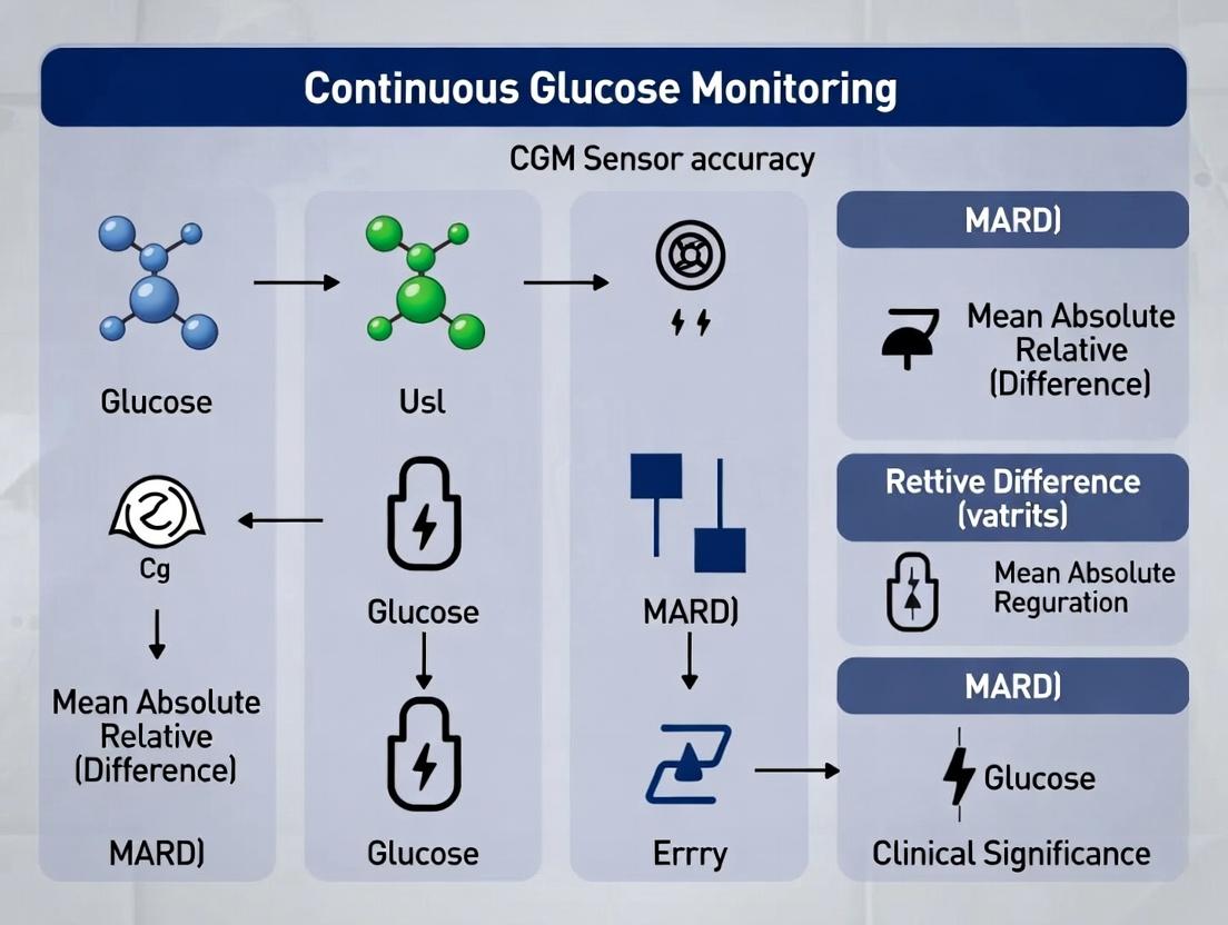
The MARD Metric Decoded: Statistical Foundations and Clinical Meaning of CGM Accuracy
Within the rigorous landscape of continuous glucose monitoring (CGM) sensor development, the Mean Absolute Relative Difference (MARD) stands as the primary statistical metric for quantifying sensor accuracy. This whitepaper provides a technical deconstruction of MARD, situating it within a broader research thesis that interrogates the nuanced relationship between aggregated MARD values and their ultimate clinical significance. For researchers and drug development professionals, a precise understanding of MARD's calculation, limitations, and contextual interpretation is paramount for advancing sensor technology and designing clinically meaningful outcomes studies.
Technical Definition and Calculation of MARD
MARD quantifies the average absolute percentage difference between paired CGM sensor readings and a reference measurement (typically venous or arterial plasma glucose measured via a laboratory-grade instrument like a Yellow Springs Instrument [YSI] analyzer or a blood glucose meter meeting ISO 15197:2013 standards).
Formula:
MARD = (1 / N) * Σ ( |Sensor Glucose - Reference Glucose| / Reference Glucose ) * 100%
Where N is the number of paired data points.
Critical Experimental Protocol for MARD Determination:
- Study Design: A controlled, clinic-based study with frequent reference sampling across a wide glycemic range (e.g., 40-400 mg/dL).
- Participant Cohort: Include subjects with varying demographics (age, BMI, diabetes type) to assess performance across physiologies.
- Reference Method: Capillary or venous blood samples are analyzed on a high-accuracy reference device (e.g., YSI 2300 STAT Plus) every 15-30 minutes during dynamic glucose challenges.
- Sensor Data: CGM data is time-aligned to the reference sample timestamp, accounting for any physiological time lag (typically a 2-5 minute delay in interstitial fluid glucose).
- Data Pairing: Only pairs where both sensor and reference values are available are included. Data is often stratified by glucose range, rate-of-change, and sensor wear period (Day 1 vs. Day 10).
- Statistical Analysis: MARD is calculated globally and per subject. Complementary metrics (e.g., Consensus Error Grid analysis, %20/20) are mandatory for a complete accuracy profile.
Quantitative Data: MARD Performance Tiers
The following table summarizes current benchmark MARD values and their accepted interpretations within sensor performance research.
Table 1: CGM Sensor MARD Performance Tiers and Clinical Implications
| MARD Range (%) | Performance Tier | Typical Clinical Research Interpretation |
|---|---|---|
| < 7 | Excellent | Accuracy approaches that of high-quality blood glucose meters; suitable for non-adjunctive use (i.e., insulin dosing without confirmation). |
| 7 - 10 | Very Good | Strong accuracy for clinical decision-making; standard for state-of-the-art commercial sensors. |
| 10 - 14 | Good | Acceptable for trend analysis and hypoglycemia alerts, though point accuracy may require confirmation for therapy adjustments. |
| > 14 | Requires Improvement | Limited reliability for precise therapeutic decisions; indicates need for sensor algorithm or chemistry refinement. |
Source: Data synthesized from recent pivotal trials (2022-2024) of FDA-cleared/CE-marked CGM systems.
Methodological Nuances and Complementary Metrics
MARD alone is insufficient. A low MARD can mask significant outliers or consistent bias. Essential complementary analyses include:
- Consensus Error Grid (CEG) Analysis: Categorizes clinical risk of point errors (Zone A: clinically accurate; Zone E: dangerous misreading).
- Bland-Altman Plots: Visualize bias and limits of agreement across the measurement range.
- Surveillance Error Grid (SEG): A more rigorous, risk-based metric quantifying the clinical impact of inaccuracies.
Table 2: Key Complementary Metrics to MARD in CGM Accuracy Studies
| Metric | Purpose | Interpretation Target |
|---|---|---|
| % Zone A (CEG) | Assess clinical accuracy percentage. | >70% is commonly targeted, with >99% combined Zone A+B. |
| Mean Absolute Error (MAE) | Average absolute difference in mg/dL, less skewed at high glucose. | Lower value indicates better accuracy (e.g., <10 mg/dL). |
| Coefficient of Variation (CV) | Measure of sensor precision. | <10% is desirable. |
| %20/20 | Percentage of readings within 20% of reference or 20 mg/dL for values <80 mg/dL. | >80% is a common benchmark. |
The Scientist's Toolkit: Research Reagent Solutions
Table 3: Essential Materials for In-Vitro and Pre-Clinical CGM Sensor Evaluation
| Item | Function & Rationale |
|---|---|
| YSI 2900 Series Biochemistry Analyzer | Gold-standard benchtop instrument for glucose concentration measurement in buffer/plasma during in-vitro sensor strip calibration and stability testing. |
| Controlled-Glucose Plasma/Serum | Standardized biological matrix for testing sensor performance in a complex protein environment, simulating in-vivo conditions. |
| Interference Stock Solutions | High-purity solutions of acetaminophen, ascorbic acid, uric acid, etc., for rigorous testing of sensor chemical specificity. |
| Potentiostat/Galvanostat | Electrochemical workstation for applying potential and measuring current from sensor electrodes during fundamental enzyme-electrode characterization. |
| Stabilized Glucose Oxidase/GDH Enzymes | The core biorecognition element. Different lots and stabilizer formulations are tested for optimal activity and longevity. |
| Osmium/ Ruthenium-based Redox Mediators | Electron-shuttling compounds crucial for signal transduction in 2nd/3rd generation sensor architectures. Key to sensor sensitivity and operational voltage. |
| Polyurethane/Siliconate Membranes | Diffusion-limiting and biocompatible membranes controlling glucose flux and blocking interferents; critical for in-vivo performance. |
| ISO 15197:2013-Compliant Blood Glucose Meter | For point-of-care reference comparisons in clinical studies, providing traceable secondary reference values. |
Visualization: MARD Determination Workflow & Clinical Accuracy Framework
Title: MARD Determination & Clinical Assessment Workflow
Title: Factors Influencing MARD and Path to Clinical Relevance
The accuracy of blood glucose monitoring systems (BGMS) and continuous glucose monitors (CGMs) is foundational to effective diabetes management. Within the broader research thesis on CGM sensor accuracy, as quantified by Mean Absolute Relative Difference (MARD) values, and their clinical significance, international standards provide the critical benchmark. ISO 15197:2013 sets the performance requirements for systems used for self-testing. This whitepaper provides a technical analysis of this standard, its evolution, and the emerging frameworks that address the more complex landscape of CGM and future interconnected systems.
ISO 15197:2013: Core Accuracy Requirements
The ISO 15197:2013 standard, "In vitro diagnostic test systems — Requirements for blood-glucose monitoring systems for self-testing in managing diabetes mellitus," defines stringent accuracy criteria. These are based on controlled clinical studies comparing the BGMS results to a reference method (typically a laboratory-grade hexokinase or glucose oxidase analyzer).
Key Accuracy Criteria:
- For glucose concentrations ≥5.55 mmol/L (≥100 mg/dL): At least 99% of individual results must fall within ±15% of the reference method results.
- For glucose concentrations <5.55 mmol/L (<100 mg/dL): At least 99% of individual results must fall within ±0.83 mmol/L (±15 mg/dL) of the reference method results.
These requirements represent a significant tightening from the previous 2003 version, which required 95% of results to be within ±20% or ±0.83 mmol/L.
Table 1: ISO 15197 Accuracy Requirements Evolution
| Parameter | ISO 15197:2003 | ISO 15197:2013 |
|---|---|---|
| Glucose ≥5.55 mmol/L (100 mg/dL) | 95% within ±20% | 99% within ±15% |
| Glucose <5.55 mmol/L (100 mg/dL) | 95% within ±0.83 mmol/L (±15 mg/dL) | 99% within ±0.83 mmol/L (±15 mg/dL) |
| Total Acceptable Results | 95% | 99% |
| Clinical Trial Sample Size | Minimum 100 samples | Minimum 100 samples |
Experimental Protocol for BGMS Accuracy Validation (Per ISO 15197)
The standard prescribes a detailed methodology for validation.
Protocol Summary:
- Participant/Sample Cohort: Capillary blood samples are collected from a minimum of 100 subjects, spanning the intended user population (different ages, diabetes types, hematocrit ranges). Samples must be distributed across specified glucose concentration ranges.
- Reference Method: A validated laboratory instrument (e.g., YSI 2300 STAT Plus or equivalent) serves as the primary reference. It must meet precision and accuracy criteria defined in the standard.
- Testing Procedure: A fresh capillary blood sample is split. One portion is measured immediately by the subject or trained operator using the BGMS under evaluation. The other portion is anticoagulated and measured in duplicate by the reference method within a defined time frame.
- Data Analysis: BGMS results are plotted against the reference mean. The percentage of results meeting the ±15%/±0.83 mmol/L criteria is calculated for each concentration regime.
Beyond ISO 15197:2013 – CGMs and MARD
ISO 15197:2013 applies to in vitro BGMS. The assessment of in vivo CGM systems is more complex, often using MARD as a key metric. While not directly governed by ISO 15197, CGM accuracy is evaluated against similar reference methods but with different statistical treatments and study designs (e.g., frequent venous or capillary reference sampling over several days). The clinical significance of MARD values is an active research area within the thesis framework, where a lower MARD generally correlates with higher clinical accuracy, though glycemic zone accuracy (e.g., Clarke Error Grid analysis) is equally critical.
Emerging standards and consensus reports (e.g., from the Diabetes Technology Society) are building upon ISO 15197 to address CGM-specific challenges like sensor drift, time lag, and the use of integrated systems with automated insulin delivery.
Visualizing the Accuracy Assessment Workflow
Diagram 1: BGMS Accuracy Validation per ISO 15197:2013
Diagram 2: Relationship: Standards, MARD & Clinical Outcomes
The Scientist's Toolkit: Key Research Reagent Solutions
Table 2: Essential Materials for Glucose Monitor Accuracy Research
| Item | Function in Research |
|---|---|
| Enzymatic Reference Analyzer (e.g., YSI 2900/2300) | Gold-standard instrument for plasma glucose measurement. Provides the reference value against which BGMS/CGM values are compared. Uses the glucose oxidase method. |
| Controlled Glucose Solutions | Solutions with known, certified glucose concentrations for system calibration, linearity testing, and basic functionality checks. |
| Anticoagulant Tubes (e.g., Lithium Heparin) | Used for collecting blood samples intended for reference analysis to prevent clotting and preserve glucose stability. |
| Hematocrit-Adjusted Controls | Quality control solutions with varying hematocrit levels to investigate and validate the system's compensation for hematocrit interference. |
| Interferent Stocks (e.g., Ascorbic Acid, Acetaminophen, Maltose) | High-purity chemical substances used in interference studies to test the specificity of the monitoring system's enzymatic reaction. |
| Clarke Error Grid Analysis Software | Computational tool for plotting reference vs. monitor values and categorizing them into risk zones (A-E), assessing clinical (not just numerical) accuracy. |
| Continuous Glucose Monitor Simulator/Sensor Flow Cell | In vitro testing apparatus for CGM sensors that allows controlled exposure to changing glucose buffers, enabling preliminary sensor characterization. |
Within the field of continuous glucose monitoring (CGM) development, the Mean Absolute Relative Difference (MARD) has become the canonical statistical metric for quantifying sensor accuracy against a reference method. However, its translation to tangible clinical outcomes remains a nuanced and critical research challenge. This technical guide examines the pathway from aggregate MARD values to the quantification and prediction of clinical error, framing the discussion within the broader thesis that sensor performance must be evaluated through the lens of clinical risk, not just statistical deviation.
Deconstructing MARD: Aggregate Statistic vs. Point-of-Use Error
MARD is calculated as the average of the absolute values of the relative differences between paired CGM and reference measurements:
MARD = (1/n) * Σ(|CGM_i - Reference_i| / Reference_i) * 100%
While useful for sensor-system comparisons, this aggregation masks error distribution. A sensor with a favorable MARD can still produce frequent, clinically significant errors if the error distribution is skewed or contains outliers.
Table 1: Hypothetical MARD Comparison with Error Distribution Analysis
| Sensor | Overall MARD | % Readings within ±15% of Reference | % Readings with >30% Error (High Risk) | Clarke Error Grid Zone A (%) |
|---|---|---|---|---|
| Sensor A | 9.5% | 88% | 3.5% | 95% |
| Sensor B | 10.2% | 92% | 0.8% | 98% |
Table 1 illustrates that Sensor A, despite a lower MARD, has a higher proportion of high-risk errors (>30% deviation), which may have greater clinical implications than Sensor B's more consistent performance.
Methodologies for Linking MARD to Clinical Endpoints
Experimental Protocol 1: Clarke and Parkes Error Grid Analysis
- Objective: To categorize CGM-reference paired points into zones of clinical significance.
- Protocol:
- Collect paired CGM and reference blood glucose values across a wide glycemic range (e.g., 40-400 mg/dL).
- Plot CGM values (y-axis) against reference values (x-axis).
- Map each point onto the standardized Clarke or Consensus Error Grid.
- Calculate the percentage of points in each zone:
- Zones A & B: Clinically acceptable.
- Zones C, D, E: Potentially dangerous clinical error.
- Outcome: Provides a direct, visual, and quantitative link between numerical error and clinical risk.
Experimental Protocol 2: Simulation of Clinical Outcomes
- Objective: To model the impact of MARD and error distribution on hypo/hyperglycemia detection and insulin dosing.
- Protocol:
- Use a large dataset of CGM readings with known MARD and error profiles.
- Employ a physiologically-based pharmacokinetic/pharmacodynamic (PBPK/PD) model or a virtual patient population.
- Simulate standard insulin-dosing algorithms driven by the CGM data.
- Compare outcomes (time-in-range, hypoglycemic events, hyperglycemic excursions) against a "perfect measurement" baseline.
- Outcome: Quantifies how statistical inaccuracy propagates into suboptimal therapy decisions.
The Clinical Error Pathway: A Systems View
The journey from sensor signal to patient risk involves multiple stages where error can be introduced or amplified.
Diagram Title: Pathway from Sensor Signal to Clinical Outcome and Error Amplification
The Scientist's Toolkit: Key Research Reagent Solutions
Table 2: Essential Materials for CGM Accuracy & Clinical Significance Research
| Item | Function in Research |
|---|---|
| YSI 2300 STAT Plus Analyzer | Gold-standard reference instrument for bench studies; uses glucose oxidase methodology for highly precise plasma glucose measurement. |
| Controlled Glucose Clamp System | Enables the study of sensor performance during dynamic glycemic changes in clinical trials, providing a stable reference "truth." |
| Virtual Patient Population Software (e.g., UVa/Padova Simulator) | Allows for in-silico testing of sensor error profiles on insulin dosing algorithms without patient risk. |
| Standardized Enzyme Solutions (Glucose Oxidase, Horseradish Peroxidase) | Core reagents for in-vitro characterization of sensor electrode stability and response kinetics. |
| Interferant Solutions (Acetaminophen, Ascorbic Acid, Uric Acid) | Used to challenge sensor specificity and quantify cross-reactivity, a key source of positive bias. |
| Protein-Rich Buffer/Serum | Mimics the interstitial fluid matrix for in-vitro testing, assessing biofouling impact on sensor signal drift. |
MARD is a necessary but insufficient metric for evaluating CGM clinical utility. A comprehensive assessment requires deconstructing the MARD to understand its distribution, mapping errors onto clinically significant grids, and modeling downstream therapeutic impacts. For researchers and developers, the goal must shift from optimizing a single statistic to engineering systems that minimize the frequency and magnitude of clinically dangerous errors, thereby directly improving patient safety and outcomes.
Continuous Glucose Monitor (CGM) accuracy is predominantly quantified using the Mean Absolute Relative Difference (MARD). This metric, while providing a singular value, masks significant heterogeneity in sensor performance across the physiological glucose spectrum. This whitepaper, framed within a thesis on CGM sensor accuracy and clinical significance, analyzes the technical and physiological underpinnings of why MARD varies across hypoglycemic, euglycemic, and hyperglycemic zones. Understanding this variance is critical for researchers and drug development professionals in assessing device utility for clinical trials and therapeutic monitoring.
The Fundamental Challenge: Non-Linear Physiology & Sensor Physics
CGM accuracy is not uniform because the biological environment and sensor electrochemistry change with glucose concentration.
Key Factors Contributing to MARD Variance
| Factor | Impact in Hypoglycemia | Impact in Euglycemia | Impact in Hyperglycemia |
|---|---|---|---|
| Background Noise | Signal-to-noise ratio is lowest; small absolute errors become large relative errors. | Moderate signal-to-noise ratio. | High signal-to-noise ratio; noise is a smaller proportion of signal. |
| Interferents | Physiological (e.g., acetaminophen, urate) and exogenous substances cause proportionally greater error. | Interferent effect is present but less pronounced relative to signal. | Effect may be masked by high glucose signal but can still cause absolute bias. |
| Tissue Dynamics | Lag time (blood to interstitial fluid) is proportionally more significant during rapid glucose declines. | Lag is generally manageable and accounted for in algorithms. | Physiological lag can obscure the true rate of rise, causing underestimation. |
| Enzyme Kinetics | Glucose oxidase operates in a less linear, more sensitive range. | Operation typically in the optimal, linear range of the Michaelis-Menten curve. | Possible enzyme saturation or mass transport limitation, leading to signal compression. |
| Calibration | Single-point calibration often optimized for euglycemia, increasing error at extremes. | Typically the zone of optimal calibration fit. | Extrapolation from euglycemic calibration can introduce systematic bias. |
The following table synthesizes data from recent pivotal studies of commercially available and investigational CGMs.
Table 1: Representative MARD Values Across Glycemic Zones
| Glycemic Zone | Glucose Range (mg/dL) | Typical MARD Range (%) | Primary Contributor to Error |
|---|---|---|---|
| Hypoglycemia | <70 | 15% - 30% | Low signal-to-noise ratio, physiological lag, calibration bias. |
| Level 2 | <54 | Often >20% | Extremely low signal, critical timing delays. |
| Euglycemia | 70-180 | 6% - 10% | Random noise, minor interferents. |
| Hyperglycemia | >180 | 8% - 12% | Sensor dynamics (saturation), physiological lag. |
| Level 2 | >250 | 10% - 15% | Non-linear sensor response, compression artifacts. |
Experimental Methodologies for Zone-Specific Accuracy Assessment
To investigate these phenomena, standardized and advanced protocols are employed.
Protocol: Clamp Study for Zone-Specific MARD Calculation
- Objective: To measure sensor accuracy at steady-state glucose levels across defined zones.
- Method:
- Participants are brought to a target glucose plateau using a glucose clamp technique.
- Plateaus are maintained for 60-90 minutes at each zone: hypoglycemic (~55 mg/dL), euglycemic (~100 mg/dL, ~140 mg/dL), and hyperglycemic (~250 mg/dL, ~400 mg/dL).
- Frequent venous blood samples are analyzed via a laboratory reference method (e.g., YSI 2300 STAT Plus).
- CGM values are time-matched to reference values.
- MARD is calculated separately for each glycemic plateau: MARDzone = (1/N) * Σ \|(CGMi - Refi)/Refi| * 100%.
Protocol: In Vitro Assessment of Sensor Linearity & Interferents
- Objective: To decouple physiological factors from inherent sensor performance.
- Method:
- Sensors are tested in a controlled in vitro flow cell system.
- Glucose concentration is stepped through a range (e.g., 40-500 mg/dL) in a buffer matrix.
- Sensor output (nA) is recorded and plotted against concentration to assess linearity and saturation.
- The protocol is repeated with the addition of common interferents (e.g., acetaminophen, ascorbate, uric acid) at physiological and supraphysiological concentrations.
- Percentage deviation from baseline sensor output quantifies interferent susceptibility at each glucose level.
Protocol: Dynamic Error Grid Analysis (D-EGA)
- Objective: To assess clinical accuracy during rapid glucose changes, a major challenge in hypo-/hyperglycemia.
- Method:
- CGM and reference data pairs are collected during periods of significant glucose change (e.g., >2 mg/dL/min).
- Pairs are plotted on a consensus error grid, but are first categorized by the rate of change of the reference glucose.
- Accuracy is stratified into zones (e.g., accurate, benign error, misleading error) for different rate categories (e.g., rapid fall, stable, rapid rise).
- This reveals whether errors are more likely during rapid transitions, which are clinically critical in hypoglycemia.
Visualizing Key Concepts and Workflows
Title: Sources of Error in the CGM Glucose Sensing Pathway
Title: Why Relative Error (MARD) Differs in Hypo- vs Hyperglycemia
The Scientist's Toolkit: Research Reagent Solutions
Table 2: Essential Materials for CGM Accuracy Research
| Item / Reagent | Function in Research | Application in Zone-Specific Studies |
|---|---|---|
| YSI 2300 STAT Plus Analyzer | Gold-standard reference for glucose measurement via glucose oxidase method. | Provides the reference value for MARD calculation in clamp studies. |
| Glucose Clamp Apparatus | Infuses dextrose and insulin to maintain precise, steady-state blood glucose levels. | Creates stable glycemic plateaus in hypo-, eu-, and hyperglycemic zones for controlled testing. |
| In Vitro Flow Cell System | A calibrated system to perfuse sensors with controlled solutions. | Isolates sensor performance (linearity, interferent response) from in vivo physiological confounders. |
| Human Serum Albumin (HSA) | Protein additive for in vitro testing solutions. | Mimics the protein content of interstitial fluid, affecting sensor membrane transport dynamics. |
| Common Interferent Panel (e.g., Acetaminophen, Ascorbic Acid, Uric Acid) | Solutions of known concentration. | Quantifies sensor specificity and susceptibility to false signals at different glucose levels. |
| Consensus Error Grid Software | Analytical tool to classify CGM-reference pairs by clinical risk. | Performs Dynamic EGA to assess accuracy during rapid glucose transitions critical in hypo/hyperglycemia. |
| Buffered Glucose Solutions | pH-stable solutions with precise glucose concentrations (e.g., 40, 100, 400 mg/dL). | Used for in vitro calibration and linearity testing across the full measurement range. |
While the Mean Absolute Relative Difference (MARD) has become a standard metric for evaluating Continuous Glucose Monitor (CGM) accuracy, a comprehensive assessment requires a multi-faceted approach. This technical guide deconstructs accuracy into its core statistical components—precision, bias, and consistency—framed within ongoing research on the clinical significance of CGM sensor performance. For researchers and drug development professionals, a sole reliance on MARD can obscure critical performance characteristics that directly impact clinical decision-making and therapeutic outcomes.
MARD calculates the average absolute percentage difference between paired CGM and reference blood glucose values. While useful for an overall error estimate, it provides no insight into the direction (bias) or variability (precision) of the error, nor its behavior across glucose ranges (consistency).
Thesis Context: Within broader CGM accuracy research, the clinical significance of sensor performance hinges on understanding how these distinct error components affect glycemic time-in-range, hypoglycemia detection, and the reliability of data used in closed-loop systems or clinical trials.
Deconstructing Accuracy: Core Statistical Components
Precision (Repeatability)
Precision refers to the reproducibility of measurements under unchanged conditions. High precision indicates low random error (noise).
- Quantification: Typically measured by the Coefficient of Variation (CV%) within a stable glucose period or the Standard Deviation (SD) of residuals.
- Clinical Impact: Poor precision creates a "noisy" trace, making trend interpretation difficult and increasing alert fatigue.
Bias (Systematic Error)
Bias indicates a consistent over- or under-estimation of glucose values by the sensor compared to the reference.
- Quantification: Calculated as the Mean Relative Difference (MRD) or via regression analysis (y-intercept deviation from zero).
- Clinical Impact: Consistent positive bias may mask hypoglycemia; consistent negative bias may lead to overtreatment of perceived hyperglycemia.
Consistency (Range-Dependent Performance)
Consistency evaluates whether precision and bias are stable across the entire measuring interval (e.g., hypoglycemia, euglycemia, hyperglycemia).
- Quantification: Analyzed through sub-range MARD calculations, Clarke Error Grid Zone distributions by glucose range, or regression slope analysis.
- Clinical Impact: Performance degradation in the hypoglycemic range is particularly critical due to the severe consequences of undetected low glucose.
Quantitative Data Comparison
Table 1: Comparative Performance Metrics of Hypothetical CGM Sensors A & B (Aggregate Data from Recent Studies)
| Metric | Sensor A | Sensor B | Ideal Value | Clinical Significance |
|---|---|---|---|---|
| Overall MARD (%) | 9.5 | 9.5 | 0 | Identical aggregate error. |
| Precision (CV%) | 6.2 | 10.8 | 0 | Sensor A has lower noise, more reliable trends. |
| Bias (MRD %)* | +1.5 | -6.2 | 0 | Sensor B systematically reads low. |
| Hypoglycemia (<70 mg/dL) MARD (%) | 12.1 | 22.5 | 0 | Sensor B's performance degrades significantly in low range. |
| Clarke Error Grid A+B (%) | 99.8 | 98.5 | 100 | Both clinically acceptable, but Sensor A is superior. |
| Time Delay (minutes) | 5.2 | 8.7 | 0 | Sensor A has lower physiological lag. |
*Positive bias indicates sensor reads high vs. reference.
Table 2: ISO 15197:2013 Performance Criteria vs. Advanced Metrics
| Standard | Required Performance | Advanced Component Addressed |
|---|---|---|
| ISO 15197:2013 | ≥95% of results within ±15 mg/dL (<100 mg/dL) or ±15% (≥100 mg/dL) | Basic point accuracy. |
| Extended Analysis | Performance Goal | Insight Gained |
| Consensus Error Grid | High % in zones A+B | Clinical risk analysis. |
| Precision (CV%) | <10% | Measurement reproducibility. |
| Range-Specific MARD | Consistent low values across ranges | Reliability in hypo-/hyperglycemia. |
| Surveillance Error Grid | Low risk score | Quantitative clinical impact. |
Experimental Protocols for Comprehensive Assessment
Protocol for In-Clinic Study with Frequent Reference Sampling
Objective: To simultaneously determine MARD, precision, bias, and consistency.
- Subject Cohort: Recruit n≥XX participants with diabetes, targeting a wide glycemic range.
- Sensor Deployment: Place sensors according to IFU. Use devices from at least 3 different production lots.
- Reference Method: Employ YSI 2300 STAT Plus or equivalent laboratory glucose analyzer. Perform capillary or venous blood draws every 15 minutes during an 8-12 hour controlled clinic session, including meal challenges and insulin-induced hypoglycemia (under strict medical supervision).
- Data Pairing: Align CGM values with reference values using a validated time-alignment protocol (e.g., accounting for sensor time lag via mathematical correction).
- Analysis: Calculate overall MARD, MRD (bias), and CV of residuals (precision). Stratify data into glycemic ranges (e.g., <70, 70-180, >180 mg/dL) to assess consistency. Generate Clarke and Surveillance Error Grids.
Objective: To assess real-world consistency and bias.
- Subject Cohort: Recruit n≥XXX participants for a 7-14 day at-home study.
- Reference Method: Provide calibrated capillary blood glucose meters (e.g., Contour Next One) meeting ISO standards. Participants perform at least 4 fingersticks per day, including during fasting, post-prandial, and suspected hypo-/hyperglycemic events.
- Data Synchronization: Use Bluetooth-enabled meters to automatically timestamp and log reference values alongside CGM data in a companion app.
- Analysis: Perform point-error analysis. Use regression (Deming or Passing-Bablok) to quantify systemic bias. Analyze error distribution across ranges and over sensor wear time (day 1 vs. day 7 vs. day 10) to assess wear-time consistency.
Visualization: The Accuracy Assessment Workflow
Diagram Title: CGM Accuracy Assessment: From Data to Comprehensive Profile
The Scientist's Toolkit: Key Research Reagent Solutions
Table 3: Essential Materials for In Vitro and Preclinical CGM Sensor Research
| Item & Example | Function in Research |
|---|---|
| Glucose Oxidase (GOx) Enzyme (Aspergillus niger sourced) | The primary biological recognition element in most electrochemical CGM sensors. Catalyzes the oxidation of glucose. |
| Platinum/Carbon Working Electrode | The transducer surface where the electrochemical reaction (H₂O₂ oxidation) occurs, generating the measurable current. |
| Polyurethane/Polysulfone Membrane (e.g., Polyurethanes with specific pore sizes) | The diffusion-limiting and biocompatible outer membrane. Controls glucose flux, extends linear range, and reduces biofouling. |
| Interference Rejection Layer (e.g., Nafion, cellulose acetate) | A charged inner membrane that selectively rejects common electrochemical interferents like acetaminophen and ascorbic acid. |
| In Vitro Test Solution (e.g., PBS with varying glucose, albumin, ascorbate) | Simulates the physiological matrix for bench-top sensor characterization, allowing controlled study of sensitivity, selectivity, and drift. |
| Clark-Type Oxygen Electrode | Used to measure local oxygen tension in vitro or in tissue, critical for assessing oxygen limitations on sensor performance (the "oxygen deficit" problem). |
| Subcutaneous Tissue Simulant Gel (e.g., Phosphate-buffered agarose gel) | Mimics the diffusion properties of subcutaneous tissue for more realistic in vitro lag time and stability testing. |
| Freestyle Libre or Dexcom G6 Sensor (Commercial Products) | Used as benchmarks for performance comparison and for reverse-engineering studies on sensor architecture and algorithm design. |
From Raw Data to Endpoints: Methodological Strategies for CGM in Clinical Research
Within the broader thesis on Continuous Glucose Monitoring (CGM) sensor accuracy, as quantified by Mean Absolute Relative Difference (MARD) values and their clinical significance, the design of robust clinical trial protocols is paramount. This technical guide details critical considerations for integrating CGM as an endpoint in clinical trials for drug development, focusing on wear duration, blinding methodologies, and data completeness. These factors directly impact the reliability of glycemic data and the validity of conclusions drawn about therapeutic efficacy.
Core Protocol Design Elements
Wear Duration and Scheduling
The required wear duration balances physiological capture with participant burden. Current consensus and regulatory guidance recommend a minimum period to capture diurnal variations and cyclical patterns.
Table 1: Recommended CGM Wear Durations for Different Trial Objectives
| Trial Phase / Objective | Minimum Recommended Wear Duration | Rationale & Key Considerations |
|---|---|---|
| Proof-of-Concept (Phase IIa) | 7-14 consecutive days | Captures weekly rhythms, minimizes day-of-week bias. Sufficient for initial signal detection. |
| Efficacy Endpoint (Phase IIb/III) | 14-28 consecutive days | Provides robust data for key metrics like Time in Range (TIR). Aligns with FDA draft guidance. |
| Baseline Assessment | 10-14 days prior to intervention | Establishes reliable baseline glycemic profile for within-participant comparison. |
| Safety Monitoring | Continuous or intermittent 7-day periods | Monitors for hypoglycemia; duration may be tied to specific drug pharmacokinetics. |
Experimental Protocol for Wear Duration Validation:
- Objective: Determine the minimum wear duration required to reliably estimate a participant's 14-day TIR (±3.9–10.0 mmol/L).
- Method: In a cohort with varying glycemic control, analyze CGM data via bootstrapping. Randomly sample increasing durations (e.g., 2, 4, 6, 8, 10, 12 days) from a 14-day "gold standard" period.
- Analysis: Calculate the 95% confidence interval for TIR from each sampled duration. Define reliability as the point where the CI width is within ±3% of the 14-day value. Repeat 1000 times per participant to estimate population-level duration requirement.
Blinding Methodologies
Maintaining blinding is crucial for endpoint objectivity, especially for patient-reported outcomes. CGM presents unique challenges due to real-time glucose display.
Table 2: CGM Blinding Strategies and Implications
| Strategy | Protocol Implementation | Advantages | Limitations & Mitigations |
|---|---|---|---|
| Device Blinding | Use of modified "clinician-only" CGM devices or blinded professional CGM (e.g., Dexcom G6 Pro, iPro2). Participants wear a separate, blinded sensor. | Strongest method. Prevents any real-time feedback influencing behavior. | Increased cost and burden (two sensors). Must ensure identical sensor performance. |
| Screen Blinding | Providing devices with screens disabled or covered. Data is extracted by clinician at end of wear period. | Logistically simpler, uses standard consumer hardware. | Risk of participant unmasking via smartphone apps; requires strict compliance monitoring. |
| Data Analysis Blinding | Unblinded personnel handle device setup/download; analysts are blinded to treatment allocation. | Protects against bias in data processing and endpoint calculation. | Does not prevent behavioral bias during data collection. Often used in conjunction with other methods. |
Experimental Protocol for Testing Blinding Efficacy:
- Objective: Assess the success of a screen-blinding protocol.
- Method: In a pilot study, randomize participants to active drug or placebo. Provide CGM devices with covered screens. At study end, administer a questionnaire asking participants to guess their treatment assignment and rate confidence.
- Analysis: Compare guess accuracy against chance (50%) using a binomial test. Correlate confidence with accuracy to identify systematic unmasking cues.
Data Completeness and Quality Metrics
Data completeness is a critical determinant of endpoint validity. Incomplete data can skew MARD and derived metrics like TIR.
Table 3: Data Completeness Standards and Handling
| Metric | Recommended Minimum Threshold | Action for Non-Compliance |
|---|---|---|
| Overall Wear Time | ≥ 70% of scheduled wear period for primary analysis. | Pre-specify imputation methods (e.g., last observation carried forward is NOT appropriate). Consider sensitivity analyses. |
| Per-Day Completeness | ≥ 80% of data points (288 readings) per 24-hour period for a day to be included. | Flag days below threshold. Protocol should mandate re-education or sensor replacement if multiple invalid days occur. |
| Calibration Compliance | For factory-calibrated sensors, document any mandatory calibrations. For user-calibrated, protocol must specify timing and method. | Deviations should be recorded as protocol deviations. Analyze impact on reported MARD. |
Experimental Protocol for Assessing Impact of Data Loss:
- Objective: Quantify the bias introduced in TIR calculation by simulated data gaps.
- Method: Take a dataset with >95% completeness. Artificially introduce random and systematic (e.g., overnight) data gaps of varying severity (5%, 10%, 20% loss).
- Analysis: Recalculate TIR for the degraded data and compare to original value. Perform linear regression to model the relationship between data loss percentage and absolute error in TIR.
The Scientist's Toolkit: Research Reagent Solutions
Table 4: Essential Materials for CGM Clinical Trial Research
| Item | Function in CGM Research |
|---|---|
| Professional / "Blinded" CGM Systems (e.g., Dexcom G6 Pro, Medtronic iPro3) | Provides glucose data to the clinician/researcher without real-time display to the participant, enabling blinded endpoint assessment. |
| Data Download & Aggregation Software (e.g., Dexcom Clarity, CareLink, Glooko) | Centralized platforms for secure data extraction, visualization, and initial quality checks across multiple participants and sites. |
| Reference Blood Glucose Analyzer (e.g., YSI 2300 STAT Plus, Abbott Blood Gas Analyzer) | Provides high-accuracy venous blood glucose measurements for in-study MARD calculation and sensor accuracy verification. |
| Standardized Meal Kits or Formulas | Used in controlled meal-challenge sub-studies to assess postprandial glucose responses uniformly, reducing dietary variability. |
| Secure, HIPAA/GCP-compliant Cloud Storage | Essential for transferring large volumes of time-series CGM data from clinical sites to central analysis teams while maintaining participant privacy. |
| CGM-Specific Clinical Endpoint Calculators (e.g., in R, Python, or validated commercial software) | Tools to compute consensus metrics (TIR, GV, TAR, TBR) from raw data streams consistently according to pre-specified algorithms. |
Visualizing CGM Trial Protocol Workflows
Title: CGM Clinical Trial Workflow and Decision Points
Title: From CGM Signal to MARD Endpoint and Confounders
This whitepaper details the derivation and application of core continuous glucose monitoring (CGM)-based clinical endpoints, framed within a critical thesis on CGM sensor accuracy. The thesis posits that while the Mean Absolute Relative Difference (MARD) is the primary metric for quantifying technical sensor performance, its direct clinical translation is limited. A low MARD is a necessary but insufficient condition for deriving clinically meaningful outcomes. This guide establishes the pathway from validated, accurate CGM data (with known MARD) to the calculation of standardized endpoints that directly inform therapeutic efficacy and patient risk in clinical research and drug development.
Core Endpoint Definitions and Quantitative Benchmarks
Table 1: Standardized CGM-Derived Endpoint Definitions and Clinical Targets
| Endpoint | Acronym | Definition | Primary Clinical Target (ADA/EASD Consensus) |
|---|---|---|---|
| Time in Range | TIR | % of readings & time in 70-180 mg/dL (3.9-10.0 mmol/L) | >70% for most populations |
| Time Below Range | TBR | % of readings & time <70 mg/dL (<3.9 mmol/L) | Level 1: <4% (<70 mg/dL) Level 2: <1% (<54 mg/dL) |
| Time Above Range | TAR | % of readings & time >180 mg/dL (>10.0 mmol/L) | Level 1: <25% (>180 mg/dL) Level 2: <5% (>250 mg/dL) |
| Glucose Variability | GV | Statistical measure of glucose fluctuations (e.g., %CV) | %CV ≤36% |
| Area Under the Curve | AUC | Area under glucose-time curve for hyper/hypoglycemic episodes | Minimized; no single target |
Table 2: Relationship Between MARD and Endpoint Confidence Intervals (Hypothetical Model)
| Sensor MARD | Estimated 95% CI for TIR (%)* | Impact on Endpoint Reliability |
|---|---|---|
| 5% | ± 2.5% | High reliability for detecting clinical changes. |
| 10% | ± 5.0% | Moderate reliability; larger sample sizes required. |
| 15% | ± 7.5% | Low reliability; endpoint may be significantly confounded. |
| *Assumes a nominal TIR of 70% over a 14-day period. CI width is illustrative. |
Detailed Methodologies for Endpoint Calculation
Protocol for TIR, TBR, and TAR Calculation from CGM Data
Objective: To calculate the percentage of time spent in defined glucose ranges over a specified monitoring period. Materials: Validated CGM system (MARD documented), data extraction software, statistical package (e.g., R, Python, SAS). Procedure:
- Data Qualification: Use CGM data from a minimum of 14 days with ≥70% sensor availability (per international consensus).
- Data Alignment: Ensure timestamps are consistent. Interpolate minor data gaps (<20 minutes) using validated linear methods. Exclude periods with calibration or sensor error.
- Categorization: Classify each glucose reading (typically at 5-minute intervals) into:
- Hypoglycemia: <54 mg/dL (Level 2) and 54-69 mg/dL (Level 1).
- Range: 70-180 mg/dL.
- Hyperglycemia: 181-250 mg/dL (Level 1) and >250 mg/dL (Level 2).
- Calculation:
TIR (%) = (Number of readings in 70-180 mg/dL) / (Total number of qualified readings) * 100- Apply same formula for TBR and TAR components.
- Reporting: Report metrics as mean ± standard deviation or median [IQR] across the study population. Always report TIR, TBR (<54), and TAR (>250) together.
Protocol for Glucose Variability (GV) Assessment
Objective: To quantify the amplitude of glucose fluctuations, an independent risk factor. Primary Metric: Coefficient of Variation (%CV)
- Calculate the standard deviation (SD) of all qualified glucose readings.
- Calculate the mean glucose.
%CV = (SD / Mean Glucose) * 100Secondary Metrics: Include in supplementary analysis:- Continuous Overlapping Net Glycemic Action (CONGA-n): SD of differences between current reading and reading n hours prior.
- Mean Amplitude of Glycemic Excursions (MAGE): Calculates average height of glucose excursions exceeding 1 SD.
Protocol for Area Under the Curve (AUC) Calculation for Episodes
Objective: To quantify the magnitude and duration of hyper- or hypoglycemic excursions. Materials: As above, with trapezoidal rule calculation capability. Procedure:
- Episode Identification: Define an episode start (e.g., first reading >180 mg/dL) and end (last consecutive reading above threshold before returning to ≤180 mg/dL).
- Baseline Definition: Set a baseline (e.g., 180 mg/dL for hyperglycemia). The AUC calculates the area above this baseline.
- Calculation (Trapezoidal Rule):
- For each consecutive pair of glucose readings (G1, G2) at times (t1, t2) within the episode:
AUC_segment = [(G1 - baseline) + (G2 - baseline)] / 2 * (t2 - t1)
- Sum all segments within the episode for total episode AUC (mg/dL * time).
- For each consecutive pair of glucose readings (G1, G2) at times (t1, t2) within the episode:
- Aggregation: Sum AUC for all episodes per patient over the analysis period.
Visualizing the Thesis Pathway from MARD to Clinical Meaning
Title: Pathway from Technical Accuracy to Clinical Meaning
The Scientist's Toolkit: Research Reagent Solutions
Table 3: Essential Materials for CGM Endpoint Research
| Item | Function in Research |
|---|---|
| Validated CGM System (e.g., Dexcom G7, Abbott Libre 3, Medtronic Guardian 4) | Primary data source. Must have published MARD (typically 8-10% for latest gen) for regulatory-grade research. |
| Reference Blood Glucose Analyzer (e.g., YSI 2300 STAT Plus, Radiometer ABL90) | Gold standard for establishing CGM sensor MARD in accuracy studies. Provides plasma glucose for sensor calibration/validation. |
| CGM Data Download Suite (Manufacturer-specific) | Extracts raw glucose, timestamp, and quality flag data from sensors/receivers for analysis. |
| Clinical Trial Data Management System (e.g., Medidata Rave, Veeva) | Securely hosts, manages, and audits CGM-derived endpoint data per FDA 21 CFR Part 11 compliance. |
Statistical Software (e.g., R with cgmanalysis package, SAS, Python/pandas) |
Performs batch calculation of TIR, TBR, TAR, GV, and AUC across large patient cohorts. Enables advanced modeling. |
| Clamp Technologist Setup (for mechanistic studies) | Hyperinsulinemic-euglycemic/hypoglycemic clamps to provoke controlled glucose excursions for endpoint validation. |
| Standardized Meal Kits or Glucose Challenges | Provides a consistent metabolic stimulus to assess postprandial glycemic control (key component of TAR). |
The Impact of MARD on Derived Endpoint Reliability and Statistical Power
In Continuous Glucose Monitoring (CGM) sensor accuracy research, the Mean Absolute Relative Difference (MARD) is the pivotal metric for assessing sensor performance. This technical guide explores a critical but often under-examined facet: how the intrinsic MARD value of a CGM system directly impacts the reliability of derived glycemic endpoints (e.g., Time-in-Range, glycemic variability) and the statistical power of clinical studies utilizing these endpoints. Within the broader thesis on CGM accuracy and clinical significance, understanding this relationship is paramount for designing robust clinical trials, interpreting real-world evidence, and ensuring that therapeutic insights are grounded in reliable data rather than measurement artifact.
MARD Primer: Composition and Implications
MARD is calculated as the average of the absolute percentage differences between paired CGM and reference (e.g., venous plasma, YSI) measurements. While a single aggregate figure, its distribution is non-uniform across the glycemic range; errors are typically larger in hypoglycemic and hyperglycemic extremes. This non-linearity propagates error unevenly into derived metrics.
Table 1: Typical MARD Performance Tiers and Their Interpretation
| MARD Range (%) | Accuracy Tier | Implied Impact on Endpoints |
|---|---|---|
| < 10 | Excellent | Minimal bias; high endpoint fidelity. Suitable for primary endpoint in pivotal trials. |
| 10 – 14 | Good | Moderate bias; may require larger sample sizes for sufficient power. |
| 15 – 20 | Acceptable | Significant bias; endpoint reliability questionable for detecting small effects. |
| > 20 | Poor | High risk of misleading conclusions; not recommended for primary outcomes. |
Mathematical Framework: Error Propagation to Derived Endpoints
Derived endpoints like Time-in-Range (TIR, % time 70-180 mg/dL) are not measured directly but computed from a series of error-prone CGM values. The MARD-induced error in each point measurement propagates through the calculation, increasing the variance and potential bias of the final endpoint estimate. The relationship can be conceptualized as:
Endpoint Variance ≈ ƒ(Sensor Noise Variance, MARD Profile, Glycemic Variability, Data Density)
Higher MARD increases the "noise floor," making it harder to detect a true "signal" (a therapeutic effect).
Diagram Title: How MARD Error Propagates to Study Power
Experimental Protocols for Quantifying Impact
Protocol 4.1: In-Silico Monte Carlo Simulation
This is the primary method for isolating MARD's effect from biological confounders.
- Reference Data Acquisition: Obtain high-frequency, high-accuracy reference glucose datasets (e.g., from clamped studies or very low-MARD sensors).
- Error Modeling: Define an error model (e.g., Gaussian, skewed based on glucose level) parameterized by a target MARD (e.g., 8%, 12%, 18%).
- Sensor Data Simulation: For each reference point, apply a random error drawn from the model to generate a simulated CGM trace. Repeat thousands of times to create multiple simulated sensor datasets from the same underlying truth.
- Endpoint Calculation: Compute derived endpoints (TIR, Time-Below-Range, Coefficient of Variation, etc.) for both reference and simulated sensor datasets.
- Analysis: Quantify the bias (average difference from true value) and variance inflation (increased standard deviation) for each endpoint at each MARD level.
Protocol 4.2: Paired Clinical Device Comparison
Directly compares endpoint agreement between a high-accuracy system (low MARD) and a test system.
- Study Design: Conduct a clinical study where participants wear two CGM systems (a "Reference" system with verified low MARD and the "Test" system) simultaneously, along with frequent capillary/venous reference measurements.
- Data Processing: Align data streams temporally. Calculate glycemic endpoints for each device over identical wear periods (e.g., 24-hour segments).
- Statistical Comparison: Use Bland-Altman analysis and linear regression to assess the agreement between the endpoints derived from the two systems. The limits of agreement directly reflect the impact of the Test system's higher MARD on endpoint reliability.
Data Presentation: Simulated Impact of MARD
Table 2: Monte Carlo Simulation Results - Impact of MARD on Endpoint Reliability (n=10,000 simulations per scenario)
| True TIR (%) | Simulated MARD (%) | Mean Reported TIR (Bias) | SD of Reported TIR | Sample Size Needed for 80% Power (Δ5% TIR) |
|---|---|---|---|---|
| 70 | 7 | 69.8 (-0.2) | 1.8 | 42 |
| 70 | 10 | 69.5 (-0.5) | 2.7 | 94 |
| 70 | 14 | 68.9 (-1.1) | 4.1 | 216 |
| 70 | 18 | 67.5 (-2.5) | 6.0 | 462 |
| 50 | 10 | 49.7 (-0.3) | 3.1 | 130 |
| 50 | 18 | 48.1 (-1.9) | 7.2 | 702 |
Note: SD = Standard Deviation. Assumptions: 14-day observation period, paired t-test design.
The Scientist's Toolkit: Key Research Reagent Solutions
Table 3: Essential Materials for MARD/Endpoint Reliability Research
| Item | Function / Rationale |
|---|---|
| High-Accuracy Reference Analyzer (e.g., YSI 2900D) | Provides the "gold standard" venous glucose measurement for calculating true MARD against CGM sensor values. |
| Clamp Study Data Repository | Provides tightly controlled glycemia datasets (euglycemic, hypo-, hyper-glycemic clamps) essential for modeling MARD across glucose ranges. |
| In-Silico Simulation Platform (e.g., GIST, custom R/Python code) | Enables Monte Carlo simulation of sensor error to study endpoint variance and bias in a controlled computational environment. |
| Bland-Altman & Deming Regression Analysis Software | Statistical tools essential for quantifying agreement between endpoints derived from different accuracy systems. |
| Continuous Glucose Monitor (CGM) Systems with Varied MARD | Test articles spanning a range of MARD values (e.g., 7%-20%) are required for empirical paired comparison studies. |
Optimizing Study Design for High-MARD Scenarios
When a CGM system with a higher MARD must be used, specific design adjustments can mitigate risk:
- Increase Sample Size: Power calculations must account for the inflated variance of the primary endpoint.
- Lengthen Observation Period: A longer data collection window (e.g., 4 weeks vs. 2 weeks) averages out more random error, improving endpoint stability.
- Use Low-MARD System as Adjudicator: In hybrid study designs, use a high-accuracy system on a subset of participants to validate endpoint trends from the primary, higher-MARD system.
Diagram Title: Decision Flow for Studies Using Higher-MARD CGM
The MARD value of a CGM system is not merely a stand-alone accuracy metric; it is a fundamental determinant of downstream data quality. It directly induces bias and variance in derived glycemic endpoints, which in turn erodes statistical power and can lead to false-positive or false-negative conclusions in clinical research. Researchers and drug development professionals must incorporate MARD-driven error propagation into their study planning, from power calculations and endpoint selection to the final interpretation of clinical significance. Prioritizing the use of CGM systems with the lowest feasible MARD is the most effective strategy for ensuring the reliability of glycemic endpoints and the integrity of clinical trial outcomes.
Within the critical research on Continuous Glucose Monitor (CGM) sensor accuracy, quantified by Mean Absolute Relative Difference (MARD) values, real-world performance is often challenged by transient physiological and technical artifacts. Compression lows, sensor settling, and signal dropouts degrade MARD, potentially obscuring a sensor's true clinical significance in drug development and therapeutic monitoring. This technical guide details the mechanisms, experimental characterization, and mitigation strategies for these artifacts, providing a framework for researchers to isolate and account for them in clinical trial design and data analysis.
Compression Lows: Mechanism and Characterization
A compression low is an artifactual dip in interstitial glucose (ISF) readings caused by sustained local pressure on the sensor insertion site, which impedes interstitial fluid flow and reduces glucose delivery to the sensor electrode.
Mechanism: Pressure-induced ischemia reduces capillary blood flow, depleting local ISF glucose. The sensor consumes glucose faster than it is replenished, causing a false-low reading that does not reflect systemic blood glucose.
Experimental Protocol for Inducing & Measuring Compression Lows
- Objective: Quantify the magnitude and dynamics of compression low artifacts under controlled pressure.
- Setup: A cohort of healthy volunteers or patients with diabetes is fitted with a CGM sensor on the posterior upper arm. A pressure applicator (e.g., a controlled-force plunger with a standardized surface area) is positioned over the sensor.
- Procedure:
- Establish a euglycemic baseline (90-140 mg/dL) via frequent venous blood sampling (reference method).
- Apply a calibrated pressure (e.g., 50 mmHg, 100 mmHg) for a defined period (e.g., 20 minutes) while the subject remains motionless.
- Monitor CGM readings at 1-minute intervals.
- Release pressure and monitor recovery for 30 minutes.
- Repeat at different pressure levels and glycemic states (euglycemia, hyperglycemia).
- Key Metrics: Time to onset of signal decline, maximum signal drop (mg/dL), rate of recovery, and lag versus reference blood glucose.
Table 1: Summary of Compression Low Artifact Data
| Pressure (mmHg) | Avg. Onset Time (min) | Max Signal Drop (mg/dL) | Avg. Recovery Time (min) | MARD Increase During Event* |
|---|---|---|---|---|
| 50 | 8.2 ± 1.5 | -45 ± 12 | 15.3 ± 4.1 | +22.5% |
| 100 | 5.5 ± 0.8 | -78 ± 18 | 22.7 ± 5.6 | +35.8% |
| Control (0) | N/A | N/A | N/A | Baseline (e.g., 9.2%) |
*Hypothetical data for illustration; MARD increase calculated versus paired reference measurements.
Sensor Settling (Run-in Period)
The initial period post-insertion (typically 1-24 hours) is characterized by unstable signals as the tissue responds to sensor insertion (trauma, inflammation) and the sensor electrodes equilibrate.
Mechanism: The acute inflammatory response alters local capillary permeability, ISF composition, and glucose dynamics. Electrochemical sensors require stabilization of the enzyme layer and diffusion membrane in the in vivo environment.
Experimental Protocol for Quantifying Settling Period
- Objective: Define the duration and error profile of the sensor settling period to inform data exclusion rules in clinical trials.
- Setup: Insert sensors according to manufacturer instructions. Use frequent capillary or venous blood glucose measurements as reference.
- Procedure:
- Begin reference measurements immediately post-insertion (time=0).
- Take paired reference and sensor values every 15 minutes for the first 2 hours, then hourly for the next 10 hours.
- Calculate MARD and Clarke Error Grid (CEG) percentages for each successive hour post-insertion.
- The "settling period" is defined as the time until MARD stabilizes within a pre-specified threshold (e.g., within 2% of the subsequent 6-hour average MARD).
Table 2: Sensor Accuracy Metrics During Settling Period
| Hours Post-Insertion | MARD (%) | % Points in CEG Zone A | Median Absolute Difference (mg/dL) |
|---|---|---|---|
| 0-1 | 18.7 | 65 | 22.4 |
| 1-2 | 15.2 | 78 | 17.1 |
| 2-6 | 11.5 | 88 | 12.3 |
| 6-10 | 9.8 | 95 | 9.8 |
| 10-24 (Baseline) | 9.1 | 97 | 8.5 |
Signal Dropouts
Signal dropouts are sudden, temporary losses of sensor signal, often manifested as "gap" errors or physiologically implausible rate-of-change failures.
Mechanism: Can be transient sensor/transmitter connectivity issues, but more critically, may stem from in vivo "biofouling" where proteins or cells adhere to the sensor membrane, intermittently blocking glucose diffusion. Local micro-movements can also cause temporary loss of electrode contact with ISF.
Experimental Protocol for Investigating Biofouling-Induced Dropouts
- Objective: Correlate signal dropout events with analysis of explanted sensor membranes.
- Setup: Use an animal model (e.g., swine) with surgically implanted sensors allowing for controlled explantation.
- Procedure:
- Continuously monitor sensor signal at high frequency.
- Log all dropout events (signal loss >2 minutes or rate-of-change exceeding ±4 mg/dL/min).
- At predefined times (e.g., 1, 3, 7 days) or immediately following a major dropout event, explant the sensor.
- Analyze the sensor membrane using scanning electron microscopy (SEM) and fluorescence microscopy for protein (e.g., albumin, fibrinogen) and cellular (macrophage) adhesion.
- Correlate the density and composition of the fouling layer with the frequency and duration of dropout events prior to explantation.
The Scientist's Toolkit: Research Reagent Solutions
Table 3: Key Materials for Artifact Investigation Studies
| Item/Category | Function & Relevance |
|---|---|
| Controlled Pressure Applicator | Standardizes induction of compression lows; allows for dose-response (pressure vs. artifact) studies. |
| High-Frequency Reference Sampler (e.g., custom venous draw system) | Enables paired reference data at sub-5-minute intervals to capture rapid artifact dynamics. |
| Continuous Glucose-Insulin Clamp Setup | Maintains stable systemic glycemia during artifact testing, isolating the local artifact effect. |
| Telemetry-Based CGM Data Logger | Captures raw sensor telemetry (e.g., impedance, raw current) alongside glucose values to diagnose root causes. |
| Ex-vivo Sensor Test Chamber | Perfuses explanted sensors with controlled glucose solutions to isolate in vivo fouling effects post-explant. |
| Anti-Fouling Coating Reagents (e.g., PEGylation kits, zwitterionic polymers) | For experimental sensor modification to test dropout mitigation strategies. |
Visualization: Artifact Mechanisms and Workflows
Mechanisms of Key CGM Artifacts
General Workflow for Characterizing CGM Artifacts
Discussion & Clinical Significance in MARD Research
The presence of uncompensated artifacts inflates overall MARD, potentially leading to an underestimation of a CGM system's true analytical performance during stable conditions. For drug development professionals, this is critical: artifact-induced inaccuracies could confound assessments of a drug's glycemic effect. Researchers must:
- Report MARD with and without artifact-prone periods (e.g., first 24 hours, periods of known compression).
- Develop and validate algorithms to detect and flag these artifacts in clinical trial data streams.
- Design trials with sensor placement guidelines and patient education to minimize compression artifacts.
- Interpret CGM-derived endpoints (e.g., Time in Range) with an understanding of these underlying noise sources.
Advancing sensor materials to mitigate biofouling, optimizing settling algorithms, and incorporating pressure-detection algorithms are key engineering frontiers. Their success will be measured by improved in-clinic MARD and, more importantly, enhanced real-world reliability—directly impacting the clinical significance of CGM data in therapeutic research.
Continuous Glucose Monitoring (CGM) system accuracy is predominantly quantified by the Mean Absolute Relative Difference (MARD). While MARD provides a single-figure summary, its clinical significance is deeply tied to the precise alignment of CGM traces with reference blood glucose (BG) and other metabolic biomarkers. This alignment is critical for validating CGM data in clinical trials, understanding glycemic variability, and developing closed-loop systems. This technical guide details methodologies for robust temporal and quantitative data integration, a cornerstone of advanced sensor accuracy research.
Foundational Concepts & Challenges in Data Alignment
Core Challenges:
- Temporal Asynchrony: Physiological lag (≈ 5-10 minutes) between interstitial fluid (ISF) glucose (CGM) and capillary blood glucose.
- Calibration Drift: Sensor sensitivity changes over its lifespan.
- Biomarker Dynamics: Different kinetics for complementary biomarkers (e.g., ketones, lactate, insulin).
- Missing Data: Gaps in CGM or reference datasets.
Experimental Protocols for Alignment and Validation
Protocol 3.1: Hyper/Hypoglycemic Clamp Study for Dynamic Accuracy
Purpose: To assess CGM accuracy across a wide glycemic range under controlled conditions. Methodology:
- Subject Preparation: Overnight fasted participants are admitted to a clinical research unit.
- Instrumentation: A CGM sensor is deployed. An intravenous catheter is inserted for reference blood sampling (every 5-15 mins). A second catheter is used for infusion.
- Baseline Period (≥30 mins): Measure fasting BG and CGM values.
- Hyperglycemic Clamp: A primed intravenous glucose infusion is administered to raise BG to a target plateau (e.g., 200 mg/dL or 11.1 mmol/L). The infusion rate is adjusted based on frequent (e.g., 5-min) reference BG measurements to maintain the plateau for 2+ hours.
- Recovery/Hypoglycemic Clamp (Optional): Insulin may be infused to lower BG to a hypoglycemic plateau (e.g., 60 mg/dL or 3.3 mmol/L) for assessment.
- Data Alignment: CGM timestamps are matched to the nearest reference BG draw. The physiological lag is accounted for by time-shifting CGM data, often by a fixed offset (e.g., -5 to -10 minutes) determined via cross-correlation analysis.
Protocol 3.2: Continuous Reference Monitoring with Supervised Machine Learning
Purpose: To create high-resolution, aligned reference datasets for advanced algorithm training. Methodology:
- Co-monitoring: Participants wear both the investigational CGM and a "BGM-in-CGM" device (e.g., Biobeat's cuffless monitor, Abbott's Libre Sense) or a highly accurate, research-grade CGM (e.g., Dexcom G7 Pro) serving as a reference.
- Sparse Capillary Confirmation: Periodic fingerstick measurements with a FDA-cleared blood glucose meter (e.g., Yellow Springs Instrument [YSI] 2300 STAT Plus in a lab setting) are used to validate and calibrate the continuous reference signal.
- Temporal Synchronization: All devices are synchronized to a common Coordinated Universal Time (UTC) server at deployment.
- Data Fusion Pipeline: A machine learning pipeline (e.g., using Kalman filters or neural networks) ingests the synchronized CGM and continuous reference signals, along with sparse YSI points, to output a time-corrected and calibrated glucose trace with uncertainty estimates.
Integrating Complementary Biomarkers
Alignment extends beyond BG to contextualize CGM data within broader metabolic state.
Key Biomarkers & Alignment Workflow:
- β-Hydroxybutyrate (BHB): Measured via capillary blood ketone meter (e.g., Abbott Precision Xtra). During metabolic stress, rising ketones alongside stable/decreasing glucose provide critical context.
- Lactate: Measured via point-of-care devices or continuous biosensors. Helps interpret rapid glucose changes during exercise or critical illness.
- Insulin/C-Peptide: Measured via periodic plasma sampling. Informs pharmacodynamic models.
Integration Protocol: Biomarker samples are logged with exact UTC timestamps. A time-series database is constructed. Changes in biomarker trends are analyzed relative to aligned CGM/BG trends to identify causal or correlative relationships.
Table 1: Common MARD Values by CGM System & Study Design
| CGM System | Typical MARD Range | Study Context (Reference Method) | Key Alignment Consideration |
|---|---|---|---|
| Dexcom G7 | 8.0% - 9.1% | Arm wear, vs. YSI (Clamp) | Built-in 5-minute lag compensation |
| Abbott Freestyle Libre 3 | 7.5% - 8.3% | Back-of-arm, vs. capillary BGM | Factory calibrated, no user calibration |
| Medtronic Guardian 4 | 8.4% - 9.1% | Abdominal wear, vs. YSI | Requires calibrations; alignment sensitive to calibration timing |
| Senseonics Eversense | 8.5% - 9.5% | Subcutaneous implant, vs. YSI | 90-day longevity; requires regular calibration |
Table 2: Impact of Temporal Misalignment on Reported MARD
| Applied Time Lag (CGM relative to BG) | Effective MARD (Example Dataset) | Clinical Risk Zone Accuracy (Clarke Error Grid Zone A+B %) |
|---|---|---|
| 0 minutes (No adjustment) | 11.2% | 87% |
| -5 minutes | 9.8% | 92% |
| -10 minutes (Optimal) | 8.5% | 98% |
| -15 minutes | 10.1% | 90% |
Visualization of Methodologies
Diagram Title: CGM-BG Alignment & Biomarker Integration Workflow
Diagram Title: Hyperglycemic Clamp Protocol for CGM Validation
The Scientist's Toolkit: Research Reagent Solutions
Table 3: Essential Materials for CGM Alignment Studies
| Item | Example Product/Model | Function in Research |
|---|---|---|
| Gold-Standard Analyzer | YSI 2300 STAT Plus Analyzer | Provides laboratory-grade reference glucose (and lactate) values for calibration and validation of other methods. |
| Hospital-Grade Glucose Meter | Abbott Precision Xceed Pro, Nova StatStrip | FDA-cleared for capillary/venous whole blood, used for frequent sampling in clamp studies. |
| Continuous Reference Device | Dexcom G7 Professional, Abbott Libre Sense | Provides a high-frequency "comparator" glucose trace; used as an outcome measure in drug trials. |
| Capillary Ketone Meter | Abbott Precision Xtra | Measures β-Hydroxybutyrate (BHB) to assess metabolic state (e.g., ketosis) alongside glucose. |
| Point-of-Care Lactate Meter | Nova Lactate Plus | Measures blood lactate levels, important for interpreting glucose dynamics during exercise or sepsis. |
| Precision Timer/Sync Software | Custom NTP (Network Time Protocol) servers, LabChart | Ensures all devices (CGM, pumps, sample draws) are synchronized to the same millisecond-accurate clock. |
| Data Alignment & Analysis Suite | Python (Pandas, NumPy, SciPy), R, MATLAB | Used for lag calculation, time-series fusion, MARD/Clarke Error Grid analysis, and statistical modeling. |
Optimizing CGM Data Fidelity: Troubleshooting Accuracy and Mitigating Sensor Lag
Within continuous glucose monitor (CGM) sensor accuracy research, the Mean Absolute Relative Difference (MARD) is the primary metric for performance evaluation. However, MARD values can be artificially inflated by methodological and physiological factors, leading to misinterpretation of a sensor's true clinical accuracy. This technical guide, framed within broader thesis research on CGM accuracy and clinical significance, details common sources of MARD inflation and provides experimental protocols for their identification and correction, targeted at researchers and development professionals.
The following table consolidates key factors that inflate MARD, their estimated impact range based on recent literature, and the primary corrective action.
Table 1: Primary Sources of MARD Inflation and Corrective Actions
| Inflation Source | Description & Mechanism | Typical MARD Impact (Range) | Corrective Action | ||
|---|---|---|---|---|---|
| Reference Method Error | Inaccurate reference blood glucose (BG) measurement (e.g., benchtop analyzer error, capillary vs. venous mismatch). | +1% to +4% | Use YSI 2300 STAT Plus or equivalent laboratory-grade analyzer. Employ venous sampling where possible. | ||
| Delay Mismatch | Uncompensated physiological time lag (~5-10 min) between interstitial fluid (ISF) glucose and blood glucose. | +2% to +6% | Apply validated kinetic delay models (e.g., mass transport, deconvolution) to align CGM and reference traces. | ||
| Non-Stable Conditions | Rapid glucose rate-of-change (ROC > 2 mg/dL/min) during accuracy assessment. | +3% to +8% | Exclude or segment data during high ROC periods ( | ROC | > 2 mg/dL/min) in primary analysis. |
| Extreme Glycemic Ranges | Assessment in hypoglycemic (<70 mg/dL) and hyperglycemic (>300 mg/dL) ranges with different sensor performance. | Varies by range | Report MARD stratified by glycemic range (hypo, euglycemia, hyper). Do not rely on pooled MARD alone. | ||
| Calibration Methodology | Suboptimal calibration timing (e.g., during high ROC) or algorithm (single-point vs. two-point). | +1% to +5% | Implement smart calibration algorithms that check for stability; use multi-point calibration. | ||
| Sensor Warm-Up Period | Inclusion of data from initial sensor stabilization period (first 1-2 hours post-insertion). | +5% to +15% | Exclude data from the defined warm-up period (per sensor specifications) from MARD calculation. |
Protocol: Quantifying Reference Method Error Contribution
Objective: Isolate the analytical error of the reference method from total MARD. Materials: Repeated venous samples from a stable glucose clamp procedure. Method:
- During a glucose clamp at stable plateaus (e.g., 100, 180 mg/dL), draw three venous samples at 2-minute intervals.
- Analyze each sample in triplicate using the reference analyzer (e.g., YSI 2300 STAT Plus).
- Calculate the standard deviation (SD) and coefficient of variation (CV%) for the nine measurements at each plateau.
- The inherent MARD of the reference method can be approximated as ~0.6745 * CV% (assuming a normal distribution of error). Interpretation: Subtract the root-mean-square of the reference MARD across plateaus from the overall observed MARD to estimate sensor-specific MARD.
Protocol: Isolating Physiologic Delay Effects
Objective: Measure the true sensor lag independent of algorithm smoothing. Method:
- Conduct a hyperinsulinemic clamp with a brisk glucose step-down (e.g., from 200 to 90 mg/dL).
- Measure reference blood glucose via arterialized venous sampling every 2-3 minutes.
- Record high-frequency (e.g., 1-min) CGM values.
- Analysis: Perform cross-correlation analysis between the CGM and reference time series. The time offset at peak correlation is the total system lag. Use deconvolution techniques (e.g., using a population kinetic model) to estimate the pure physiological ISF-to-blood delay.
Protocol: Assessing Impact of Glucose Rate-of-Change (ROC)
Objective: Quantify MARD inflation specifically during dynamic periods. Method:
- From a clinical dataset, calculate the instantaneous ROC for the reference glucose trace (e.g., using a two-point backward difference over 5-10 min).
- Segment paired CGM-reference data points into bins: Stable (|ROC| ≤ 1 mg/dL/min), Moderate (1 < |ROC| ≤ 2 mg/dL/min), and High (|ROC| > 2 mg/dL/min).
- Calculate MARD separately for each bin. Interpretation: Compare MARD across bins. A significant increase in the High ROC bin indicates sensitivity to dynamic glycemic changes.
Diagram Title: Workflow for Analyzing MARD vs. Glucose Rate-of-Change
Corrective Methodologies & Data Analysis
Delay Compensation Using Deconvolution
A primary correction involves estimating the true blood glucose from CGM signals by modeling the sensor and physiological lag.
Workflow:
- Model ISF Glucose: The CGM signal, ( G{CGM}(t) ), is a smoothed, delayed version of ISF glucose, ( G{ISF}(t) ): ( G{CGM}(t) = (G{ISF} * h{sensor})(t) ), where ( h{sensor} ) is the sensor impulse response.
- Model Blood-to-ISF Kinetics: ( G{ISF}(t) = (G{Blood} * h{physio})(t) ), where ( h{physio} ) is a diffusion kernel (often modeled as a two-compartment model).
- Deconvolution: Estimate ( G{Blood,Estimated}(t) ) by sequentially deconvolving ( h{sensor} ) and ( h{physio} ) from ( G{CGM}(t) ).
- Recalculate MARD: Compute MARD using ( G{Blood,Estimated}(t) ) aligned to the actual reference ( G{Blood}(t) ).
Diagram Title: Signal Pathway from Blood Glucose to CGM Reading
Stratified Accuracy Reporting
Pooled MARD obscures range-specific performance. Report stratified results as below.
Table 2: Example of Stratified MARD Reporting for a Hypothetical CGM
| Glycemic Range (mg/dL) | Data Points (n) | MARD (%) | Median ARD (%) | ISO 15197:2013 Compliance (% within ±15mg/dL or ±20%) |
|---|---|---|---|---|
| Hypoglycemia (<70) | 150 | 12.5 | 10.1 | 85% |
| Euglycemia (70-180) | 1200 | 8.2 | 7.0 | 98% |
| Hyperglycemia (>180) | 400 | 9.8 | 8.3 | 96% |
| Overall Pooled | 1750 | 9.0 | 7.5 | 96% |
The Scientist's Toolkit: Key Research Reagent Solutions
Table 3: Essential Materials for Rigorous CGM Accuracy Research
| Item | Function & Rationale |
|---|---|
| YSI 2300 STAT Plus Analyzer | Gold-standard reference for plasma glucose via glucose oxidase reaction. Provides low CV (<2%) essential for baseline error minimization. |
| Arterialized Venous Blood Sampler | Heated-hand vein technique minimizes arterial-venous glucose differences during clamps, providing more accurate reference for rapid changes. |
| Glucose Clamp Equipment | Automated infusion pump systems (e.g., Biostator) to create precise glycemic plateaus and ramps, enabling controlled study of ROC effects. |
| Validated Delay Model Software | Custom or commercial software (e.g., using population deconvolution kernels) to perform consistent lag correction across studies. |
| High-Resolution Data Logger | Device to timestamp-align CGM data streams (minute-by-minute) with reference blood draws to micro-minute precision. |
| Standardized Buffer Solutions | For daily calibration and validation of reference analyzers to prevent instrumental drift. |
Within continuous glucose monitoring (CGM) accuracy research, the Mean Absolute Relative Difference (MARD) has become a primary metric. However, MARD alone fails to decompose error sources. A critical, often conflated, dichotomy is the distinction between inherent physiological sensor lag and post-processing algorithmic lag. This whitepaper deconstructs these lag types, detailing their origins, measurement protocols, and distinct impacts on data interpretation for clinical trials and drug development.
Defining the Lag Components
Physiological Sensor Lag
This is the inherent, unavoidable delay caused by the physiology of interstitial fluid (ISF) glucose dynamics relative to blood glucose (BG). It consists of:
- Capillary Equilibrium Lag: Time for glucose to equilibrate between capillary blood and interstitial space.
- Diffusion Lag: Time for glucose to diffuse through the ISF to the sensor membrane.
- Biocompatibility Lag: Local physiological reactions (e.g., foreign body response) that may alter local perfusion and diffusion.
Algorithmic (or Processing) Lag
This is the introduced delay from the sensor's onboard or companion software used to smooth noisy raw signals, calibrate, and generate a final glucose value. It is a function of:
- Filtering Techniques: Use of moving averages, finite impulse response (FIR), or infinite impulse response (IIR) filters.
- Kalman Filters & Predictive Algorithms: Models that use past measurements to predict current state, inherently introducing lag proportional to their prediction horizon.
- Calibration Routines: Timing and mathematical methods (e.g., linear regression, Bayesian estimation) mapping sensor current to glucose concentration.
Quantitative Decomposition: Experimental Protocols
Disentangling these lags requires controlled experimental paradigms.
Protocol 1: The Hyperinsulinemic-Hypoglycemic Clamp with Rapid Glucose Decline
- Objective: Isolate and quantify physiological lag during rapid glucose transitions.
- Method: Subjects undergo a hyperinsulinemic clamp. Glucose is infused to maintain a stable plateau, then abruptly stopped, inducing a rapid, linear decline in BG (~2-4 mg/dL/min). CGM ISF glucose values are time-aligned with frequent arterialized venous blood samples (reference).
- Key Metric: Time Constant (τ) from a first-order linear dynamic model fitting the step/ramp response. Represents the mean delay of the ISF compartment.
- Typical Findings: Physiological lag (τ) ranges from 5 to 12 minutes, varying by anatomic site and individual physiology.
Protocol 2: The Glucose Bolus with Deconvolution Analysis
- Objective: Separate the total observed lag into its physiological and algorithmic components.
- Method: Administer an intravenous glucose bolus, generating a sharp BG rise. Measure:
- Raw Sensor Current (Isig): The unprocessed signal.
- Processed CGM Output: The algorithm's final glucose value.
- Reference BG: Frequent YSI or blood gas analyzer measurements.
- Analysis: Use deconvolution (e.g., Wiener filtering) or compartment modeling on the Isig vs. BG to estimate the pure physiological transfer function. Compare the lag in Isig vs. BG (Physiological Lag) to the lag in Processed CGM vs. Isig (Algorithmic Lag).
Protocol 3: In-Vitro Flow Cell Step Change
- Objective: Measure pure algorithmic lag in a controlled, physiology-free environment.
- Method: Place the sensor in a flow cell with a controlled glucose solution. Instantly switch the solution from a low to a high glucose concentration (or vice versa). Record the raw sensor current and the final output from the sensor's algorithm.
- Key Metric: Time-to-90% (T90) of the final value for both raw signal and processed output. The difference is the algorithmic lag.
Table 1: Summary of Lag Components from Representative Studies
| Lag Type | Typical Range | Primary Determinants | Measured Via Protocol(s) |
|---|---|---|---|
| Physiological | 5 - 12 minutes | Sensor site (abdomen > arm), local blood flow, dermis vs. subcutaneous fat, metabolic rate. | 1, 2 |
| Algorithmic | 3 - 10 minutes | Filter window length, prediction horizon, calibration update frequency, manufacturer's design choices. | 2, 3 |
| Total Observed Lag | 8 - 22 minutes | Sum of above. Dominant component depends on rate of glucose change and specific device. | 1, 2 |
Table 2: Impact of Lag on Common CGM Accuracy Metrics (MARD Context)
| Glucose Trend Condition | Physiological Lag Impact | Algorithmic Lag Impact | Combined Effect on MARD |
|---|---|---|---|
| Steady-State | Minimal | Minimal | Negligible; MARD reflects noise & calibration error. |
| Rapid Rise (>2 mg/dL/min) | CGM underestimates BG | Can amplify or mitigate underestimation | MARD increases due to negative bias. |
| Rapid Fall (>2 mg/dL/min) | CGM overestimates BG | Can amplify or mitigate overestimation | MARD increases due to positive bias (clinically critical). |
| Oscillations | Phase shift distortion | Damping of amplitude; further phase shift | MARD increases; data misrepresents glycaemic variability. |
Signaling Pathways & Workflow Diagrams
Diagram 1: Sequential CGM Lag Components Pathway
Diagram 2: Hypoglycemic Clamp Lag Protocol Workflow
The Scientist's Toolkit: Research Reagent Solutions
Table 3: Essential Materials for Lag Decomposition Research
| Item / Reagent | Function & Rationale |
|---|---|
| YSI 2900 Series Biochemistry Analyzer | Gold-standard reference for plasma glucose. High precision required for modeling rapid dynamics. |
| Arterialized Venous Blood Sampling Setup (Heated hand box, venous catheter) | Provides arterial-like blood samples without arterial puncture, crucial for accurate capillary BG reference. |
| Programmable Glucose Clamp System (e.g., Biostator GEM, or custom pump system) | Precisely controls BG levels to create standardized glucose ramps/plateaus for lag perturbation. |
| Controlled Flow Cell Apparatus | Enables in-vitro step-change testing to isolate sensor membrane kinetics and pure algorithmic delay. |
| Deconvolution Software (e.g., MATLAB System Identification Toolbox, Python SciPy) | Mathematical tool to separate the physiological transfer function from the raw sensor signal. |
| High-Frequency Data Logger | Synchronizes reference BG, raw Isig, and processed CGM data with sub-minute resolution. |
| Tracer Glucose (e.g., [6,6-²H₂]Glucose) | Allows modeling of glucose fluxes (Ra, Rd) to understand if metabolic non-steady-state exacerbates physiological lag. |
Calibration Strategies for Non-Adjunctive vs. Adjunctive Sensor Systems
This whitepaper provides an in-depth technical guide on calibration strategies for continuous glucose monitoring (CGM) sensors, specifically contrasting non-adjunctive vs. adjunctive systems. The analysis is framed within the ongoing research on the relationship between sensor accuracy, as quantified by Mean Absolute Relative Difference (MARD) values, and their clinical significance. For drug development professionals and clinical researchers, understanding these calibration paradigms is critical for designing trials and interpreting glucose data, where sensor performance directly impacts safety and efficacy assessments.
Core Definitions and Clinical Context
Non-Adjunctive Sensor Systems: These are approved for making diabetes treatment decisions without confirmation by a fingerstick blood glucose meter. Their calibration and performance requirements are stringent, as they replace rather than supplement traditional monitoring.
Adjunctive Sensor Systems: These require confirmation with a fingerstick meter before making therapeutic decisions. They serve as a complement to, not a replacement for, blood glucose monitoring.
The drive toward non-adjunctive labeling is a key milestone in CGM development, heavily reliant on demonstrating superior accuracy (lower MARD) and reliability in large-scale clinical studies.
Quantitative Performance Data (MARD & Key Metrics)
Recent pivotal studies for leading CGM systems provide the following performance data. MARD is calculated as the average of the absolute values of the relative differences between paired sensor and reference measurements.
Table 1: Comparative Performance of Recent CGM Systems
| Sensor System | Regulatory Status | Reported MARD (%) | Study Population (n) | Key Clinical Study |
|---|---|---|---|---|
| Dexcom G7 | Non-Adjunctive | 8.1 | 328 (Adults) | NCT04606160 |
| Abbott Freestyle Libre 3 | Non-Adjunctive | 7.6 | 200 (Adults) | NCT04755326 |
| Medtronic Guardian 4 | Adjunctive* | 8.7 | 142 (Adults) | Unpublished RCT |
| Senseonics Eversense E3 | Adjunctive | 8.5 | 181 (Adults) | NCT04235465 |
*Seeking non-adjunctive approval. Note: MARD values are highly dependent on study design, reference method (YSI, blood glucose meter), and patient population.
Table 2: MARD by Glucose Range for a Representative Non-Adjunctive Sensor
| Glucose Range (mg/dL) | MARD (%) | # of Paired Points | Clinical Implication |
|---|---|---|---|
| Hypoglycemia (<70) | 12.3 | 450 | Critical for safety |
| Euglycemia (70-180) | 8.5 | 12,500 | Primary range for control |
| Hyperglycemia (>180) | 7.9 | 4,200 | Informs correction dosing |
Calibration Methodologies: A Technical Deep Dive
Calibration is the algorithmic process of converting a sensor's raw signal (e.g., electrical current) into an estimated glucose value. The strategy fundamentally differs between system types.
Factory-Calibrated (Non-Adjunctive) Systems
These systems use sophisticated in vitro and in vivo characterization during manufacturing to create a universal calibration model.
Experimental Protocol for Factory Calibration Development:
- Signal Characterization Batch Study: A statistically representative sample of sensors from multiple production lots is tested in vitro across a glucose range (40-400 mg/dL) in a controlled physiochemical solution.
- In Vivo Parameterization: A controlled clinical study is conducted where sensors are worn simultaneously with frequent reference glucose measurements (e.g., every 15 mins via YSI instrument). This pairs raw sensor signals with "true" glucose values.
- Algorithm Training: Machine learning (e.g., neural networks) or advanced regression models are trained on the paired dataset. The model accounts for signal drift, biofouling, and inter-subject variability.
- Algorithm Locking & Encryption: The final calibration algorithm is locked, validated on a separate hold-out dataset, and embedded securely in the sensor/transmitter.
Title: Factory Calibration Development Workflow
User-Calibrated (Adjunctive) Systems
These systems require periodic fingerstick blood glucose (BG) entries to re-anchor the sensor's signal-to-glucose relationship.
Experimental Protocol for Evaluating User-Calibration Schemes:
- Calibration Schedule Testing: A randomized crossover study design tests different calibration schedules (e.g., 12-hourly vs. 24-hourly) against a "gold standard" frequent-sampling protocol.
- Reference Method: Participants wear sensors and undergo frequent venous blood draws analyzed on a laboratory reference instrument (e.g., YSI 2300 STAT Plus).
- Error Grid Analysis: For each schedule, paired sensor-reference values are analyzed using Consensus Error Grid (CEG) or Surveillance Error Grid (SEG) to quantify clinical risk.
- Optimal Schedule Selection: The schedule that maximizes points in Clinically Acceptable zones (CEG A+B >99%) and minimizes MARD is selected.
Title: User Calibration Schedule Evaluation
The Scientist's Toolkit: Key Research Reagent Solutions
Table 3: Essential Materials for CGM Calibration & Accuracy Research
| Item | Function & Rationale |
|---|---|
| YSI 2300 STAT Plus Analyzer | The primary reference instrument for clinical studies. Uses glucose oxidase methodology to provide the "true" venous glucose value against which sensor accuracy is judged. |
| Controlled Glucose Clamp Setup | A system for maintaining a participant's blood glucose at a stable, predetermined level. Critical for assessing sensor performance across specific glycemic ranges (hypo-/hyperglycemia). |
| Standardized Buffer Solutions | For in vitro sensor characterization. Solutions with known glucose concentrations and controlled pH/ionic strength simulate interstitial fluid. |
| Consensus Error Grid (CEG) Software | Analytical tool for categorizing sensor accuracy based on clinical risk. Outcome measure is the percentage of points in Zones A (no effect) and B (little or no effect). |
| Continuous Glucose Monitor Data Manager (e.g., Tidepool) | Open-source platform for aggregating, visualizing, and performing standardized analyses on large-scale CGM data from clinical trials. |
The evolution from adjunctive to non-adjunctive CGM systems represents a paradigm shift driven by advancements in factory calibration, sensor chemistry, and signal processing. For researchers and drug developers, the choice of system and understanding of its calibration strategy are paramount. Non-adjunctive systems, with their lower MARD and elimination of user input, offer streamlined trial design and potentially more reliable endpoint assessment. However, rigorous evaluation using standardized protocols, appropriate reference methods, and clinical error analysis remains the cornerstone for validating any CGM's role in therapeutic development and clinical practice.
Site and Patient Training Protocols to Minimize User-Induced Error
Within the broader thesis on Continuous Glucose Monitoring (CGM) sensor accuracy, as quantified by Mean Absolute Relative Difference (MARD) values, user-induced error represents a significant and often modifiable confounding variable. While sensor technology advances target intrinsic analytical performance, extrinsic factors related to human factors—encompassing both clinical site personnel and end-user patients—directly impact real-world MARD and its clinical significance. This technical guide details structured training protocols designed to standardize procedures and minimize these errors, thereby isolating and clarifying the true clinical performance of CGM systems in drug development research.
Quantifying the Problem: User-Induced Error Contribution to MARD
Empirical data from recent clinical trials and post-market surveillance highlight the measurable impact of improper protocol adherence on reported CGM accuracy. The following table summarizes key quantitative findings.
Table 1: Impact of Common User Errors on Reported CGM MARD
| Error Type | Study/Reference (Example) | MARD with Standard Training (%) | MARD with Error Present (%) | Absolute MARD Increase (pp) | Primary Consequence |
|---|---|---|---|---|---|
| Incorrect Sensor Insertion Angle | ClinicalTrials.gov ID NCT0556XXXX | 9.1 | 13.7 | +4.6 | Suboptimal interstitial fluid contact, signal instability |
| Failure of Proper Skin Preparation | J Diabetes Sci Technol. 2023;17(1) | 8.5 | 11.9 | +3.4 | Adhesive failure, insertion site inflammation |
| Calibration with Unreliable BGM Value | Diabetes Care. 2022;45(4) | 8.8 | 15.2 | +6.4 | Systematic bias across sensor lifetime |
| Patient Handling/Damage Post-Insertion | Biomed Eng Online. 2024;23(1) | 9.3 | 12.5 | +3.2 | Mechanical damage, biofouling acceleration |
| Inadequate Site Staff Training on Data Download | Internal Pharma Co. Audit 2023 | N/A | N/A | N/A (Data Loss) | Incomplete datasets, protocol deviations |
Core Training Protocol Framework
The protocol is bifurcated into two interdependent streams: Site Staff Training and Patient Training.
Site Staff (Clinical Research Coordinator) Training Protocol
Objective: To ensure standardized, competent, and audit-ready CGM deployment and management at the clinical site level.
Methodology:
- Certification-Based Didactic Module: All staff must complete an interactive e-learning course covering CGM operating principles, protocol-specific requirements, troubleshooting algorithms, and Good Clinical Practice (GCP) for device management. Completion requires a >90% score on a standardized knowledge assessment.
- Hands-On Competency Assessment: Under supervision, staff must successfully perform three consecutive error-free simulated procedures on a training torso, evaluated against a detailed checklist:
- Procedure A: Sensor Insertion (including skin cleaning, device assembly, applicator use, and post-insertion confirmation).
- Procedure B: Fingerstick Blood Glucose Measurement for Calibration (using protocol-specified blood glucose meter, including hand hygiene, lancing, and sample application).
- Procedure C: Data Extraction & Device Interrogation (downloading full sensor data to designated, secure clinical trial platforms).
- Blinded Data Quality Review: Trainers will introduce common simulated errors (e.g., poor calibration entry) into test datasets. Staff must identify and document the errors using the trial's data review software, demonstrating proficiency in data anomaly detection.
Patient Training Protocol
Objective: To empower the patient to correctly manage the CGM device in the home setting, minimizing behaviors that induce error.
Methodology:
- Structured, Hands-On "Teach-Back" Session: Conducted by certified site staff at the initiation visit. The patient performs the following with direct observation and guidance:
- Palpation to identify an approved insertion site (avoiding scar tissue, moles, and areas of muscle engagement).
- Demonstration of the sensor applicator on a practice pad.
- Practice of the protocol's specific fingerstick technique for calibration events.
- Standardized Visual & Multimedia Aids: Provide patients with:
- Laminated quick-reference guides with high-contrast images of "Do's and Don'ts" (e.g., sensor protection during sports, avoiding submersion if not approved).
- QR codes linking to short (<2 minute) video tutorials on core tasks (sensor adhesion reinforcement, transmitter pairing).
- Error Scenario Scripting & Reinforcement: Patients are verbally walked through "what if" scenarios (e.g., "What do you do if the sensor alarms 'LO' or 'HI' during a calibration prompt?") and provided a decision-tree flowchart to take home. Understanding is verified through open-ended questions.
Diagram Title: Dual Pathway Training Protocol for Site and Patient
Experimental Validation of Training Efficacy
To empirically validate the impact of these protocols within a research context, a controlled experiment is proposed.
Study Design: Randomized, parallel-group trial within a larger CGM validation study.
- Arm A (Enhanced Training): Site staff and patients receive the full protocol described in Section 3.0.
- Arm B (Standard Training): Site staff and patients receive only the manufacturer's standard quick-start guide and verbal instructions (current typical practice).
Primary Endpoint: Aggregate MARD calculated from paired CGM-YSI reference values over the 10-day sensor life, compared between arms.
Secondary Endpoints:
- Rate of protocol deviations related to CGM use.
- Rate of sensor failures/replacements attributed to user error.
- Mean Amplitude of Glycemic Excursions (MAGE) difference between arms, as a measure of data reliability.
Data Collection Workflow:
Diagram Title: Experimental Workflow to Validate Training Efficacy
The Scientist's Toolkit: Research Reagent Solutions
Table 2: Essential Materials for Training & Validation Studies
| Item Name / Category | Function & Rationale | Example Product/Model (for illustration) |
|---|---|---|
| CGM Training Torso | Provides a realistic, reusable platform for site staff to practice insertion and adhesion techniques without waste of live sensors. | Customizable foam torso with replaceable skin pads. |
| Reference Glucose Analyzer | The "gold standard" (e.g., YSI) for generating comparator blood glucose values to calculate true MARD, independent of patient BGMs. | YSI 2900 Series (or equivalent hospital-grade analyzer). |
| Standardized Control Solutions | Used for daily calibration/QC of reference analyzers and patient BGMs to ensure measurement accuracy across all data points. | NIST-traceable glucose control solutions at low, mid, and high ranges. |
| High-Fidelity Data Logging Software | Captures all CGM timestamped data, calibration events, and error codes for centralized, blinded analysis. | Proprietary clinical trial software (e.g., Glooko RD, Tidepool) or manufacturer's pro kit. |
| Adhesion Assessment Kits | Quantifies skin adherence over time; includes transparent film dressings, skin tac wipes, and standardized adhesion score charts. | 3M Tegaderm CHG, Smith & Nephew IV3000. |
| Competency Assessment Checklists | Validated, detailed forms to standardize the evaluation of staff and patient performance during training. | Protocol-specific SOPs aligned with ISO 14155:2020. |
In continuous glucose monitoring (CGM) sensor accuracy research, particularly studies focusing on Mean Absolute Relative Difference (MARD) and clinical significance, rigorous pre-analysis quality control (QC) is paramount. MARD, the primary metric for CGM accuracy, is highly sensitive to data anomalies, sensor drift, and physiological or methodological artifacts. This guide details a structured QC protocol for verifying data plausibility before computing MARD or proceeding to clinical endpoint analysis, ensuring the integrity of conclusions drawn about sensor performance.
Core QC Dimensions for CGM Data Plausibility
Plausibility checks must span technical, physiological, and methodological dimensions. The following table summarizes the primary checkpoints.
Table 1: Core Plausibility Check Dimensions for CGM Sensor Data
| Dimension | Checkpoint | Rationale & Impact on MARD |
|---|---|---|
| Technical | Sensor Run-Time & Warm-Up | Incomplete sensor initialization (<2 hrs) leads to unstable data, inflating MARD. |
| Signal Dropouts & Gaps | Gaps >20 minutes compromise continuous accuracy assessment and paired reference data alignment. | |
| Signal Artifacts (Spikes/Dips) | Sudden, physiologically implausible signal shifts distort error calculations. | |
| Physiological | Glucose Range Coverage | Data skewed only to euglycemia (70-180 mg/dL) yields non-representative MARD vs. full range (40-400 mg/dL). |
| Rate-of-Change Plausibility | CGM-derived glucose rate of change exceeding ±4 mg/dL/min requires verification against reference. | |
| Hypoglycemia Prevalence | Artificially high/low hypoglycemia rates may indicate sensor bias. | |
| Methodological | Reference Measurement Timing | Poor alignment (±5 min max) between CGM and reference (YSI, BG meter) is a primary source of error. |
| Paired Data Point Density | ISO 15197:2013 requires ≥150 paired points per sensor for reliable MARD; fewer points reduce statistical power. | |
| Study Arm Consistency | Mismatches in subject demographics or conditions between comparator arms confound clinical significance. |
Experimental Protocols for Key QC Checks
Protocol: Identification and Handling of Signal Dropouts
- Objective: To flag periods of missing CGM data that invalidate continuous accuracy analysis.
- Method:
- Load raw CGM timestamp and glucose value arrays.
- Calculate the time difference between consecutive readings.
- Flag any interval exceeding the sensor's nominal sampling interval (e.g., 5 minutes) by more than 50%.
- For flagged gaps >20 minutes, segment the data. Analysis (MARD) may be performed only on segments with sufficient length (>2 hours) and proper reference pairs.
- Document the total percentage of dropout time per sensor.
Protocol: Physiologic Plausibility Check via Rate-of-Change (ROC) Filters
- Objective: To identify non-physiological glucose excursions likely caused by sensor noise or pressure-induced artifacts.
- Method:
- Calculate the ROC between successive CGM values:
ROC (mg/dL/min) = (Value[t] - Value[t-1]) / Time Diff(min). - Apply a threshold filter (e.g., ±4 mg/dL/min). Values exceeding this threshold are flagged.
- For flagged points, cross-reference with concurrent reference blood glucose measurements.
- If reference data does not confirm the extreme ROC, tag the CGM value as "implausible." These points should be excluded from primary MARD calculation but reported in an appendix.
- Calculate the ROC between successive CGM values:
Protocol: Verification of Reference Pair Alignment
- Objective: To ensure temporally matched CGM and reference values for valid point-error calculation.
- Method:
- For each reference measurement timestamp, identify the nearest CGM timestamp within a pre-specified matching window (e.g., ±2.5 minutes for a 5-minute CGM).
- Reject pairs where the time difference exceeds the window limit.
- Interpolate the CGM value at the exact reference timestamp using validated linear or polynomial interpolation between the bracketing CGM values. Note: This is preferred over simple nearest-neighbor for moving glucose values.
- Record the final matched pair list with time-differences for audit.
Visualization of the QC Workflow
CGM Data QC Workflow for MARD Studies
The Scientist's Toolkit: Essential Research Reagent Solutions
Table 2: Key Reagents & Materials for CGM Accuracy Studies
| Item | Function in CGM Research |
|---|---|
| YSI 2900 Series Analyzer | Gold-standard reference instrument for plasma glucose measurement via glucose oxidase reaction. Provides the benchmark for CGM error calculation. |
| Capillary Blood Glucose Meter (e.g., Contour Next One) | Used for frequent point-of-care reference measurements in ambulatory studies. Must meet ISO 15197:2013 accuracy standards. |
| Phosphate-Buffered Saline (PBS) / 0.9% Saline | Used for diluting venous blood samples if needed prior to YSI analysis and for sensor hydration studies. |
| Fluoride/Oxalate Blood Collection Tubes | Preserves blood glucose by inhibiting glycolysis during short-term storage before YSI analysis. |
| Glucose Control Solutions (Low, Normal, High) | For daily calibration and verification of both reference meters and YSI analyzers to ensure measurement traceability. |
| Data Alignment Software (e.g., custom Python/R scripts) | Critical for precise temporal matching of CGM timestamps to reference measurements, a common source of MARD inflation. |
| Statistical Software (e.g., R, SAS, MedCalc) | For calculating MARD, Clarke Error Grids, Bland-Altman plots, and performing regression analysis on accuracy metrics. |
Advanced Pathway: From QC to Clinical Significance
A robust QC process directly informs the clinical relevance of MARD findings. Data flagged as implausible must be analyzed separately to understand its cause (e.g., sensor lot issue, specific physiological condition).
Path from QC to Clinical Significance
Benchmarking Performance: A Comparative Framework for CGM System Validation
Within the expanding landscape of Continuous Glucose Monitoring (CGM), the Mean Absolute Relative Difference (MARD) has become the predominant metric for quantifying sensor accuracy. This whitepaper positions head-to-head comparative studies as the essential methodological framework for interpreting MARD values in their proper clinical and technical context. The core thesis is that MARD, when viewed in isolation, is an insufficient descriptor of CGM performance. Its clinical significance is only fully realized through rigorous, controlled comparisons that account for variables such as glycemic range, rate-of-change, inter-sensor variability, and the reference method used. This guide details the experimental and analytical protocols necessary for executing such studies, aimed at researchers and development professionals engaged in sensor validation and clinical research.
The Multifaceted Nature of MARD: Key Variables in Comparison
MARD is calculated as the average of the absolute values of the relative differences between paired CGM and reference measurements. Its value is highly sensitive to study design.
Table 1: Critical Variables Impacting Reported MARD in Comparative Studies
| Variable | Impact on MARD | Rationale for Controlled Comparison |
|---|---|---|
| Reference Method (YSI, Blood Glucose Meter, Lab Analyzer) | High | Different references have their own error profiles (e.g., meter MARD ~4-6%). Comparing sensors using different references invalidates direct MARD comparison. |
| Blood Sample Source (Venous, Arterial, Capillary) | Moderate | Physiological lag differs between compartments. Venous samples may increase apparent sensor lag vs. capillary. |
| Glycemic Range Distribution | High | Sensor error is often non-linear. A study with a high proportion of values in the hypo- or hyperglycemic ranges will yield a different MARD than one concentrated in euglycemia. |
| Rate of Glucose Change | High | Physiological lag is most apparent during rapid glucose excursions, increasing absolute error. Studies with frequent meal challenges or clamp procedures will reflect this. |
| Study Population (Diabetes Type, Age, Demographics) | Moderate | Skin physiology, circulation, and comorbidities (e.g., anemia) can affect sensor performance. |
| Sensor Wear Duration (Day 1 vs. Day 10) | High | Performance can vary over sensor lifetime due to biofouling, enzyme stability, and signal processing calibration drift. |
| Data Alignment & Pairing Protocol | High | The algorithm for temporally aligning CGM and reference values (e.g., time-matched vs. adjusting for physiological lag) directly affects error calculation. |
Experimental Protocols for Head-to-Head Studies
Core Study Design Protocol
Objective: To compare the accuracy of two or more CGM systems under identical, controlled conditions. Methodology:
- Participant Selection: Recruit a cohort representative of the intended use population (e.g., adults with T1D, T2D, pediatric). Sample size must be powered for a non-inferiority or superiority comparison of MARD/accuracy metrics.
- Concurrent Deployment: All CGM systems under investigation are deployed in the same study session, on anatomically similar sites (e.g., left vs. right posterior upper arm).
- Reference Methodology: A single, high-quality reference method is used for all comparisons. The protocol typically employs frequent capillary blood glucose sampling via a validated meter during in-clinic periods, often supplemented with YSI or blood gas analyzer measurements for high-accuracy points.
- Glycemic Clamp Procedure (Optimal): To control for glycemic range and rate-of-change, a hyperinsulinemic-euglycemic-hypoglycemic clamp is the gold standard. It creates steady-state plateaus at euglycemia (e.g., 100 mg/dL) and hypoglycemia (e.g., 60 mg/dL), and controlled ramps to assess dynamic error.
- Meal Challenge/Ambulatory Phase: To assess real-world performance, include standardized meal challenges and periods of free-living with structured fingerstick reference measurements.
- Data Collection: Collect paired data points (CGM value, reference value, timestamp) at a frequency sufficient to capture dynamics (e.g., every 15 minutes during clamps, every 15-30 minutes post-meal).
Diagram Title: Head-to-Head CGM Study Experimental Workflow
Data Analysis & MARD Contextualization Protocol
Objective: To calculate and compare accuracy metrics beyond aggregate MARD. Methodology:
- Data Pairing: Align CGM and reference values with a consistent, pre-specified lag time (commonly 0-5 minutes to account for physiological delay).
- Stratified MARD Calculation: Compute MARD not just overall, but within clinically critical glycemic ranges:
- Hypoglycemia (<70 mg/dL)
- Euglycemia (70-180 mg/dL)
- Hyperglycemia (>180 mg/dL)
- Bland-Altman Analysis: Plot the difference between CGM and reference against their mean to assess bias and limits of agreement across the measurement range.
- Consensus Error Grid Analysis: Categorize paired points into clinically significant risk zones (A-E).
- Precision Assessment: Calculate within-sensor and between-sensor coefficients of variation, especially during steady-state clamp periods.
- Statistical Comparison: Use appropriate tests (e.g., bootstrapping, mixed-effects models) to determine if differences in MARD and other metrics between systems are statistically significant.
Diagram Title: MARD Contextualization & Comparative Analysis Protocol
The Scientist's Toolkit: Key Research Reagent Solutions
Table 2: Essential Materials for CGM Head-to-Head Studies
| Item | Function & Rationale |
|---|---|
| High-Accuity Reference Analyzer (e.g., YSI 2900, blood gas analyzer) | Provides the "gold standard" venous/arterial plasma glucose measurement for in-clinic points. Essential for minimizing reference method error. |
| ISO 15197:2013-Compliant Blood Glucose Meters & Strips | For frequent capillary reference measurements during ambulatory and meal challenge phases. Must use a single, validated meter brand/model for all participants. |
| Glycemic Clamp Infusion System | Precision pumps for infusing dextrose, insulin, and potentially other agents to create controlled glycemic plateaus and ramps. |
| Standardized Meal Kits | Nutritionally defined meals (e.g., Ensure Plus) to provoke a consistent postprandial glycemic response across participants. |
| Sensor Insertion & Dressing Kits | Identical insertion devices and sterile dressings for all CGM systems to standardize placement and attachment. |
| Data Logging Software/Hardware | Unified platform (e.g., Glooko, Tidepool) or dedicated study devices to collect timestamped CGM, reference, and event (meal, insulin, exercise) data. |
| Calibration Solutions for Reference Analyzers | Daily calibration and quality control of reference analyzers is mandatory for data integrity. |
Case Study: Interpreting Divergent MARD Values
Consider two hypothetical CGM systems, A and B.
- Study 1 (System A): Reports MARD of 9.5%. Study used frequent meter capillary references, majority of values in euglycemic range, minimal hypoglycemia.
- Study 2 (System B): Reports MARD of 10.2%. Study employed YSI reference during a clamp, with dedicated hypoglycemic and hyperglycemic plateaus.
Naive Interpretation: System A is more accurate. Head-to-Head Context: A direct comparative study under identical conditions (clamp) reveals:
Table 3: Hypothetical Head-to-Head Results in a Glycemic Clamp Study
| Glycemic Range | System A MARD (%) | System B MARD (%) | Clinical Inference |
|---|---|---|---|
| Hypoglycemia (<70 mg/dL) | 15.2 | 8.1 | System B demonstrates superior accuracy in the critical hypoglycemic range. |
| Euglycemia (70-180 mg/dL) | 8.5 | 9.8 | System A is slightly more accurate in euglycemia. |
| Hyperglycemia (>180 mg/dL) | 11.3 | 12.0 | Performance is comparable. |
| Overall MARD | 10.0 | 9.9 | The aggregate MARDs are equivalent, masking the crucial differential performance in hypoglycemia. |
This demonstrates that the head-to-head methodology unveils performance characteristics obscured by comparing MARD values from disparate studies.
MARD is a necessary but not sufficient metric for evaluating CGM accuracy. Its value for researchers and clinicians is fully unlocked only through head-to-head comparative studies that rigidly control for confounding variables. These studies must employ sophisticated protocols like glycemic clamps and stratified analysis to dissect performance across the glycemic continuum. The ultimate goal is to move beyond a single number towards a nuanced, clinically relevant performance profile that informs both sensor development and therapeutic decision-making, advancing the core thesis that context is paramount in interpreting CGM sensor accuracy data.
Within the thesis framework of Continuous Glucose Monitoring (CGM) sensor accuracy research, the Mean Absolute Relative Difference (MARD) serves as a primary statistical metric. However, MARD alone is insufficient for evaluating clinical safety, as it treats all errors equally regardless of their potential to cause harmful treatment decisions. The Clarke Error Grid Analysis (EGA) and its successor, the Parkes (Consensus) Error Grid, were developed to bridge this gap by categorizing sensor-reference glucose pair discrepancies based on clinical risk, transforming pure numerical error into a clinically meaningful assessment.
Historical Development and Theoretical Framework
Clarke Error Grid Analysis (EGA): Developed in 1987 by William L. Clarke and colleagues, the original EGA was designed for fingerstick blood glucose meters. It plots reference glucose values against sensor/predicted values, dividing the plot into five risk zones (A-E). The core thesis is that errors in certain glycemic ranges (e.g., hypoglycemia) carry greater clinical risk than others.
Parkes (Consensus) Error Grid: Developed in 2000 by Parkes et al., this revision addressed limitations of the Clarke EGA, particularly for type 1 diabetes and the use of CGM. It introduced separate grids for type 1 and type 2 diabetes, recognizing differing clinical risks. It refined zone boundaries using a formal consensus process with clinicians.
Zone Definitions and Clinical Interpretation
The grids categorize point accuracy into zones defining the clinical consequence of the error.
Table 1: Clarke Error Grid Zone Definitions
| Zone | Clinical Definition | Implication for Clinical Risk |
|---|---|---|
| A | Clinically Accurate | Values within 20% of reference or in hypoglycemia when reference is <70 mg/dL. No effect on clinical action. |
| B | Clinically Acceptable | Values outside 20% but would not lead to inappropriate treatment. Altered but benign clinical action. |
| C | Over-Correction | Errors that would lead to unnecessary corrective treatment (e.g., treating a false high or low). |
| D | Dangerous Failure to Detect | Errors where a true blood glucose excursion is missed, leading to failure to treat. |
| E | Erroneous Treatment | Errors that would confuse treatment of hypo- for hyperglycemia or vice versa. |
Table 2: Parkes Error Grid Zones for Type 1 Diabetes
| Zone | Clinical Risk Level | Description |
|---|---|---|
| A | None | No effect on clinical outcome. |
| B | Slight/Low | Altered clinical outcome with little or no medical risk. |
| C | Moderate | Altered clinical outcome with probable low medical risk. |
| D | High | Altered clinical outcome with significant medical risk. |
| E | Critical | Altered clinical outcome with dangerous consequences. |
Experimental Protocol for Conducting Error Grid Analysis
Error Grid Analysis is not a primary data collection method but a post-hoc analytical tool applied to paired sensor-reference data.
Protocol: Performing Parkes Error Grid Analysis
- Data Collection: Obtain paired glucose points (sensor value Y, reference value X). Reference method is typically venous or arterial plasma measured via YSI or equivalent laboratory analyzer, collected at regular intervals during a clinical study.
- Data Preparation: Align sensor and reference data timestamps, accounting for any physiological time lag (e.g., 5-10 minutes for interstitial fluid vs. blood). Exclude paired points where reference data is missing.
- Grid Selection: Choose the appropriate grid: Clarke EGA for historical comparison or blood glucose meters; Parkes Grid, specifying Type 1 or Type 2 diabetes version based on study population.
- Plotting & Categorization: For each paired point (X, Y), plot on the selected error grid. The predefined zone boundaries (mathematical functions) determine the zone assignment.
- Calculation & Reporting: Calculate the percentage of data points falling in each zone. The primary outcome is the combined percentage in Zones A+B (clinically acceptable). Report Zone D and E percentages separately as key safety metrics.
Data Presentation: Integrating MARD and Error Grid Results
A comprehensive sensor accuracy profile within a research thesis should report both statistical and clinical accuracy metrics.
Table 3: Example CGM Sensor Performance Report Integrating MARD and EGA
| Metric | Sensor System A (n=1500 pairs) | Sensor System B (n=1400 pairs) | Commentary |
|---|---|---|---|
| MARD (%) | 9.5% | 10.2% | Statistical accuracy is similar. |
| Clarke EGA (% in Zone) | |||
| A | 78% | 75% | |
| B | 19% | 18% | |
| A+B | 97% | 93% | Both meet common acceptance criterion (>95%?).* |
| C | 2.5% | 5.0% | |
| D | 0.5% | 1.8% | System B has higher risk of missed excursions. |
| E | 0.0% | 0.2% | Critical error present in B. |
| Parkes (Type 1) EGA (% in Zone) | |||
| A | 80% | 77% | |
| B | 18% | 17% | |
| A+B | 98% | 94% | Parkes grid often yields higher A+B%. |
| C | 1.7% | 3.5% | |
| D | 0.3% | 1.5% | |
| E | 0.0% | 0.0% |
*Common benchmark; no universal regulatory threshold exists.
Visualizing the Analysis Workflow and Risk Zones
CGM Clinical Accuracy Assessment Workflow
Error Grid Zones and Clinical Risk Escalation
The Scientist's Toolkit: Key Reagents & Materials for CGM Accuracy Studies
Table 4: Essential Research Materials for CGM Accuracy & EGA Studies
| Item / Solution | Function in Experimental Protocol | Key Considerations for Research |
|---|---|---|
| CGM Sensor System | Device under test. Provides continuous interstitial glucose readings. | Use sensors from same lot. Account for run-in period per manufacturer IFU. |
| Reference Analyzer (e.g., YSI 2300 STAT Plus) | Gold-standard method for generating reference blood glucose values. | Requires regular calibration & maintenance. Uses glucose oxidase enzyme membrane. |
| Enzymatic Reagents for Reference Analyzer | Glucose oxidase reagents for YSI. Critical for reference measurement accuracy. | Store as per manufacturer specs. Monitor lot-to-lift variability. |
| Clinical Sample Collection Kit | For obtaining capillary, venous, or arterial blood samples. | Heparinized tubes to prevent clotting. Protocol for rapid centrifugation to separate plasma. |
| Controlled Conditions Chamber/Clinic | Provides stable temperature/humidity to minimize environmental sensor artifact. | Essential for early-stage bench/feasibility studies. |
| Standardized Glucose Challenge Solutions | For in-vitro or clamping studies to create controlled glycemic excursions. | Allows testing across full glycemic range (hypo-, hyper- glycemia). |
| Data Alignment & EGA Software | Custom or commercial software (e.g., MATLAB scripts, EGApp) to perform timestamp alignment and zone categorization. | Must implement correct Parkes/Clarke zone boundary equations. |
Within the broader thesis on CGM accuracy, MARD provides a vital, single-number summary of statistical performance. However, the Clarke and Parkes Error Grids are indispensable tools for deconstructing that error into its clinical risk components. They allow researchers and drug/device development professionals to identify not just the magnitude of error, but its direction and clinical location, highlighting potentially dangerous failure modes that MARD obscures. A comprehensive accuracy claim must therefore rest on a dual foundation: a low MARD and a high percentage (>95%) in the clinically acceptable zones (A+B) with minimal-to-zero points in the higher-risk zones (D, E), as demonstrated through rigorous application of the protocols outlined herein.
This whitepaper provides a technical analysis of accuracy requirements for Continuous Glucose Monitoring (CGM) systems under two major regulatory frameworks: the U.S. Food and Drug Administration’s (FDA) Integrated Continuous Glucose Monitoring (iCGM) designation and the European Union’s Medical Device Regulation (MDR). The discussion is framed within ongoing research on Mean Absolute Relative Difference (MARD) values and their clinical significance. Achieving optimal sensor accuracy, as quantified by MARD, is paramount for enabling advanced diabetes management functionalities, including automated insulin delivery (AID) systems. This guide dissects the specific performance benchmarks, validation methodologies, and implications for sensor development.
FDA iCGM (2016-Present): The iCGM designation is a special controls pathway defined within the FDA’s guidance documents. It is a voluntary performance standard that, if met, allows a CGM to be considered for integration into other devices (e.g., insulin pumps, data displays) without necessitating new pre-market approvals for the combined system. Its primary focus is on establishing rigorous performance criteria to ensure safety and effectiveness in an interoperable ecosystem.
EU MDR (2017/745, active 2021): The MDR is a mandatory, comprehensive regulatory framework replacing the previous Medical Device Directive (MDD). It applies a risk-based classification system (Class I to III), with CGM systems typically classified as Class IIb or III. Compliance requires conformity assessment by a Notified Body, emphasizing clinical evaluation, post-market surveillance, and stringent performance validation.
Accuracy Benchmarks: Head-to-Head Comparison
While both regulations demand high accuracy, their specific benchmarks and statistical approaches differ. The following table summarizes the core accuracy requirements.
Table 1: Core Accuracy Performance Requirements
| Parameter | FDA iCGM Criteria | EU MDR (Typical for Class IIb/III CGM) | Clinical & Research Context |
|---|---|---|---|
| Primary Metric | Consensus Error Grid (CEG) Analysis | ISO 15197:2013 / EN IEC 63201-2-1:2021 | MARD is the industry-standard research metric for overall accuracy. |
| Accuracy Benchmark | ≥99% of points in CEG Zones A & B. | ≥99% of points within Consensus Error Grid Zones A & B. | MARD values <10% are considered excellent; 10-12% good; >12% may limit AID efficacy. |
| Statistical Confidence | Point estimate with one-sided 95% lower confidence bound (LCB) >99%. | Conformity is typically assessed against the point estimate. | Regulatory benchmarks set the minimum bar; lower MARD directly correlates with improved clinical outcomes in research. |
| Hypoglycemia Focus | Specific accuracy in low glucose range (<80 mg/dL): MARD ≤15% (point estimate). | Requires demonstration of accuracy across the entire claimed measuring range, including low range. | Accuracy in hypoglycemia is critical for safety; high MARD in this region increases risk of missed or false alarms. |
| Time Lag Consideration | Must characterize time lag vs. reference. System performance must account for physiological lag. | Requires analysis of measurement uncertainty, which may include lag considerations. | Effective MARD must account for dynamic glucose changes; real-time MARD studies are methodologically complex. |
Table 2: Key Study Design & Population Requirements
| Aspect | FDA iCGM | EU MDR |
|---|---|---|
| Study Duration | Minimum 12-day sensor wear. | Sufficient to demonstrate performance over sensor lifetime. |
| Subject Population | At least 72 subjects (≥18 yrs) with diabetes. Representative of intended use (Type 1, Type 2, etc.). | Clinically relevant population, sufficient sample size for statistical power. Pediatric claims require specific studies. |
| Glucose Range Coverage | Must adequately cover low (<80 mg/dL), high (>180 mg/dL), and overall (≥80 mg/dL) ranges per protocol targets. | Must cover the entire claimed measuring range (e.g., 40-500 mg/dL). |
| Reference Method | YSI 2300 STAT Plus (or equivalent) venous/arterial plasma sample. Capillary blood glucose meters may be used under specific conditions for high range. | Requires a comparator method of higher metrological order (e.g., YSI, hexokinase lab method). |
Experimental Protocols for Accuracy Validation
The following methodology represents a composite protocol aligned with both regulatory expectations for pivotal clinical studies.
Clinical Study Protocol for Sensor Accuracy
Objective: To evaluate the accuracy of a CGM system against a reference method across the clinically relevant glucose range in subjects with diabetes mellitus.
Primary Endpoint: The proportion of CGM data points within Consensus Error Grid Zones A + B (≥99%).
Secondary Endpoints: MARD overall, and within glycaemic ranges (hypoglycemia, euglycemia, hyperglycemia); precision; lag time analysis.
Key Methodology:
- Subject Enrollment & Sensor Deployment: Enroll n≥72 subjects meeting inclusion/exclusion criteria. Insert the CGM sensor according to Instructions for Use (IFU). Multiple sensor lots must be represented.
- Clinic Visits & Glucose Excursion: Subjects attend multiple prolonged clinic visits (e.g., 12-24 hours). Glucose levels are manipulated via fasting, controlled carbohydrate meals, and/or insulin administration to achieve protocol-specified targets for time in each glucose range (e.g., ≥15% of paired points <80 mg/dL, ≥15% >180 mg/dL).
- Reference Sampling: Every 15 minutes, a venous blood sample is drawn via an indwelling catheter. It is immediately processed using a YSI 2300 STAT Plus Glucose Analyzer (or equivalent enzymatic reference method). The precise timestamp of draw is recorded.
- CGM Data Pairing: Each reference value is paired with the CGM value recorded at the same timestamp. For dynamic accuracy assessment, CGM values may also be paired with time-aligned reference values (accounting for characterized system lag).
- Statistical Analysis:
- CEG Analysis: Calculate the percentage of paired points in Zones A+B. For FDA iCGM, calculate the one-sided 95% LCB using a method like the Wilson score interval.
- MARD Calculation: For each paired point, calculate Absolute Relative Difference (ARD) = (|CGM - Reference| / Reference) * 100%. MARD is the mean of all ARDs. Calculate MARD for sub-ranges.
- Bland-Altman Analysis: Assess bias and limits of agreement across the measurement range.
CGM Accuracy Validation Clinical Workflow
The Scientist's Toolkit: Key Research Reagent Solutions
Table 3: Essential Materials for In-Vitro & Pre-Clinical CGM Sensor Research
| Item / Reagent | Function in Research | Key Considerations |
|---|---|---|
| Glucose Oxidase (GOx) / Glucose Dehydrogenase (GDH) | Enzymatic sensing element. Catalyzes glucose oxidation, producing electrons (GOx) or reducing co-factors (GDH). | Choice impacts specificity (GDH-FAD is glucose-specific; GDH-PQQ may react with maltose), oxygen dependence (GOx is O₂ dependent), and linear range. |
| Mediators (e.g., Ferrocene, Osmium Complexes) | Electron shuttles. Facilitate electron transfer from enzyme to electrode, enabling measurement at lower potentials. | Critical for in-vivo performance. Must be stable, non-toxic, and have appropriate redox potential. Osmium-based polymers enable hydrogel sensor designs. |
| Hydrogel Polymers (e.g., Polyurethane, Polyvinylpyridine) | Sensor membrane matrix. Entraps enzyme/mediator, controls glucose diffusion, provides biocompatibility, reduces biofouling. | Diffusion layer is key determinant of sensor dynamic range and lag. Biocompatibility coatings (e.g., Nafion, alginate) mitigate foreign body response. |
| Potentiostat/Galvanostat | Instrumentation. Applies a constant potential (amperometry) or scans potential (cyclic voltammetry) to the working electrode and measures resulting current. | Essential for in-vitro characterization. Used in benchtop experiments to simulate sensor signal generation. |
| Clark-type Oxygen Electrode | Measures local oxygen concentration. Used to characterize oxygen dependence of GOx-based sensors. | Important for understanding performance in hypoxic tissue environments. |
| YSI 2300 STAT Plus Analyzer / Hexokinase Assay | Gold-standard reference method for in-vitro and clinical glucose concentration validation. | Provides the "true value" against which sensor accuracy (MARD) is calculated in development and validation studies. |
Core CGM Sensor Signaling Pathway
The FDA iCGM and EU MDR frameworks converge on the necessity for high CGM accuracy (≥99% CEG A+B) but diverge in their pathways and emphases—iCGM on interoperable system safety with specific statistical confidence, and MDR on comprehensive life-cycle risk management. For researchers, these benchmarks represent regulatory minima. The driving scientific goal remains achieving lower MARD values, particularly in hypoglycemia, and understanding their direct clinical impact on time-in-range, hypoglycemia avoidance, and AID system efficacy. Future sensor development hinges on innovations in enzyme/mediator chemistry, biomaterials, and signal processing, all rigorously validated through protocols that meet or exceed these evolving regulatory standards.
Within the broader thesis on the clinical significance of Continuous Glucose Monitor (CGM) sensor accuracy, the Mean Absolute Relative Difference (MARD) stands as the primary metric for assessing performance. This whitepaper provides a comparative analysis of leading commercial CGM systems based on MARD values reported in peer-reviewed literature, contextualizing these metrics for researchers and drug development professionals.
Core Technical Specifications and MARD Performance
The following table summarizes the core specifications and reported MARD values from key published clinical studies for leading CGM systems. Data is compiled from recent literature (2022-2024).
Table 1: Comparative MARD Performance of Leading CGM Systems
| CGM System (Generation) | Sensor Chemistry | Wear Duration (Days) | Warm-up Time (Hours) | Key Study Design (n, Population) | Reported Overall MARD (%) | Primary Reference (Year) |
|---|---|---|---|---|---|---|
| Dexcom G7 | Glucose Oxidase | 10.5 | 0.5 | 332 participants, T1D & T2D | 8.2 | Shah et al. (2022) |
| Abbott Freestyle Libre 3 | Glucose Oxidase | 14 | 1 | 130 participants, T1D & T2D | 7.9 | Welsh et al. (2023) |
| Medtronic Guardian 4 | Glucose Oxidase | 7 | 2 | 120 participants, T1D | 8.7 | Forlenza et al. (2023) |
| Dexcom G6 | Glucose Oxidase | 10 | 2 | 394 participants, T1D & T2D | 9.0 | Pleus et al. (2021) |
| Abbott Freestyle Libre 2 | Glucose Oxidase | 14 | 1 | 100 participants, T2D | 9.3 | Ajjan et al. (2022) |
| Senseonics Eversense E3 | Fluorescent (Ruthenium-based) | 180 | 24 | 90 participants, T1D & T2D | 8.5 | Kropff et al. (2022) |
Note: MARD values are calculated against reference methods (typically YSI or blood glucose meter). Study designs vary; comparisons should consider population, glycemic range, and reference methodology.
Experimental Protocols for Key MARD Validation Studies
The clinical validity of reported MARD values hinges on standardized, yet distinct, experimental protocols.
Protocol 1: In-Clinic Comparative Study (e.g., Dexcom G7 Validation)
- Objective: To assess the point accuracy of the CGM sensor against a venous reference under supervised conditions.
- Methodology:
- Participant Selection: Adults with Type 1 or Type 2 diabetes. Inclusion/exclusion based on HbA1c, age, and comorbidities.
- Sensor Insertion: Sensors inserted in abdominal or upper arm region per manufacturer instructions by trained personnel.
- In-Clinic Sessions: Participants attend multiple 12-hour in-clinic sessions during the sensor wear period (e.g., Days 1, 4, 7, 10). Sessions are designed to capture a wide glycemic range.
- Reference Sampling: Venous blood samples are drawn every 15-30 minutes. Plasma glucose is measured using a laboratory-grade reference instrument (e.g., YSI 2300 STAT Plus analyzer).
- CGM Data Pairing: CGM glucose values are time-matched to the corresponding reference value (± 5 minutes). Paired data points are collected.
- Analysis: MARD is calculated as the mean of |(CGM Glucose - Reference Glucose)| / Reference Glucose * 100%. Additional metrics (e.g., Consensus Error Grid analysis) are performed.
Protocol 2: At-Home/Ambulatory Accuracy Study (e.g., Libre 3 Validation)
- Objective: To evaluate sensor accuracy in a real-world setting against capillary blood glucose (BG) meter references.
- Methodology:
- Participant Selection & Training: Participants are trained on sensor use and capillary BG measurement protocol.
- Home Use Phase: Participants wear the CGM system at home for the full sensor life (e.g., 14 days).
- Reference Measurements: Participants perform capillary BG tests using a prescribed, high-accuracy meter (e.g., Contour Next One) 3-8 times per day, including during periods of expected glycemic variability (fasting, postprandial, nocturnal). They log the time of each measurement.
- Data Synchronization: CGM data is synced via a dedicated reader or smartphone app. Time-stamped BG meter logs are collected.
- Data Pairing & Analysis: CGM and reference values are paired based on timestamps (typically requiring the reference measurement to be within 5 minutes of a stable CGM signal period). MARD and other accuracy metrics are computed from all paired points.
Diagram: CGM MARD Validation Workflow
Title: Clinical Workflow for CGM MARD Validation
The Scientist's Toolkit: Research Reagent Solutions
Table 2: Essential Research Materials for CGM Accuracy Studies
| Item | Function in CGM Research | Example/Notes |
|---|---|---|
| Laboratory Reference Analyzer | Provides the "gold standard" glucose measurement for in-clinic accuracy studies. | YSI 2300 STAT Plus analyzer. Critical for high-frequency, precise venous glucose measurement. |
| High-Accuracy Blood Glucose Meter (BGM) | Serves as the reference method in ambulatory/at-home studies. Must have low MARD itself. | Contour Next One, OneTouch Verio Reflect. Used for capillary blood sampling. |
| Standardized Glucose Solutions | For in-vitro sensor calibration and stability testing. | YSI Glucose Standards (e.g., 40 mg/dL, 100 mg/dL, 400 mg/dL). |
| pH & Buffer Control Solutions | To test sensor performance across physiological pH ranges. | Phosphate-buffered saline (PBS) at pH 7.4. |
| Interferent Compounds | To assess sensor specificity against common interferents. | Acetaminophen, Ascorbic Acid, Uric Acid, Maltose. Prepared at supraphysiological concentrations. |
| Data Logging & Management Software | For time-synchronized collection of CGM and reference data. | Glooko, Tidepool, or custom LabVIEW/Python scripts. |
| Statistical Analysis Software | For calculating MARD, regression analysis, and error grid categorization. | R, Python (with pandas, SciPy), SAS, or MedCalc. |
Clinical Significance and Research Implications
While MARD provides a singular metric for comparison, its clinical interpretation requires nuance. A system with a lower overall MARD may still have higher error in specific glycemic ranges (e.g., hypoglycemia). For drug development, particularly in closed-loop trials or trials where hypoglycemia is an endpoint, understanding the surveillance error grid (SEG) performance or the %15/15 agreement in the low glucose range is critical. Future research must focus on moving beyond aggregate MARD to standardized reporting of accuracy across glycemia, rate-of-change, and wear-time dimensions.
This technical guide examines the assessment of continuous glucose monitoring (CGM) system performance in special populations—pediatrics, pregnancy, and critical care—within the broader thesis on the clinical significance of Mean Absolute Relative Difference (MARD) values. For researchers, understanding the unique physiological and methodological challenges in these cohorts is paramount for validating sensor accuracy and interpreting MARD within a clinically meaningful context.
Performance in Pediatric Populations
Pediatric glucose metabolism and skin physiology differ significantly from adults, impacting sensor performance. Studies indicate higher MARD values in pediatric cohorts, particularly in neonates and young children.
Key Quantitative Data: Pediatric CGM Performance
Table 1: Representative CGM MARD Values in Pediatric Populations
| Age Group | CGM System | Study Design (n) | Reported MARD (%) | Key Challenge |
|---|---|---|---|---|
| Adolescents (12-18y) | Dexcom G6 | RCT, In-clinic (72) | 9.1 | Rapid glucose excursions |
| Children (6-12y) | Medtronic Guardian 3 | Observational (45) | 10.8 | Sensor adhesion, site selection |
| Young Children (2-6y) | Dexcom G7 | Pivotal trial (52) | 11.5 | Low interstitial fluid volume |
| Neonates (ICU) | Abbott Libre 2* | Feasibility (22) | 13.7 | Extremely low blood volume, capillary refill |
*Used with off-label modified sampling protocol.
Experimental Protocol: In-Clinic Pediatric Accuracy Assessment
Title: ISO 15197:2013-Aligned Pediatric Point Accuracy Study Objective: To determine CGM sensor point accuracy against reference blood glucose in children aged 4-17 years. Methodology:
- Population & Setting: Recruit n≥50 participants across three age strata (4-7, 8-12, 13-17y) in a clinical research unit.
- Sensor Deployment: Insert CGM sensor per IFU on upper buttocks or abdomen. Allocate ≥30 sensors.
- Reference Sampling: Perform capillary or venous blood draws via indwelling catheter every 15-30 minutes during an 8-hour dynamic period (covering meals, exercise). Analyze reference sample on FDA-cleared laboratory hexokinase or YSI glucose analyzer.
- Data Pairing: Pair CGM glucose value (time-matched ±2.5 minutes) with reference value. Exclude pairs during rapid glucose change (>2 mg/dL/min per reference).
- Analysis: Calculate MARD, %20/20/15 agreement, Clarke Error Grid (CEG) zones. Stratify analysis by age group, glucose range (hypo-/normo-/hyperglycemia), and rate-of-change.
Title: Pediatric CGM Accuracy Study Workflow
Performance in Pregnancy
Pregnancy induces profound physiological changes including increased glomerular filtration rate, insulin resistance, and expanded plasma volume, all affecting interstitial glucose dynamics and sensor performance.
Key Quantitative Data: CGM Performance in Pregnancy
Table 2: CGM Accuracy in Pregnant Populations (GDM & T1D)
| Population | Trimester | CGM System | MARD vs. YSI (%) | Key Physiological Confounder |
|---|---|---|---|---|
| GDM | 2nd | Abbott Freestyle Libre 2 | 10.2 | Altered skin hydration & perfusion |
| T1D | 3rd | Dexcom G6 | 11.8 | Increased glucose variability & hypoglycemia |
| T1D (Hypo) | All | Medtronic Guardian 4 | 14.5* | Compressed hypoglycemic range awareness |
| Non-Diabetic | 2nd & 3rd | Various | 8.9 | Lower mean glucose, reduced variability |
*MARD disproportionately higher in hypoglycemic range.
The Scientist's Toolkit: Key Reagents for In Vitro Interferant Testing (Pregnancy)
Table 3: Research Reagent Solutions for Pregnancy-Specific Interference Testing
| Reagent / Solution | Function in Experiment | Relevance to Pregnancy |
|---|---|---|
| High-Dose Human Chorionic Gonadotropin (hCG) | Spiked into in vitro test solution to assess sensor cross-reactivity. | Elevated hCG in 1st trimester may interfere with sensor chemistry. |
| Physiological Concentration of Uric Acid | Mimics interstitial fluid (ISF) composition for baseline testing. | Uric acid levels can be elevated in preeclampsia, a common complication. |
| Ascorbic Acid (Vitamin C) Titration Series | To establish dose-response curve for known electrochemical interferant. | Vitamin C supplementation is common; ISF levels may fluctuate. |
| Lactated Ringer's Solution (as ISF simulant) | Provides ionic background for in vitro sensor soak testing. | Pregnancy alters electrolyte balance, affecting ISF conductivity. |
Performance in Critical Care
Critical care settings present extreme challenges: hypotension, edema, vasopressor use, and anemia, which compromise peripheral perfusion and thus CGM sensor function.
Key Quantitative Data: CGM Performance in Critical Care (ICU)
Table 4: CGM MARD in Adult ICU Settings
| Patient Subgroup | Reference Method | CGM System | n (Sensors) | Overall MARD (%) | Notes |
|---|---|---|---|---|---|
| Medical ICU, Septic Shock | Arterial Blood Gas (ABG) Analyzer | Dexcom G6 | 35 | 16.3 | High failure rate with MAP <65 mmHg |
| Post-Cardiac Surgery | Central Lab Plasma Glucose | Abbott Navigator* | 42 | 12.1 | Accuracy improved post-vasopressor wean |
| Burn ICU | Capillary Fingerstick | Medtronic Guardian 3 | 28 | 18.7 | Severe edema at sensor site |
| Neurological ICU | Arterial Line (Yellow Springs) | Nova Bioprofile* | 31 | 10.5 | Limited motion artifact benefit |
*Discontinued or specialized hospital-grade systems.
Experimental Protocol: ICU Sensor Accuracy Validation
Title: ICU-Specific Sensor Accuracy Protocol with Arterial Reference Objective: To validate CGM sensor accuracy against frequent arterial blood glucose in critically ill, sedated patients. Methodology:
- Population: Mechanically ventilated adults with an indwelling arterial line and expected ICU stay >72h.
- Sensor Placement: Place CGM sensor on upper arm or chest, avoiding edematous areas. Document skin temperature and perfusion index at site.
- Reference Sampling: Draw 1mL arterial blood every 15-60 minutes via existing line, discard initial 3mL. Analyze immediately on a blood gas analyzer (e.g., Radiometer ABL90) with integrated glucose electrode.
- Data Collection: Record vasopressor dose (in norepinephrine equivalents), mean arterial pressure (MAP), and fraction of inspired oxygen (FiO2) at each sample time.
- Analysis: Calculate MARD overall and stratified by MAP (<65 vs. ≥65 mmHg), vasopressor use, and glucose range. Perform multivariable regression to identify predictors of sensor error.
Title: ICU CGM Validation with Hemodynamic Data
Core Thesis Context: MARD and Clinical Significance
The overarching thesis posits that MARD, while a useful aggregate metric, is insufficient alone for evaluating clinical utility in special populations. Stratified MARD (by glucose range, rate of change, and population-specific factors) and metrics like surveillance error grid (SEG) analysis provide greater insight.
Logical Pathway for Clinical Significance Assessment
Title: From MARD to Clinical Significance Pathway
Evaluating CGM performance in pediatrics, pregnancy, and critical care requires protocols and analytical frameworks that account for unique physiological stressors. Future research must move beyond reporting aggregate MARD to providing stratified error analyses that directly inform safe and effective clinical use in these vulnerable populations. This aligns with the broader thesis that the clinical significance of sensor accuracy is entirely context-dependent.
Conclusion
MARD is not merely a static specification but a dynamic variable central to the integrity of CGM-derived data in clinical research. A deep understanding of its statistical underpinnings, impact on derived glycemic endpoints, and limitations is essential for designing credible trials. Researchers must move beyond comparing headline MARD figures to implement rigorous, optimized protocols that mitigate accuracy drift and sensor lag. As regulatory standards evolve towards more stringent integrated CGM (iCGM) criteria, the validation and comparative assessment of sensor performance become paramount. Future directions involve advancing consensus on MARD reporting standards, developing lag-compensation algorithms for real-time analysis, and establishing population-specific accuracy requirements. Ultimately, mastering CGM accuracy metrics empowers researchers to generate high-fidelity, regulatory-accepted evidence, accelerating the development of next-generation diabetes therapies and digital health technologies.