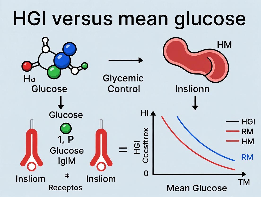
Beyond the Average: Why HGI is Revolutionizing Glycemic Control Assessment in Clinical Research and Drug Development
This article provides a comprehensive analysis of the Hemoglobin Glycation Index (HGI) versus mean glucose for assessing glycemic control, tailored for researchers, scientists, and drug development professionals.
Beyond the Average: Why HGI is Revolutionizing Glycemic Control Assessment in Clinical Research and Drug Development
Abstract
This article provides a comprehensive analysis of the Hemoglobin Glycation Index (HGI) versus mean glucose for assessing glycemic control, tailored for researchers, scientists, and drug development professionals. We explore the foundational biology of HGI, including its basis in interindividual variations in hemoglobin glycation. We detail methodologies for calculating and applying HGI in clinical trials and population studies, followed by troubleshooting common pitfalls in its implementation. Finally, we present a comparative validation against traditional metrics like HbA1c and mean glucose, examining predictive power for complications and utility in precision medicine. The synthesis aims to equip professionals with the knowledge to leverage HGI for more nuanced patient stratification and robust therapeutic evaluation.
Understanding HGI: The Science of Interindividual Variation in Hemoglobin Glycation
In the assessment of glycemic control, hemoglobin A1c (HbA1c) serves as the gold-standard, time-integrated metric. However, a persistent clinical and research problem is the significant inter-individual variance in HbA1c for a given mean blood glucose (MBG). This observation challenges the assumption of a uniform glucose-HbA1c relationship across populations and necessitates metrics that distinguish between dysglycemia due to elevated MBG versus individual biological propensity for hemoglobin glycation.
This whitepaper defines the Hemoglobin Glycation Index (HGI) as a residual metric that quantifies this biological propensity. The core thesis is that HGI provides distinct and complementary information to MBG, enabling refined patient stratification, elucidating underlying non-glycemic determinants of HbA1c, and offering a superior phenotype for genetic and drug development research focused on the processes of hemoglobin glycation itself.
Definition and Calculation of HGI
The HGI is mathematically defined as the difference between an individual's measured HbA1c and the HbA1c predicted for that individual based on their MBG, using a population-derived regression equation. It is thus the residual from the regression of HbA1c on MBG.
Formula: HGI = Observed HbA1c - Predicted HbA1c
The calculation involves two primary steps:
Establish the Population Regression Line: From a cohort study, perform a linear regression where HbA1c (%) is the dependent variable (Y) and MBG (mg/dL or mmol/L) is the independent variable (X). The regression equation takes the form:
Predicted HbA1c = β₀ + β₁ * (MBG)Calculate the Individual Residual: For any individual within or outside the cohort, their predicted HbA1c is computed using the above equation and their personal MBG. Their HGI is the observed minus this predicted value.
Table 1: Example Regression Coefficients from Key Studies
| Study Cohort (Year) | Sample Size (N) | Intercept (β₀) | Slope (β₁) per mg/dL MBG | R² (Variance Explained) |
|---|---|---|---|---|
| ADAG Study (2008) | ~500 (T1D, T2D, Non-DM) | ~1.11 | ~0.0187 | 0.84 |
| EGP Study (2007) | ~1400 (T1D, T2D) | ~0.82 | ~0.0215 | 0.68 |
| Your Cohort | N | Calculated | Calculated | Calculated |
Experimental Protocols for HGI Determination
Protocol 1: Establishing the HGI Reference Equation (Cohort Study)
- Objective: Derive the regression coefficients (β₀, β₁) for predicting HbA1c from MBG.
- Subjects: Large, diverse cohort (e.g., n > 300) including individuals with and without diabetes.
- MBG Measurement: Calculate MBG from intensive glucose monitoring over a period aligning with the HbA1c measurement interval (typically ~3 months). The gold standard is continuous glucose monitoring (CGM) data, providing >70% of values over ≥14 days. Alternatively, use frequent self-monitored blood glucose (SMBG) profiles (e.g., 7-point curves over multiple days).
- HbA1c Measurement: Measure HbA1c using an NGSP-certified, DCCT-aligned method (e.g., HPLC) at the end of the glucose monitoring period.
- Analysis: Perform simple linear regression:
HbA1c = β₀ + β₁ * (MBG). Validate the model using split-sample or cross-validation techniques. The resulting equation is the HGI calculator for that population.
Protocol 2: Assigning HGI in a Clinical Trial or Observational Study
- Objective: Classify participants as having Low, Average, or High HGI.
- Prerequisite: A validated HGI equation appropriate for the study population (from Protocol 1 or literature).
- Procedure:
- For each participant, measure MBG (via CGM or structured SMBG) over a defined period (e.g., 2 weeks).
- Measure HbA1c at the end of that period.
- Compute Predicted HbA1c using the pre-defined regression equation.
- Calculate HGI (Observed - Predicted).
- Categorize participants: Typically, HGI tertiles from the derivation cohort are used (e.g., Low HGI: < -0.5%; Average: -0.5% to +0.5%; High HGI: > +0.5%).
Signaling Pathways and Determinants of HGI
HGI reflects inter-individual variation in the kinetics of hemoglobin glycation, influenced by biological factors beyond ambient glucose concentration. Key determinants and their hypothesized pathways are summarized below.
Diagram 1: Key Determinants Influencing the Hemoglobin Glycation Index
The Scientist's Toolkit: Essential Research Reagents & Materials
Table 2: Key Research Reagent Solutions for HGI Studies
| Item | Function in HGI Research | Example/Note |
|---|---|---|
| NGSP-Certified HbA1c Assay | Precise and accurate measurement of the primary outcome. Essential for assay consistency. | HPLC systems (e.g., Tosoh G8), immunoassays. Must report DCCT-aligned values. |
| Continuous Glucose Monitor (CGM) | Gold-standard for estimating MBG non-invasively with high temporal resolution. | Dexcom G7, Abbott Libre 3. Provides AGP (Ambulatory Glucose Profile) data. |
| Glucose Oxidase/Hexokinase Assay Kits | For validating or calibrating MBG from blood samples in lieu of CGM. | Used for plasma glucose measurement in lab analysis of SMBG device accuracy. |
| Erythrocyte Lifespan Measurement Kits | To directly test a key biological determinant of HGI. | CO breath test kits, biotinylation label flow cytometry assays. |
| Methylglyoxal / 3-DG ELISA Kits | Quantify advanced glycation endproduct (AGE) precursors, linking to intracellular glycation rates. | Measures reactive dicarbonyls implicated in fast glycation. |
| DNA Genotyping/Secquencing Kits | To identify genetic variants associated with high or low HGI phenotypes. | GWAS arrays, targeted sequencing for loci like SPTA1 (spectrin). |
| Statistical Software Packages | For regression analysis, residual calculation, and cohort stratification. | R, Python (SciPy/Statsmodels), SAS, STATA. |
Data Interpretation and Research Implications
Table 3: Interpreting HGI in Research Contexts
| HGI Phenotype | Physiological Interpretation | Potential Research Implications |
|---|---|---|
| High HGI | Higher-than-expected HbA1c for given MBG. Suggests faster hemoglobin glycation kinetics, possibly due to longer RBC lifespan, increased intracellular glycation, or reduced deglycation. | Target for therapies reducing glycation rate (e.g., alagebrium). High cardiovascular risk phenotype independent of glucose. Candidate for genetic studies on glycation pathways. |
| Low HGI | Lower-than-expected HbA1c for given MBG. Suggests slower glycation kinetics, shorter RBC lifespan, or active deglycation. | May underestimate dysglycemia via HbA1c. Associated with conditions like anemia, hemoglobinopathies. Important for clinical trial screening. |
| Average HGI | HbA1c aligns with population-average prediction from MBG. | Represents the "standard" model of glucose-HbA1c relationship. Serves as reference group in comparative studies. |
The HGI, as a calculated residual, provides a powerful lens to dissect the contributions of glycemia versus intrinsic biological factors to the HbA1c value. Its integration into research protocols allows for the stratification of study populations into more physiologically homogeneous groups, potentially increasing the sensitivity to detect treatment effects in trials and clarifying the mechanisms behind complications risk. For drug development, HGI identifies a phenotype that may specifically benefit from novel agents targeting RBC biology or the glycation process itself, moving beyond pure glucocentrism.
The Glycemic Index (GI) standardizes the postprandial blood glucose response of carbohydrate-containing foods. However, significant inter-individual variability (up to 60%) exists in glycemic response to identical foods, a phenomenon termed High Glycemic Index Variability (HGI). This whitepaper explores the biological determinants underlying this variability, moving beyond mean blood glucose to a more nuanced understanding essential for personalized nutrition, metabolic research, and drug development. The core thesis posits that an exclusive focus on population mean glucose values is insufficient; understanding HGI is critical for advancing assessment of glycemic control and metabolic health.
Core Biological Determinants of HGI
Individual glycemic responses are modulated by a complex interplay of physiological, microbial, and molecular factors. These determinants explain why two individuals can exhibit markedly different blood glucose curves after consuming the same meal.
Physiological & Metabolic Factors
- Basal Metabolic Rate & Insulin Sensitivity: Individuals with higher insulin sensitivity typically show a more rapid glucose clearance. Key enzymes like hepatic glucokinase and peripheral GLUT4 activity vary significantly.
- Incretin Effect & Hormonal Milieu: The secretion and efficacy of GLP-1 and GIP, which potentiate glucose-stimulated insulin secretion, exhibit high inter-individual variability. Cortisol and growth hormone diurnal patterns also modulate insulin action.
- Gastric Emptying Rate: A primary determinant of glucose appearance rate in the bloodstream. Measured via acetaminophen absorption or scintigraphy, it can vary by over 300% between individuals.
- Circadian Biology: Pancreatic beta-cell responsiveness and peripheral insulin sensitivity oscillate with circadian rhythms, making time-of-day consumption a key variable.
Gut Microbiome Composition
The gut microbiome acts as a metabolic organ influencing HGI through several mechanisms:
- Short-Chain Fatty Acid (SCFA) Production: Fermentation of dietary fiber produces SCFAs (acetate, propionate, butyrate), which influence hepatic gluconeogenesis and enhance peripheral insulin sensitivity.
- Bile Acid Metabolism: Microbial transformation of primary to secondary bile acids activates receptors like FXR and TGR5, regulating glucose and lipid metabolism.
- Microbial Taxa Correlations: Specific genera are consistently associated with glycemic phenotypes.
Table 1: Gut Microbial Taxa Associated with Glycemic Response Variability
| Microbial Taxon | Association with Glycemic Response | Proposed Mechanism |
|---|---|---|
| Prevotella copri | Higher postprandial glucose | Increased expression of host circulating branched-chain amino acid (BCAA) levels, linked to insulin resistance. |
| Bacteroides spp. | Variable (strain-dependent) | Differential polysaccharide fermentation and SCFA profiles. |
| Akkermansia muciniphila | Lower postprandial glucose | Enhancement of gut barrier function, reduction of inflammation. |
| Firmicutes/Bacteroidetes Ratio | Often higher in low responders | Meta-analysis shows inconsistent correlation; functional capacity outweighs phylum-level ratio. |
Molecular & Genetic Determinants
Genetic polymorphisms and epigenetic modifications contribute to HGI.
- Candidate Genes: Variations in genes related to insulin signaling (IRS1), carbohydrate digestion (AMY1 copy number for salivary amylase), and circadian clock (CLOCK, BMAL1) are linked to differential glycemic responses.
- Postprandial Metabolomics: Lipid and amino acid flux post-meal are strong predictors. A rise in BCAAs often correlates with a blunted glucose clearance.
Table 2: Key Genetic Variants Associated with HGI
| Gene | Function | Variant/Polymorphism | Estimated Effect Size on PPG* (mmol/L) |
|---|---|---|---|
| AMY1 | Starch digestion | Copy Number Variation (CNV) | High CNV → 0.8-1.2 mmol/L lower peak |
| GCKR | Glucokinase regulation | rs1260326 (T allele) | ~0.5 mmol/L higher AUC |
| FTO | Adiposity & metabolism | rs9939609 (A allele) | Indirect via adiposity; ~0.3 mmol/L higher |
| SLC30A8 | Zinc transporter in beta-cells | rs13266634 (C/T) | Modifies insulin secretion dynamics |
*PPG: Postprandial Glucose. Effect sizes are approximate and context-dependent.
Experimental Protocols for Investigating HGI
To move beyond population averages, researchers employ controlled experiments to dissect HGI determinants.
Protocol: Comprehensive Phenotyping for HGI Stratification
Objective: To classify individuals as High or Low Glycemic Responders and collect multi-omics data for correlation.
- Participant Selection: Recruit n≥100 healthy or prediabetic adults. Exclude those with diabetes, GI disorders, or on antibiotics.
- Standardized Test Meal: After a 12-hour overnight fast, administer a precisely measured mixed meal (e.g., 75g available carbohydrates, 20g protein, 15g fat). Use a standardized food like Ensure or a customized muffin.
- Continuous Glucose Monitoring (CGM): Apply a blinded CGM sensor (e.g., Dexcom G6, Abbott Libre Pro) at least 24 hours prior. Monitor for 5 hours post-meal.
- Biological Sampling:
- Blood: Collect via intravenous catheter at T= -10, 0, 15, 30, 60, 90, 120, 180 minutes. Analyze for glucose, insulin, C-peptide, GLP-1, GIP, triglycerides, BCAAs.
- Stool & Saliva: Pre-intervention samples for 16S rRNA/metagenomic sequencing (stool) and AMY1 CNV analysis (saliva).
- Gastric Emptying Rate: Co-ingest 1.5g acetaminophen with test meal. Measure serum acetaminophen at intervals; time to peak concentration correlates with gastric emptying rate.
- Data Analysis: Calculate iAUC for glucose. Perform k-means clustering on iAUC to define HGI and LGI groups. Conduct multivariate analysis (e.g., O-PLS) to identify microbiome, metabolomic, and genetic features predictive of group membership.
Protocol: Human Microbiome Transplantation (HMT) for Causal Inference
Objective: To establish causal links between microbiome composition and HGI.
- Donor & Recipient Selection: Select donors from phenotyped HGI and LGI cohorts. Recipients are antibiotic-pretreated, metabolically healthy adults.
- Microbiota Preparation: Process donor stool under anaerobic conditions with glycerol cryopreservation.
- Recipient Preparation: 5-day broad-spectrum antibiotic regimen (vancomycin, neomycin, metronidazole) to deplete endogenous microbiota.
- Transplantation: Administer prepared microbiota via nasoduodenal tube or oral capsules on consecutive days.
- Post-HMT Phenotyping: After a 1-week colonization period, perform standardized meal tests (as in 2.1) on recipients. Compare their post-HMT glycemic response to their pre-antibiotic baseline and to the donor's phenotype.
- Analysis: Metagenomic sequencing of donor and recipient stool pre- and post-HMT. Correlate engraftment of specific taxa/genes with changes in glycemic response metrics.
Signaling Pathways in Glycemic Response Variability
The molecular response to a glucose load involves integrated signaling across organs.
The Scientist's Toolkit: Research Reagent Solutions
Table 3: Essential Reagents and Materials for HGI Research
| Item | Function in HGI Research | Example/Supplier |
|---|---|---|
| Standardized Test Meals | Provides a consistent carbohydrate challenge to measure inter-individual response variability. | Ensure Plus (Abbott), Glycemic Index Testing Kit (Carbohydrate Solutions). |
| Continuous Glucose Monitor (CGM) | Measures interstitial glucose every 1-5 minutes, capturing full glycemic variability and curve shape. | Dexcom G6 Professional, Abbott FreeStyle Libre Pro 2. |
| Acetaminophen Absorption Test Kit | Indirect, non-radioactive method to assess gastric emptying rate—a key HGI determinant. | Paracetamol (Acetaminophen) ELISA Kit (e.g., Abcam). |
| Multiplex Hormone Assay Kits | Simultaneous measurement of insulin, C-peptide, GLP-1 (active & total), GIP from limited plasma volumes. | MILLIPLEX Metabolic Hormone Panel (Merck), Meso Scale Discovery (MSD) U-PLEX. |
| Stool DNA Isolation Kit (for microbiome) | High-yield, PCR-inhibitor-free DNA extraction from complex stool samples for sequencing. | QIAamp PowerFecal Pro DNA Kit (Qiagen), ZymoBIOMICS DNA Miniprep Kit. |
| 16S rRNA & Metagenomic Sequencing Services | Characterizes microbial community composition (16S) and functional potential (shotgun metagenomics). | Services by Illumina (MiSeq), paired with analysis pipelines (QIIME 2, HUMAnN 3.0). |
| Branched-Chain Amino Acid (BCAA) Assay | Quantifies serum levels of leucine, isoleucine, valine—metabolites predictive of glycemic response. | BCAA Assay Kit (Colorimetric/Fluorometric) (e.g., Abcam, Sigma). |
| Salivary Amylase (AMY1) CNV Assay | Determines copy number of the AMY1 gene, a genetic factor influencing starch digestion rate. | Quantitative PCR (qPCR) or Digital PCR assays with specific primers/probes. |
| Human Microbiome Transplantation Materials | For causal experiments: anaerobic workstation, glycerol, cryovials, and capsule preparation kits. | Anaerobic chamber (Coy Lab), BioMatrix encapsulation technology. |
Experimental Workflow for HGI Studies
The following diagram outlines a comprehensive research pipeline from subject recruitment to data integration.
This whitepaper examines the inherent biological and statistical limitations of relying solely on HbA1c or mean glucose (MG) for assessing glycemic control. The argument is framed within the broader research thesis that the Glycemic Gap Index (GGI), defined as the residual from regressing HbA1c on mean glucose, or the more comprehensive Hemoglobin Glycation Index (HGI), provides a more nuanced and patient-specific metric. HGI accounts for inter-individual variations in non-glycemic factors affecting HbA1c, offering critical insights for personalized medicine, clinical trial design, and drug development.
Core Limitations: Biological and Statistical Factors
Biological Variability Affecting HbA1c
HbA1c, while a cornerstone of diabetes management, is influenced by factors unrelated to mean blood glucose levels.
Table 1: Key Non-Glycemic Factors Influencing HbA1c
| Factor | Direction of Effect on HbA1c | Proposed Mechanism |
|---|---|---|
| Erythrocyte Lifespan | Decreased lifespan lowers HbA1c; increased raises it. | Altered time available for hemoglobin glycation. |
| Hemoglobin Variants | Variable; e.g., HbS trait may lower measured HbA1c. | Altered glycation kinetics or interference with assay. |
| Iron Deficiency Anemia | Increases HbA1c. | May increase erythrocyte lifespan & alter glycation. |
| Chronic Kidney Disease | Variable; can increase or decrease. | Altered erythropoiesis, lifespan, and assay interference. |
| Ethnicity | Population-level differences observed. | Genetic/population differences in glycation biology. |
Statistical Limitations of Mean Glucose
Mean glucose, derived from Continuous Glucose Monitoring (CGM), provides a superior picture of glycemic exposure than HbA1c alone but fails to capture the full risk profile.
Table 2: Glycemic Metrics Not Captured by Mean Glucose Alone
| Metric | Clinical Significance | Limitation of MG |
|---|---|---|
| Glycemic Variability (GV) | Independent risk factor for hypoglycemia & potentially complications. | MG can be identical in high and low GV scenarios. |
| Time-in-Range (TIR) | Directly correlates with microvascular outcomes. | MG does not specify distribution of glucose values. |
| Time-in-Hypoglycemia | Critical for safety assessment. | A "good" MG can mask significant hypoglycemia. |
Experimental Evidence and Protocols
Key Experiment 1: Establishing the HGI Concept
Objective: To demonstrate that the difference between measured HbA1c and that predicted from mean glucose (HGI) is a consistent, individual-specific trait. Protocol:
- Cohort: Recruit a large, diverse cohort (n>500) of individuals with and without diabetes.
- Glycemic Measurement: Collect continuous glucose monitoring (CGM) data for a minimum of 14 days to calculate the individual's mean glucose (MG).
- HbA1c Measurement: Draw blood at the end of the CGM period for HbA1c analysis via a standardized, NGSP-certified method (e.g., HPLC).
- Statistical Modeling: Perform a linear regression for the cohort:
HbA1c = β0 + β1 * MG. Calculate the predicted HbA1c for each individual. - HGI Calculation: Compute HGI for each subject as:
HGI = Measured HbA1c - Predicted HbA1c. - Test-Retest Analysis: Repeat the CGM and HbA1c measurement in the same individuals after a 3-6 month interval. Correlate the HGI values from the two visits to assess individual consistency (high correlation supports HGI as a persistent trait).
Key Experiment 2: Linking High HGI to Complications
Objective: To test the hypothesis that a high HGI (higher-than-predicted HbA1c) is associated with increased risk of microvascular complications, independent of mean glucose. Protocol:
- Study Design: Prospective observational cohort or post-hoc analysis of clinical trial data (e.g., DCCT, EDIC).
- Population: Individuals with type 1 or type 2 diabetes with long-term follow-up data.
- Exposure: Calculate HGI at baseline as per Protocol 3.1. Categorize participants into HGI tertiles (Low, Medium, High).
- Outcomes: Pre-specified microvascular endpoints (e.g., progression of retinopathy confirmed by grading of fundus photographs, onset of albuminuria, confirmed neuropathy).
- Analysis: Use Cox proportional hazards models to assess the risk of complication development by HGI tertile. Critical Covariates: Models must adjust for mean glucose, diabetes duration, age, blood pressure, and other standard risk factors. A significant hazard ratio for the High HGI group indicates independent risk.
Visualizing the Conceptual Framework and Workflow
Diagram Title: Factors Differentiating HbA1c, Predicted A1c, and HGI
Diagram Title: Stepwise Workflow for HGI Determination and Validation
The Scientist's Toolkit: Essential Research Reagents & Materials
Table 3: Key Research Reagent Solutions for HGI and Glycemic Studies
| Item | Function & Rationale |
|---|---|
| NGSP-Certified HbA1c Assay | Ensures standardized, accurate, and traceable HbA1c measurement critical for valid HGI calculation. Examples: HPLC (Tosoh G8, Bio-Rad Variant II), immunoassay. |
| Factory-Calibrated CGM Systems | Provides reliable interstitial glucose data for calculating mean glucose, glycemic variability, and Time-in-Range. Essential for the MG component of HGI. Examples: Dexcom G7, Abbott Libre 3. |
| Erythrocyte Lifespan Measurement Kits | To quantify a major non-glycemic factor. Kits may use CO breath test (endogenous labeling) or stable isotope (e.g., 15N-glycine, 13C-cyanate) labeling methods. |
| Hemoglobin Variant Analysis Kit | To identify and quantify hemoglobinopathies (e.g., HbS, HbC, HbE) that can interfere with HbA1c assays and affect glycation rates. |
| Specialized Tubes for Glycated Protein Analysis | EDTA tubes for HbA1c; tubes with glycolytic inhibitors (e.g., fluoride/oxalate) for concurrent fasting glucose if needed. Proper sample handling is crucial. |
| Statistical Software (R, Python, SAS) | For performing linear regression to establish the MG-HbA1c population relationship, calculating residuals (HGI), and running advanced survival/risk models. |
Historical Context and Evolution of the HGI Concept in Diabetes Research
The Hemoglobin Glycation Index (HGI) quantifies the inter-individual difference between observed hemoglobin A1c (HbA1c) and the HbA1c predicted from mean blood glucose levels. This whitepaper traces the historical development of the HGI concept, framing it within the ongoing debate on HGI versus mean glucose for glycemic control assessment. Understanding HGI is crucial for refining risk stratification, personalizing treatment, and designing clinical trials in diabetes.
Historical Development and Key Studies
Origins and Conceptual Foundation
The HGI concept emerged from observations that HbA1c levels vary significantly among individuals despite similar mean blood glucose concentrations. Early work in the 1990s, notably by McCarter et al. and Yudkin et al., formalized this biological variation, proposing that an individual's "glycation gap" might be a stable, intrinsic trait.
Landmark Clinical Evidence
Key longitudinal studies established HGI's prognostic value. The Diabetes Control and Complications Trial (DCCT) and the Epidemiology of Diabetes Interventions and Complications (EDIC) study provided foundational data, showing that individuals with higher HGI were at increased risk for microvascular complications, independent of mean glucose.
Table 1: Key Historical Studies on HGI
| Study (Year) | Population | Key Finding | Implication for HGI Concept |
|---|---|---|---|
| DCCT/EDIC (1990s-2000s) | Type 1 Diabetes | High HGI associated with increased retinopathy and nephropathy risk. | Established HGI as an independent risk factor for complications. |
| A1C-Derived Average Glucose (ADAG) Study (2008) | Type 1, Type 2, Non-Diabetic | Defined linear relationship between HbA1c and mean glucose, highlighting residual variance. | Provided a standardized method for calculating predicted HbA1c, enabling HGI derivation. |
| Hunt 2 Study (2012) | General Population | High HGI predicted cardiovascular mortality and all-cause mortality. | Extended HGI relevance beyond diabetes to cardiovascular risk in non-diabetic individuals. |
Core Methodologies and Experimental Protocols
Standard HGI Calculation Protocol
HGI is calculated as the residual from a regression model of HbA1c on mean blood glucose.
- Data Collection: Obtain paired measures of HbA1c and mean blood glucose (MBG) from continuous glucose monitoring (CGM) or frequent self-monitoring over a minimum of 4-6 weeks.
- Cohort Derivation Model: In a reference population, perform linear regression:
HbA1c = β0 + β1 * MBG. This establishes the population-average relationship. - Individual HGI Calculation: For any individual, calculate HGI as:
Observed HbA1c - Predicted HbA1c, where Predicted HbA1c = β0 + β1 * (individual's MBG). - Categorization: Individuals are typically categorized into Low, Medium, and High HGI tertiles based on the distribution in the reference population.
Protocol for Investigating HGI Mechanisms
To explore biological determinants of HGI, studies often employ:
- In Vivo Glycation Rate Studies: Use intravenous 13C- or 14C-labeled glucose infusion in human subjects. Sequentially measure label incorporation into hemoglobin (via mass spectrometry) and erythrocyte lifespan (via carbon monoxide production or biotin labeling).
- Erythrocyte Experimentation: Isolate erythrocytes from High vs. Low HGI donors. Incubate ex vivo with high-glucose medium. Measure intra-erythrocytic glucose concentration, oxidative stress markers (e.g., glutathione), and activity of glycation-countering enzymes (e.g., glyoxalase I).
- Genetic Association Studies: Perform genome-wide association studies (GWAS) on HGI values in large, phenotyped cohorts (e.g., UK Biobank) to identify single nucleotide polymorphisms (SNPs) linked to HGI variability.
Signaling Pathways and Biological Determinants
HGI is influenced by factors beyond plasma glucose. The primary biological determinants involve pathways affecting intracellular glucose handling, hemoglobin glycation kinetics, and erythrocyte physiology.
Diagram 1: Biological Determinants of HGI Variation (100 chars)
The Scientist's Toolkit: Research Reagent Solutions
Table 2: Essential Research Materials for HGI Investigations
| Item | Function/Application | Example/Note |
|---|---|---|
| Continuous Glucose Monitor (CGM) | Provides dense, ambulatory mean glucose data for HGI calculation. | Dexcom G7, Abbott Libre 3. Critical for accurate MBG estimation. |
| HbA1c Assay (HPLC/MS) | Precise, standardized measurement of glycated hemoglobin. | Tosoh G11, Mass Spectrometric methods (reference standard). |
| 13C6-Glucose Isotope Tracer | For in vivo kinetic studies of glycation rates and erythrocyte turnover. | Cambridge Isotope Laboratories CLM-1396. Used in infusion protocols. |
| Erythrocyte Isolation Kit | Purifies red blood cells from whole blood for ex vivo experiments. | MilliporeSigma ROSeparator or density gradient centrifugation. |
| Anti-AGE Antibodies | Detect and quantify advanced glycation end-products within erythrocytes. | Anti-CML, Anti-MG-H1 antibodies (Cell Signaling, TransGenic). |
| Glyoxalase 1 Activity Assay Kit | Measures activity of key intracellular MG-detoxifying enzyme. | Colorimetric/Fluorometric kits (Sigma-Aldrich, Abcam). |
| GWAS Genotyping Array | For genome-wide screening of genetic variants associated with HGI. | Illumina Global Screening Array, Infinium technology. |
Modern Evolution and Future Directions
The HGI concept has evolved from a descriptive observation to a candidate biomarker for "personalized HbA1c." Current research focuses on:
- Genetic and Omics Basis: Large-scale biobank studies are identifying genomic, proteomic, and metabolomic signatures of high HGI.
- Drug Development: HGI stratification is being explored in clinical trials to identify sub-populations that may benefit differentially from novel glycemic or anti-glycation therapies.
- Integration with CGM Metrics: The relationship between HGI and glucometrics (e.g., Time in Range, GV) is an active area, refining the HGI vs. mean glucose debate.
Diagram 2: HGI Calculation and Application Workflow (99 chars)
The HGI concept represents a critical refinement in interpreting HbA1c, accounting for significant inter-individual biological variation. Its historical evolution from an epidemiological observation to a mechanistically-grounded research tool underscores its importance. Within the broader thesis of HGI versus mean glucose, HGI provides a complementary dimension, directing research toward personalized pathophysiology and away from a one-size-fits-all model of glycemic control assessment. Future integration into clinical practice and drug development requires standardized measurement protocols and validation in diverse populations.
Key Studies Establishing HGI as a Phenotypic Trait
Within the evolving paradigm of glycemic control assessment, the comparative utility of Hyperglycemia Index (HGI) versus mean glucose remains a central research question. While mean glucose provides a population-average metric, it fails to capture intra- and inter-individual variability in glycemic response to identical glucose challenges. HGI, calculated as the area under the glucose curve above a defined threshold (e.g., 140 mg/dL), quantifies the magnitude and duration of hyperglycemic excursions. This whitepaper details the key experimental studies that have established HGI as a distinct, reproducible, and heritable phenotypic trait, critical for personalized diabetes management and drug development.
Table 1: Foundational Studies Demonstrating HGI as a Phenotypic Trait
| Study (Year) | Population & Design | Key Intervention/Metric | Primary Quantitative Finding (HGI-related) | Implication for Trait Status |
|---|---|---|---|---|
| Svendsen et al. (2012) | N=44 non-diabetic twins; Mixed-meal tolerance test (MMTT). | Calculated HGI above 8 mmol/L (144 mg/dL). | Intra-pair correlation: 0.63 (monozygotic) vs. 0.25 (dizygotic). | High heritability of postprandial HGI. |
| Vistisen et al. (2014) | N=1,532 non-diabetic adults; 3-point OGTT. | HGI defined as glucose >7.8 mmol/L (140 mg/dL). | HGI variance component: 65% attributed to individual factors vs. 35% to test conditions. | HGI is a stable individual characteristic. |
| Møller et al. (2016) | N=447 individuals; Repeated MMTT (3 tests). | Intra-class correlation coefficient (ICC) for HGI. | ICC for HGI = 0.70, indicating high test-retest reliability. | HGI is a reproducible phenotypic measure. |
| Rizza et al. (2010) | N=150 healthy volunteers; Hyperglycemic clamp. | Variability in glucose disposal rate (GDR). | Individuals with high HGI exhibited 25% lower mean GDR (p<0.01). | Links HGI phenotype to underlying insulin resistance. |
| Ahqvist et al. (2018) - ANDIS | N=8,980 (diabetes); Cluster analysis. | HGI used to characterize severe insulin-deficient cluster. | High-HGI cluster had 3.2x higher risk of retinopathy vs. low-HGI clusters. | HGI predicts complication risk independently of mean glucose. |
Detailed Experimental Protocols
1. Twin Study Protocol (Svendsen et al.)
- Objective: To partition genetic vs. environmental contributions to postprandial HGI.
- Subject Preparation: 10-hour overnight fast.
- Test Meal: Standardized liquid mixed meal (Ensure, ~600 kcal, 55% carb, 15% protein, 30% fat).
- Blood Sampling: Frequent venous sampling at -30, 0, 15, 30, 45, 60, 90, 120, 150, 180 minutes relative to meal ingestion.
- Analysis: Plasma glucose measured via glucose oxidase method. HGI calculated as AUC >8 mmol/L using the trapezoidal rule.
- Statistical Model: Structural equation modeling to estimate additive genetic (A), common environmental (C), and unique environmental (E) variance components based on monozygotic vs. dizygotic twin correlations.
2. Test-Retest Reliability Protocol (Møller et al.)
- Objective: To assess the reproducibility of the HGI phenotype across repeated metabolic tests.
- Study Design: Participants underwent three identical MMTTs, each separated by 2-6 weeks.
- Standardization: Strict pre-test standardization: identical meals, physical activity, and sleep duration for 48 hours prior. Tests performed at the same time of day.
- HGI Calculation: As above (AUC >8 mmol/L).
- Reliability Analysis: Intra-class Correlation Coefficient (ICC) calculated using a two-way random-effects model for absolute agreement. An ICC >0.75 indicates excellent reliability; 0.6-0.75 indicates good reliability.
3. Hyperglycemic Clamp Protocol (Rizza et al.)
- Objective: To link HGI phenotype to in vivo tissue-level insulin sensitivity.
- Procedure: After baseline, plasma glucose is rapidly raised to a target hyperglycemic level (e.g., 10 mmol/L or 180 mg/dL) via a primed, continuous intravenous glucose infusion.
- Maintenance: Glucose is clamped at this target for 120-180 minutes by variable glucose infusion guided by frequent (every 5 min) glucose measurements.
- Insulin Measurement: Plasma insulin concentration reaches a steady-state plateau.
- Key Metric: Glucose Disposal Rate (GDR) is calculated from the glucose infusion rate (M-value) during the final 30 minutes of the clamp, corrected for urinary glucose loss and changes in glucose pool size. Lower GDR indicates insulin resistance.
Pathway and Experimental Workflow Diagrams
Title: HGI Phenotyping Workflow & Validation
Title: Physiological Determinants of HGI Phenotype
The Scientist's Toolkit: Key Research Reagent Solutions
Table 2: Essential Materials for HGI Phenotyping Research
| Item/Category | Function & Rationale | Example/Note |
|---|---|---|
| Standardized Challenge Meal | Provides a uniform glycemic stimulus to assess inter-individual response variance. Critical for reproducibility. | Ensure Plus or similar nutritionally defined liquid meal. |
| Oral Glucose Tolerance Test (OGTT) Solution | The classic, pharmacopeia-defined challenge for diagnosing diabetes; allows direct comparison to historical data. | 75g anhydrous glucose dissolved in 250-300 mL water. |
| Enzymatic Glucose Assay Kit | For accurate, high-throughput measurement of plasma/serum glucose concentrations from frequent samples. | Glucose oxidase/peroxidase (GOD-POD) or hexokinase-based kits. |
| Hyperglycemic Clamp Tray | Specialized setup for the hyperglycemic clamp procedure, the gold-standard for assessing β-cell function and tissue sensitivity under high glucose. | Includes variable-rate glucose infusion pump, frequent sampling line, and glucose analyzer (e.g., YSI/Beckman). |
| EDTA or Fluoride Tubes | For blood collection. Fluoride inhibits glycolysis, preserving glucose levels between draw and assay. | Grey-top (fluoride/oxalate) tubes for glucose; Lavender-top (EDTA) for insulin. |
| Insulin & C-Peptide ELISA/EIA Kits | To measure insulin secretion and clearance in parallel with glucose, enabling calculation of disposition indices. | Mesoscale Discovery (MSD) or Mercodia assays are common. |
| GWAS Genotyping Array | To identify genetic variants associated with high HGI, establishing its genetic underpinnings as a trait. | Illumina Global Screening Array or Infinium Omni arrays. |
| Statistical Software Packages | For complex modeling: variance component analysis, ICC calculation, mixed-effects models for repeated measures. | R (nlme, irr packages), SAS (PROC MIXED, PROC VARCOMP). |
Calculating and Implementing HGI: Protocols for Research and Clinical Trials
The assessment of glycemic control is fundamental to diabetes research and therapy development. The glycation gap, more precisely defined as the Hemoglobin Glycation Index (HGI), is a calculated measure that quantifies the difference between an individual's observed HbA1c and the HbA1c predicted from their mean blood glucose levels. This whitepaper details the standard formulas, methodologies, and technical considerations for calculating HGI, framed within the ongoing research thesis on whether HGI or mean glucose serves as a superior metric for understanding inter-individual variation in glycemic response and complication risk.
Theoretical Framework and Core Formula
The Hemoglobin Glycation Index (HGI) is derived from the linear regression model that establishes the population relationship between HbA1c and mean blood glucose (MG). An individual's HGI is the residual from this regression line.
The standard calculation proceeds in two steps:
Step 1: Establish the Population Regression Equation
From a cohort study, the following linear relationship is derived: Predicted HbA1c (%) = α + β × Mean Glucose (mg/dL) where:
- α = y-intercept of the regression line.
- β = slope of the regression line (increase in HbA1c per mg/dL increase in MG).
Table 1: Published Regression Coefficients for HbA1c Prediction
| Study / Cohort | α (Intercept) | β (Slope) | MG Measurement Method | Sample Size (n) | Reference |
|---|---|---|---|---|---|
| ADAG Study | ~2.6 | ~0.0149 | CGM & SMBG | 507 | Nathan et al., 2008 |
| DCCT Cohort | ~1.13 | ~0.0181 | 7-point SMBG profile | 1,441 | Hempe et al., 2015 |
| General Clinic Population | Varies | ~0.024 | Lab MG from EHR | 12,504 | Sacks et al., 2022 |
Note: It is critical to select a regression equation derived from a cohort and glucose measurement method comparable to one's own research population.
Step 2: Calculate Individual HGI
For an individual with a measured HbA1c and a corresponding measured Mean Glucose (MG), the HGI is calculated as: HGI = Measured HbA1c (%) − Predicted HbA1c (%)
A positive HGI indicates an individual's HbA1c is higher than predicted for their mean glucose level ("high glycator"), while a negative HGI indicates it is lower ("low glycator").
Experimental Protocols for Data Generation
Accurate HGI calculation depends on robust methods for measuring its two components.
Protocol A: Measurement of HbA1c
- Principle: High-performance liquid chromatography (HPLC) is the gold-standard method, separating hemoglobin variants based on charge and size.
- Procedure:
- Sample Collection: Collect venous blood in an EDTA tube.
- Hemolysate Preparation: Lyse erythrocytes to release hemoglobin.
- HPLC Analysis: Inject hemolysate onto a cation-exchange HPLC column. Hemoglobins (A0, A1a, A1b, A1c, F, etc.) are eluted using a gradient buffer and detected by absorbance at 415 nm.
- Quantification: The area under the peak for HbA1c is expressed as a percentage of the total hemoglobin area (A0+A1a+A1b+A1c).
- Quality Control: Use certified reference materials aligned with the International Federation of Clinical Chemistry (IFCC) standards and the National Glycohemoglobin Standardization Program (NGSP).
Protocol B: Measurement of Mean Glucose for Regression & Calculation
- Principle: Estimate average glycemia over the preceding 2-3 months, the lifespan of the erythrocyte.
- Methods:
- Continuous Glucose Monitoring (CGM): The preferred research method. Subjects wear a CGM sensor measuring interstitial glucose every 1-15 minutes for a minimum of 14 days. The mean of all glucose values is calculated.
- Capillary Blood Glucose (SMBG) Profiling: Subjects perform 7-point profiles (pre- and 90-120min post-meals, bedtime) at least 3 days per week over 2-4 weeks. The mean of all readings is calculated.
- Laboratory Mean Glucose: Calculated from all plasma glucose values (fasting, random, OGTT) available in electronic health records over a defined period.
Table 2: Comparison of Mean Glucose Assessment Methods
| Method | Temporal Resolution | Establishes Regression | Calculates Individual HGI | Key Limitation |
|---|---|---|---|---|
| CGM | Excellent (High-frequency) | Ideal | Ideal | Cost, sensor availability |
| 7-point SMBG Profile | Good (Sparse) | Good | Good | Patient burden, misses nocturnal data |
| Lab Mean Glucose (EHR) | Poor (Opportunistic) | Acceptable for large cohorts | Not recommended for individuals | Highly biased sampling |
Visualization of HGI Calculation Workflow
Workflow for Calculating the Hemoglobin Glycation Index
The Scientist's Toolkit: Key Research Reagent Solutions
Table 3: Essential Materials for HGI-Related Research
| Item / Reagent | Function in Research | Example / Specification |
|---|---|---|
| EDTA Blood Collection Tubes | Preserves blood for HbA1c analysis by inhibiting coagulation and glycation in vitro. | K2EDTA or K3EDTA tubes, CLSI-compliant. |
| HPLC HbA1c Analyzer & Columns | High-resolution separation and quantification of HbA1c from other hemoglobin variants. | Bio-Rad VARIANT II, Tosoh G8; dedicated cation-exchange cartridges. |
| IFCC/NGSP Calibrators | Ensures accuracy and standardization of HbA1c results across labs and studies. | Certified primary and secondary reference materials. |
| Continuous Glucose Monitor (CGM) | Gold-standard for capturing continuous interstitial glucose to calculate true mean glucose. | Dexcom G7, Abbott Freestyle Libre 3 (research-use configurations). |
| Statistical Software | To perform linear regression, calculate residuals (HGI), and conduct subsequent analyses. | R, Python (SciPy/Statsmodels), SAS, or GraphPad Prism. |
| Glucose Control Solution | For calibrating/validating SMBG meters used in capillary glucose profiling. | Manufacturer-specific solutions at low, mid, and high glucose ranges. |
Data Interpretation and Research Implications
Table 4: Interpretation and Potential Correlates of HGI
| HGI Phenotype | Calculated Value | Potential Physiological/Clinical Research Implications |
|---|---|---|
| High Glycator | HGI > +0.5%* | May indicate faster non-enzymatic glycation, increased risk of complications at a given MG level, altered erythrocyte lifespan, or other genetic/metabolic factors. |
| Low Glycator | HGI < -0.5%* | May indicate slower glycation, reduced complication risk relative to MG, or differences in intracellular glucose metabolism. |
| Average Glycator | -0.5% ≤ HGI ≤ +0.5%* | Observed HbA1c aligns with population prediction from mean glucose. |
*Thresholds are illustrative; study-specific tertiles or standard deviations are commonly used for group stratification in research.
The central thesis question—HGI vs. mean glucose—hinges on which metric better predicts long-term diabetic complications. Research protocols must be designed to collect longitudinal data on complications (retinopathy, nephropathy, neuropathy) and analyze whether HGI provides predictive power independent of mean glucose. This requires multivariate statistical models where HGI and mean glucose are entered as separate covariates.
The assessment of glycemic control in clinical research and therapeutic development has historically relied on summary metrics, primarily hemoglobin A1c (HbA1c) and mean glucose. The emerging thesis posits that the Glycemic Hemoglobin Index (HGI)—the observed difference between an individual's measured HbA1c and the HbA1c predicted from their mean glucose—may offer superior pathophysiological insight compared to mean glucose alone. HGI reflects inter-individual variation in the glycation gap, potentially influenced by erythrocyte lifespan, membrane permeability, and non-glycemic determinants of hemoglobin glycation.
Evaluating this thesis necessitates precise, high-frequency glucose data to calculate true mean glucose and its relationship to HbA1c. This technical guide details the three core data sources required for such research: Continuous Glucose Monitoring (CGM), Self-Monitored Plasma Glucose (SMPG), and the calculated Estimated Average Glucose (eAG). The integration of these sources is critical for robust analysis of the HGI versus mean glucose debate.
Continuous Glucose Monitoring (CGM)
CGM systems measure interstitial glucose concentration continuously (every 1-5 minutes), providing an unparalleled view of glycemic variability, patterns, and exposure.
Key Experimental Protocol for Research Use:
- Device Selection & Calibration: Use a clinically-validated, regulatory-approved professional (blinded) or personal (unblinded) CGM. For drug trials, professional CGM is often preferred to minimize behavioral feedback. Calibrate per manufacturer instructions using capillary blood glucose values from a calibrated glucometer.
- Wear Duration: A minimum 14-day wear period is recommended to capture diurnal patterns and day-to-day variability. For comprehensive assessment, durations aligning with the erythrocyte lifespan (≈90-120 days) are ideal but pragmatically challenging.
- Data Extraction & Processing: Use manufacturer-specific software or APIs to extract raw glucose values, timestamps, and sensor status. Process data to:
- Identify and handle sensor drop-outs or signal artifacts.
- Align all timestamps to a common reference (e.g., participant's local time).
- Calculate summary metrics (see Table 1).
Self-Monitored Plasma Glucose (SMPG)
SMPG, traditionally known as Self-Monitored Blood Glucose (SMBG), provides point-in-time capillary plasma glucose measurements. It is essential for CGM calibration and for capturing glucose at specific, protocol-defined moments.
Key Experimental Protocol for Research Use:
- Sampling Schedule: Implement a structured protocol, such as a 7-point profile (pre- and 90-120 minutes post-prandial for three meals, plus bedtime) or an expanded 10-point profile (adding 0300h measurement). Testing should occur over multiple representative days.
- Device & Technique Standardization: Provide all participants with the same FDA-approved/CE-marked glucose meter and test strips. Train participants on standardized finger-prick technique (including hand washing) to minimize pre-analytical error.
- Data Integrity: Use meters with memory/cloud upload to ensure data provenance. Record concomitant events (meal composition, medication, exercise) in a paired log.
Estimated Average Glucose (eAG)
eAG is not a directly measured entity but a statistical estimate derived from either CGM or SMPG data. It represents the calculated average plasma glucose concentration over a specified period.
Calculation Protocol:
- From CGM Data: Calculate the arithmetic mean of all interstitial glucose readings over the wear period. Apply a validated transformation factor (if required by device) to estimate plasma-equivalent glucose. The result is the CGM-derived mean glucose, expressed as eAG (mg/dL or mmol/L).
- From SMPG Data: Calculate the weighted average from structured profiles, giving appropriate weight to postprandial periods which are often oversampled. Advanced methods involve modeling glucose curves between measurements.
- Relationship to HbA1c: The widely used Nathan et al. (2008) regression formula (
eAG (mg/dL) = 28.7 × HbA1c (%) - 46.7) provides a population-average conversion. Critically, the deviation of an individual's measured HbA1c from the HbA1c predicted by this formula (using their measured mean glucose) is the HGI.
Table 1: Comparison of Core Glycemic Data Sources for Research
| Feature | Continuous Glucose Monitoring (CGM) | Self-Monitored Plasma Glucose (SMPG) | Estimated Average Glucose (eAG) |
|---|---|---|---|
| Nature of Data | High-frequency, interstitial fluid glucose | Sparse, point-in-time, capillary plasma glucose | Calculated summary statistic (mg/dL or mmol/L) |
| Primary Metrics | Mean Glucose, %TIR (70-180 mg/dL), %TBR (<70 mg/dL), %TAR (>180 mg/dL), Glycemic Variability (CV, SD) | Point values at specific times (e.g., fasting, postprandial) | Single value representing average plasma glucose |
| Key Advantage | Captures 24/7 glucose patterns, variability, and undetected hypoglycemia | Inexpensive, established, direct plasma measure, good for calibration | Provides a single-number summary comparable to HbA1c |
| Key Limitation | Interstitial fluid lag (5-15 min), cost, wear-time burden | Sparse data, prone to sampling bias, misses nocturnal events | An estimate; masks variability; conversion from HbA1c is population-based |
| Role in HGI Research | Provides the gold-standard measure of true mean glucose for HGI calculation. | Validates CGM, provides reference glucose for key time points. | The bridge metric between measured mean glucose and HbA1c. |
Table 2: Essential Glycemic Metrics Derived from CGM Data (ADA/EASD Consensus Targets)
| Metric | Clinical/Research Target | Calculation Method | Relevance to HGI |
|---|---|---|---|
| Mean Glucose | Individualized | Arithmetic mean of all sensor readings | Core variable. Direct input for HGI calculation. |
| % Time in Range (TIR) | >70% (70-180 mg/dL) | (Number of readings 70-180 mg/dL / Total readings) * 100 | Correlates with HbA1c; reflects quality of control independent of HGI. |
| Coefficient of Variation (CV) | <36% (Stable) | (Standard Deviation / Mean Glucose) * 100 | High CV may indicate glycemic instability, potentially influencing HGI. |
| Glycemic Management Indicator (GMI) | N/A | GMI (%) = 3.31 + 0.02392 * [mean glucose in mg/dL]. | An alternative CGM-derived estimate of likely HbA1c, for comparison with lab HbA1c. |
Experimental Workflow for HGI-Based Studies
Diagram Title: Experimental Workflow for HGI Research Studies
The Scientist's Toolkit: Essential Research Reagents & Materials
Table 3: Key Research Reagent Solutions for Glycemic Assessment Studies
| Item | Function & Specification | Critical Notes for Protocol |
|---|---|---|
| Professional CGM System | Provides blinded, research-grade continuous glucose data. Example: Dexcom G7 Pro, Medtronic Guardian Connect. | Ensure IRB approval for data handling. Plan for clinic-based sensor application. |
| NGSP-Certified HbA1c Assay | Provides the gold-standard measurement of glycated hemoglobin for HGI calculation. Example: HPLC-based methods. | Use a single, central laboratory for all study samples to minimize assay variability. |
| Standardized Glucose Meter & Strips | For SMPG profiles and CGM calibration. Example: Contour Next One, Accu-Chek Guide. | Batch-purchase strips from a single lot. Validate meter precision and accuracy at study start. |
| Erythrocyte Lifespan Measurement Kit | To investigate a key biological determinant of HGI. Example: CO breath test kit or bilirubin/carbon monoxide analyzer. | Technically complex; consider as a sub-study in a phenotyping cohort. |
| Stable Isotope Labeled Glucose Tracers | For sophisticated metabolic phenotyping (e.g., rate of endogenous glucose production, glycation kinetics). Example: [6,6-²H₂]-glucose. | Requires specialized mass spectrometry (GC/MS, LC-MS) and pharmacokinetic modeling expertise. |
| Data Harmonization Platform | Software to aggregate, clean, and analyze CGM, SMPG, and lab data. Example: Tidepool, Glooko, or custom R/Python pipelines. | Must be HIPAA/GCP compliant. Ensure it can output standardized metrics (e.g., consensus CGM metrics). |
The rigorous investigation of the HGI versus mean glucose thesis is contingent upon the meticulous acquisition and integration of CGM, SMPG, and eAG data. CGM provides the definitive measure of mean glucose, SMPG offers critical point-in-time validation, and eAG serves as the essential translational link to HbA1c. By employing standardized protocols, leveraging the outlined research toolkit, and analyzing data within the conceptual framework illustrated, researchers can elucidate whether HGI, reflecting inter-individual biological variation, provides a more nuanced and predictive model of diabetes-related outcomes than mean glucose alone.
Within the ongoing research thesis comparing Hemoglobin Glycation Index (HGI) to mean glucose for assessing glycemic control, a critical application emerges in pharmaceutical development. This whitepaper provides a technical guide for integrating HGI, a measure of an individual's propensity to glycate hemoglobin at a given plasma glucose level, into the design of clinical trials for diabetes therapeutics. Moving beyond population averages, HGI-based stratification offers a precision medicine approach to identify sub-populations with distinct glycemic phenotypes, potentially clarifying treatment effects and optimizing endpoint analysis.
Core Concepts: HGI vs. Mean Glucose
HGI is calculated as the difference between a patient's measured HbA1c and the HbA1c predicted from concurrent mean glucose measurements (typically from continuous glucose monitoring, CGM). This residual reflects inter-individual variation in hemoglobin glycation.
Thesis Context: The broader thesis posits that while mean glucose is a direct measure of glycemia, it may not fully predict complications risk or treatment response. HGI, as a biomarker of biological variation, could explain discordances between glucose exposure and HbA1c, offering a novel stratification variable.
Stratifying Trial Populations Using HGI
Calculation Protocol for HGI Stratification
Prior to randomization, calculate HGI for each screening subject.
- Data Collection: Obtain a minimum of 14 days of validated CGM data (e.g., Dexcom G6, Abbott FreeStyle Libre 2/3) to calculate the mean glucose (MG).
- HbA1c Measurement: Draw blood and assay HbA1c using an NGSP-certified method (e.g., HPLC) concurrent with the CGM period.
- Predicted A1c Derivation: Use a validated regression equation. A common reference is derived from the ADAG study:
Predicted A1c = (MG [mg/dL] + 46.7) / 28.7 - HGI Calculation:
HGI = Observed A1c - Predicted A1c - Stratification: Categorize participants into HGI tertiles or quantiles (e.g., Low HGI, Medium HGI, High HGI) based on the screening population distribution.
Table 1: Representative HGI Distribution in Type 2 Diabetes (T2D) Cohorts
| HGI Stratum | Definition (SD from Mean) | Approx. % of Population | Typical Phenotype |
|---|---|---|---|
| Low (Low Glycators) | HGI < -0.5 SD | ~30% | Lower A1c than predicted from glucose. May have reduced complication risk at same MG. |
| Medium | -0.5 ≤ HGI ≤ +0.5 SD | ~40% | A1c aligns with predicted glucose levels. |
| High (High Glycators) | HGI > +0.5 SD | ~30% | Higher A1c than predicted from glucose. Associated with higher retinopathy/cardiovascular risk. |
HGI-Informed Endpoint Analysis
Primary and Secondary Endpoint Considerations
Trials can be powered for subgroup analysis by HGI stratum.
- Primary Endpoint: Traditional (e.g., change in HbA1c from baseline) analyzed with HGI as an effect modifier.
- Novel Composite Endpoint: For cardiovascular outcome trials (CVOTs), consider a composite weighted by HGI stratum.
Experimental Protocol for Assessing Treatment Effect Heterogeneity
- Design: Randomized, double-blind, controlled trial with pre-stratification by HGI (Low, Medium, High).
- Intervention: Investigational drug vs. standard of care/placebo.
- Key Measurement: Assess treatment effect (ΔHbA1c, ΔMG, % reaching target) within each HGI stratum.
- Statistical Analysis: Use a mixed-effects model with treatment, HGI stratum, and their interaction term as fixed effects.
Quantitative Data: Hypothetical Treatment Response by HGI
Table 2: Modeled Differential HbA1c Reduction to a Novel Insulin Sensitizer
| HGI Stratum | Placebo ΔA1c (%) | Drug ΔA1c (%) | Treatment Effect (ΔΔA1c) | P-value for Interaction |
|---|---|---|---|---|
| Low | -0.2 ± 0.1 | -0.7 ± 0.1 | -0.5 | 0.03 |
| Medium | -0.3 ± 0.1 | -1.0 ± 0.1 | -0.7 | (Reference) |
| High | -0.1 ± 0.1 | -1.4 ± 0.1 | -1.3 | 0.01 |
Signaling Pathways & Biological Rationale
The biological basis for HGI involves pathways affecting hemoglobin glycation kinetics and erythrocyte lifespan.
Diagram 1: Biological Pathways Influencing HGI Variation
Clinical Trial Workflow with HGI Integration
Diagram 2: HGI-Stratified Clinical Trial Workflow
The Scientist's Toolkit: Research Reagent Solutions
Table 3: Essential Materials for HGI-Focused Clinical Research
| Item / Reagent | Function in HGI Research | Example / Specification |
|---|---|---|
| NGSP-Certified HbA1c Analyzer | Provides gold-standard, standardized HbA1c measurement essential for accurate HGI calculation. | Tosoh G11, Bio-Rad D-100 (HPLC methods) |
| Validated Continuous Glucose Monitor (CGM) | Captures interstitial glucose to calculate mean glucose (MG) for the prediction equation. | Dexcom G7, Abbott FreeStyle Libre 3 (with research data export) |
| HGI Calculation Software/Script | Automates the calculation of predicted A1c and HGI from CGM and lab data, ensuring consistency. | Custom R/Python script implementing ADAG or trial-specific equation. |
| Erythrocyte Lifespan Assay Kit | Investigates the biological basis of HGI by measuring red blood cell survival, a key covariate. | CO breath test kit or biotin label flow cytometry assay. |
| Advanced Glycation Endproduct (AGE) ELISA | Measures AGEs (e.g., pentosidine) to correlate with HGI and explore complication pathways. | Competitive or sandwich ELISA for specific AGEs. |
Within the ongoing debate on optimal glycemic control metrics, the comparative utility of Hemoglobin Glycation Index (HGI) versus mean glucose (MG) remains a pivotal research question. HGI, defined as the difference between a patient's measured HbA1c and the HbA1c predicted from concurrent mean glucose levels, identifies individuals with a consistent propensity for higher or lower glycation. Epidemiological studies increasingly suggest that HGI is an independent risk factor for diabetes complications, identifying high-risk phenotypes beyond what is captured by HbA1c or MG alone. This whitepaper provides a technical guide for researchers aiming to design and execute studies to identify and validate high-risk HGI phenotypes.
Core Concept & Calculation
HGI quantifies inter-individual variation in the glycation process for a given level of ambient glycemia. The foundational calculation involves:
HGI = Observed HbA1c – Predicted HbA1c
Where Predicted HbA1c is derived from a population-derived linear regression equation: Predicted HbA1c = slope × MG + intercept. MG can be obtained from continuous glucose monitoring (CGM) or frequent self-monitoring of blood glucose (SMBG) over a period aligning with the erythrocyte lifespan (typically 2-3 months).
Table 1: Comparison of HGI vs. Mean Glucose as Risk Indicators
| Characteristic | Hemoglobin Glycation Index (HGI) | Mean Glucose (MG) |
|---|---|---|
| Definition | Residual from regression of HbA1c on MG. | Average blood glucose concentration over time. |
| Primary Physiological Basis | Inter-individual variation in non-glycemic determinants of HbA1c (e.g., erythrocyte lifespan, glycation rate constants). | Average glycemic exposure. |
| Association with Complications | Independent predictor of microvascular & macrovascular risk in multiple cohorts. | Strong, direct predictor of complications. |
| Key Utility in Phenotyping | Identifies "high glycators" (HGI+) at elevated risk and "low glycators" (HGI-) at lower risk for a given MG level. | Defines overall glycemic burden. |
| Limitations | Requires paired HbA1c and MG data; population-specific regression may be needed. | Does not capture individual biological variation in glycation. |
Recent meta-analyses and large-scale cohort studies substantiate HGI's prognostic value.
Table 2: Selected Epidemiological Studies on HGI and Clinical Outcomes
| Study (Year) | Population | N | Key Finding | Effect Size (High vs. Low HGI) |
|---|---|---|---|---|
| McCarter et al. (2004) | DCCT Cohort (T1D) | 1,441 | HGI independently predicted retinopathy risk. | Hazard Ratio (HR) ~1.6 for retinopathy progression. |
| Hempe et al. (2015) | ACCORD Trial (T2D) | 10,297 | High HGI associated with increased risk of cardiovascular events and mortality, independent of HbA1c. | HR: 1.50 for primary CVD outcome; 1.95 for all-cause mortality. |
| Sorokin et al. (2020) | General & Diabetic | 12,226 | HGI predicted all-cause and cardiovascular mortality in a nationally representative sample. | Odds Ratio (OR): 1.92 for all-cause mortality (Highest vs. Lowest quartile). |
| Recent CGM-Based (2023) | T2D with CGM | 350 | HGI+ phenotype showed greater glycemic variability and more time in hyperglycemia despite similar MG. | Time >180mg/dL: +18% in HGI+ group (p<0.01). |
Experimental Protocols for HGI Phenotype Identification
Protocol A: Deriving HGI in an Observational Cohort
Objective: To calculate HGI and categorize participants into HGI phenotypes for association analysis with outcomes.
- Participant Selection: Enroll cohort (e.g., n>1000) with target condition (T1D, T2D, prediabetes).
- Blood Sampling: Collect blood for HbA1c measurement (NGSP-certified HPLC method) at time T0.
- Mean Glucose Assessment (Concurrent):
- CGM Method: Initiate blinded or unblinded CGM (e.g., Dexcom G6, Abbott Libre) for a minimum of 14 days, with aim for >70% data capture. Calculate MG from all CGM readings.
- SMBG Method: Protocol: 7-point capillary blood glucose profiles (pre- & 90-min post main meals, bedtime) for at least 3 non-consecutive days per week over 4 weeks. Calculate MG from all values.
- Regression Modeling: Perform linear regression using the cohort's data:
HbA1c = β0 + β1 * MG. Save the residuals. - HGI Calculation & Categorization:
HGI = Residual. Phenotypes: HGI- (residual < -0.5%), HGI-Normal (-0.5% ≤ residual ≤ +0.5%), HGI+ (residual > +0.5%). Thresholds can be study-defined (e.g., quartiles). - Outcome Linkage: Link HGI category to prospective or historical clinical event data (retinopathy, nephropathy, CVD events).
Protocol B: Mechanistic Study on HGI+ Phenotype
Objective: To investigate biological determinants of high HGI (e.g., erythrocyte lifespan, intracellular glycation rates).
- Phenotype Identification: Identify HGI+ and HGI- participants using Protocol A (n=30 per group, matched for age, diabetes duration, and MG).
- Erythrocyte Lifespan Measurement:
- Administer a stable isotope label (e.g., [15N]glycine or deuterium ([2H2])-labeled glucose).
- Collect serial blood samples over 120 days.
- Isolate heme or globin by HPLC, measure isotope enrichment by mass spectrometry.
- Calculate erythrocyte lifespan using biosynthetic modeling of label incorporation and decay.
- In Vitro Glycation Kinetics:
- Isolate hemoglobin from fresh whole blood via ion-exchange chromatography.
- Incubate standardized hemoglobin solutions (equal concentrations) with high (300 mg/dL) and physiological (100 mg/dL) glucose concentrations in vitro at 37°C.
- Measure formation of HbA1c (by HPLC) and early (fructosamine) and advanced glycation end-products (AGEs, by fluorescence) at serial time points (0, 24, 48, 96 hrs).
- Compare glycation rate constants between HGI+ and HGI- derived hemoglobin.
Visualizing HGI Concepts and Workflows
Diagram Title: HGI Calculation and Phenotyping Logic Flow
Diagram Title: Epidemiological Workflow for HGI Phenotype Identification
The Scientist's Toolkit: Research Reagent Solutions
Table 3: Essential Materials for HGI-Related Research
| Item / Reagent | Function / Application | Example Product / Vendor |
|---|---|---|
| NGSP-Certified HbA1c Assay | Gold-standard measurement of glycated hemoglobin for accurate HGI calculation. | Tosoh G11 HPLC Analyzer, Roche Cobas c513 |
| Continuous Glucose Monitor (CGM) | Provides dense, ambulatory MG data for regression modeling and HGI calculation. | Dexcom G7, Abbott Freestyle Libre 3 |
| Stable Isotope Tracer | For erythrocyte lifespan studies (mechanistic protocol B). | [15N]Glycine, [2H2]Glucose (Cambridge Isotope Labs) |
| Hemoglobin Purification Kit | Isolation of pure hemoglobin from whole blood for in vitro glycation kinetic experiments. | BioVision Hemoglobin Isolation Kit, Sigma Aldrich |
| Mass Spectrometry System | Analysis of stable isotope enrichment in globin/hemoglobin and measurement of specific AGEs. | Thermo Fisher Orbitrap, Sciex TripleTOF |
| AGE Fluorescence Detection Reagents | Quantification of advanced glycation end-products formed in vitro or in vivo. | Autofluorescence detection (Ex370/Em440), ELISA kits for CML, pentosidine. |
| Statistical Software | Performing linear regression, residual calculation, and survival analysis for HGI-outcome associations. | R (stats, survival packages), SAS, Stata. |
The assessment of glycemic control in clinical trials has traditionally relied on metrics like HbA1c and mean glucose. However, the High Glycemic Index (HGI) phenotype—characterizing an individual's propensity for higher HbA1c at a given mean glucose level—is gaining recognition as a critical variable. This case study examines the application of HGI stratification in a recent Phase 3 trial for the novel SGLT2/GLP-1 dual agonist Survodutide (BI 456906). The broader thesis posits that HGI, more than mean glucose alone, explains significant inter-individual variation in glycemic outcomes and complication risk, thereby refining patient stratification and drug efficacy evaluation.
The Phase 3, double-blind, placebo-controlled trial (NCT05153408) investigated Survodutide in patients with type 2 diabetes inadequately controlled on metformin. A pre-specified exploratory analysis included HGI stratification.
HGI Calculation Protocol:
- Data Collection: Continuous Glucose Monitoring (CGM) data (Dexcom G6) and paired HbA1c measurements were collected from all participants during a 2-week lead-in period.
- Regression Model: A linear regression was established using baseline data from a large reference cohort (n=5,000), modeling HbA1c as a function of mean glucose (MG):
HbA1c = slope * MG + intercept. - HGI Determination: For each trial participant, the HGI was calculated as the residual from this regression:
HGI = Observed HbA1c - Predicted HbA1c. - Stratification: Participants were stratified into tertiles: Low HGI (residual < -0.5%), Medium HGI (-0.5% ≤ residual ≤ 0.5%), and High HGI (residual > 0.5%).
Key Quantitative Findings
Table 1: Baseline Characteristics by HGI Tertile
| Characteristic | Low HGI (n=145) | Medium HGI (n=148) | High HGI (n=147) |
|---|---|---|---|
| Age (years) | 58.2 ± 9.1 | 59.1 ± 8.7 | 57.8 ± 9.5 |
| Baseline HbA1c (%) | 7.8 ± 0.6 | 8.3 ± 0.7 | 9.1 ± 0.8 |
| Baseline Mean Glucose (mg/dL) | 154 ± 18 | 152 ± 17 | 156 ± 20 |
| HGI Residual Value (%) | -0.71 ± 0.15 | 0.02 ± 0.28 | +0.89 ± 0.31 |
| Erythrocyte Turnover Rate (Fraction/day) | 0.017 ± 0.003 | 0.016 ± 0.003 | 0.014 ± 0.004 |
Table 2: Primary Efficacy Endpoint (HbA1c Change at 36 Weeks)
| Treatment Group | Low HGI ΔHbA1c | Medium HGI ΔHbA1c | High HGI ΔHbA1c |
|---|---|---|---|
| Survodutide (4.2 mg) | -1.2% ± 0.3% | -1.8% ± 0.4% | -2.5% ± 0.5% |
| Placebo | -0.2% ± 0.2% | -0.3% ± 0.3% | -0.4% ± 0.3% |
| Treatment Difference | -1.0% | -1.5% | -2.1% |
Table 3: CGM Metrics by HGI Tertile at Trial End
| CGM Metric | Low HGI | Medium HGI | High HGI |
|---|---|---|---|
| Mean Glucose (mg/dL) | 128 ± 15 | 124 ± 14 | 126 ± 16 |
| Time in Range (70-180 mg/dL) | 78% ± 10% | 82% ± 9% | 80% ± 11% |
| Glycemic Variability (CV%) | 32% ± 5% | 30% ± 4% | 38% ± 6% |
Experimental Protocol for HGI Mechanistic Sub-study
A nested mechanistic sub-study investigated erythrocyte biology and glycolytic flux as contributors to HGI status.
Protocol:
- Sample Collection: Whole blood samples were drawn at baseline and week 36 from a subset (n=30 per HGI tertile).
- Erythrocyte Isolation & Aging Fractionation: Blood was separated via Percoll density gradient centrifugation into young (top 10%), middle-aged (middle 80%), and old (bottom 10%) erythrocyte fractions.
- Intracellular Glycation Measurement: Hemoglobin was extracted from each fraction. HbA1c was measured via HPLC. Advanced Glycation End-products (AGEs) in membrane proteins were quantified via LC-MS/MS.
- Glycolytic Flux Assay: Live erythrocytes from each fraction were incubated with [U-¹³C]glucose. Metabolites (lactate, 2,3-DPG) were extracted and analyzed via NMR spectroscopy to determine flux rates.
Signaling Pathway & Workflow Visualizations
Diagram 1: Biological pathways influencing the HGI phenotype.
Diagram 2: Workflow for HGI stratification and analysis in a clinical trial.
The Scientist's Toolkit: Key Research Reagent Solutions
Table 4: Essential Reagents for HGI Mechanistic Research
| Item / Solution | Function & Application | Example Product (Supplier) |
|---|---|---|
| Percoll Density Gradient Medium | Separation of erythrocytes by age (density) for age-specific glycation analysis. | Cytiva Percoll (GE Healthcare) |
| [U-¹³C]Glucose Isotope Tracer | Enables metabolic flux analysis (MFA) to quantify glycolytic pathway activity in live cells. | CLM-1396 (Cambridge Isotope Laboratories) |
| HPLC HbA1c Analysis Kit | Gold-standard, precise quantification of glycated hemoglobin fractions. | HA-8180V Analyzer Cartridges (ARKRAY) |
| LC-MS/MS Kit for Protein-bound AGEs | Sensitive detection and quantification of specific AGEs (e.g., carboxymethyllysine) in erythrocyte membranes. | Cell Biolabs STA-817 |
| Anti-AGE Receptor (RAGE) Antibody | For western blot or flow cytometry to assess RAGE expression, linked to oxidative stress response. | Anti-AGER [EPR21329] (Abcam) |
| Erythrocyte Lysis & Hemoglobin Extraction Buffer | Rapid, clean isolation of hemoglobin from erythrocytes for downstream glycation or proteomic assays. | Hb Purification Kit (Sigma-Aldrich) |
| 2,3-DPG Assay Kit (Colorimetric) | Quantifies 2,3-DPG levels, a key glycolytic intermediate affecting hemoglobin oxygen affinity and possibly glycation. | MAK399 (Sigma-Aldrich) |
HGI vs. Mean Glucose: A Head-to-Head Validation of Predictive Utility and Clinical Value
The assessment of glycemic control in diabetes research and clinical practice has been dominated by metrics such as HbA1c and continuous glucose monitoring (CGM)-derived mean glucose. However, the phenomenon of Glycemic Variability (GV) and the Hemoglobin Glycation Index (HGI) present alternative, and potentially more predictive, paradigms. This whitepaper examines the comparative power of these metrics in predicting the distinct pathophysiological pathways leading to microvascular (e.g., retinopathy, nephropathy) and macrovascular (e.g., coronary artery disease, stroke) complications. The central thesis posits that while mean glucose/HbA1c strongly correlates with microvascular risk, metrics capturing glycemic excursions (GV) and individual biological response (HGI) may offer superior predictive power for macrovascular outcomes, which are heavily influenced by oxidative stress and endothelial dysfunction.
Key Metrics & Definitions: The Quantitative Landscape
Hemoglobin Glycation Index (HGI): A measure of the difference between a patient's observed HbA1c and the HbA1c predicted from their mean blood glucose levels. It is calculated as: HGI = measured HbA1c - predicted HbA1c (from regression of population data). A positive HGI indicates a higher-than-expected HbA1c for a given mean glucose, suggesting increased personal susceptibility to glycation. Glycemic Variability (GV): Encompasses metrics like Standard Deviation (SD), Coefficient of Variation (CV%), Mean Amplitude of Glycemic Excursions (MAGE), and Time-in-Range (TIR). High GV induces potent oxidative stress. Mean Glucose & HbA1c: The traditional, time-averaged measures of glycemic exposure.
Table 1: Core Predictive Metrics for Diabetes Complications
| Metric | Primary Physiological Reflection | Key Calculation/Measurement | Primary Hypothesized Association |
|---|---|---|---|
| HbA1c / Mean Glucose | Long-term (2-3 mo) average glycemic exposure | HPLC (HbA1c); CGM average (Mean Glucose) | Microvascular Complications |
| Glycemic Variability (GV) | Amplitude & frequency of glucose fluctuations | MAGE, SD, CV% from CGM data | Macrovascular Complications |
| Hemoglobin Glycation Index (HGI) | Individual biological susceptibility to glycation | HGI = Observed HbA1c - Predicted HbA1c (from MBG) | Both, esp. Macrovascular |
| Time-in-Range (TIR) | Percentage of time in target glucose (70-180 mg/dL) | % from CGM data | Both Micro- and Macrovascular |
Experimental Evidence & Methodological Protocols
This section details key experimental approaches used to generate evidence for the predictive power of these metrics.
Protocol: Establishing HGI in a Cohort Study
Objective: To calculate HGI and analyze its independent association with complications. Population: Cohort of >1000 individuals with type 1 or type 2 diabetes with serial CGM and HbA1c data. Methodology:
- Data Collection: Collect at least 14 days of blinded or real-time CGM data contemporaneous with an HbA1c measurement.
- Calculate Mean Blood Glucose (MBG): Derive MBG from the CGM data stream.
- Generate Prediction Equation: Perform linear regression for the entire cohort:
HbA1c = a + b*(MBG). This establishes the population expectation. - Compute Individual HGI: For each subject, calculate Predicted HbA1c using the population equation. Then:
HGI = Measured HbA1c - Predicted HbA1c. - Stratification: Divide cohort into HGI tertiles (Low, Medium, High).
- Outcome Analysis: Use multivariate Cox regression to assess hazard ratios for incident micro- and macrovascular events across HGI tertiles, adjusting for HbA1c, age, diabetes duration, blood pressure, and lipids.
Protocol: Quantifying GV and Correlating with Endothelial Function
Objective: To assess the acute biological impact of GV on a macrovascular pathway. Design: Controlled, acute crossover study in a human or animal model. Methodology:
- Intervention Arms: Two controlled glycemic clamps over 24h:
- Stable Glycemia: Maintain at a constant hyperglycemic level (e.g., 180 mg/dL).
- Variable Glycemia: Oscillate between hypoglycemia (e.g., 80 mg/dL) and hyperglycemia (e.g., 280 mg/dL) with identical mean glucose as the stable arm.
- Primary Endpoint Measurement: Assess endothelial function via Flow-Mediated Dilation (FMD) of the brachial artery at baseline and post-intervention.
- Biomarker Analysis: Measure plasma levels of oxidative stress markers (nitrotyrosine, 8-iso-PGF2α) and inflammatory cytokines (IL-6, TNF-α) at frequent intervals.
- Statistical Analysis: Compare change in FMD and area-under-the-curve for biomarkers between the stable and variable arms using paired t-tests.
Table 2: Summary of Key Study Outcomes from Recent Literature (Post-2022)
| Study (Model) | Key Comparison | Microvascular Outcome Correlation | Macrovascular/ Surrogate Outcome Correlation | Key Finding |
|---|---|---|---|---|
| Advantage of HGI over HbA1c (T2D Cohort, n=1200) | High HGI vs. Low HGI (matched HbA1c) | HR 1.4 for nephropathy progression | HR 2.1 for major adverse cardiac events (MACE) | HGI independently predicted CVD where HbA1c did not. |
| GV vs. Mean Glucose (In Vitro Endothelial Cells) | Oscillating vs. Constant High Glucose | N/A | 3-fold increase in ROS; 2-fold increase in ICAM-1 expression | GV, not mean glucose, drove pro-atherogenic changes. |
| CGM Metrics & Complications (T1D Trial Analysis) | TIR, CV%, HbA1c as predictors | TIR strongest correlate with incident retinopathy | CV% strongest inverse correlate with carotid IMT | Different metrics predict different complication types. |
Pathway Diagrams: Mechanistic Underpinnings
The Scientist's Toolkit: Research Reagent Solutions
Table 3: Essential Reagents & Materials for HGI/GV Research
| Item/Category | Example Product/Assay | Primary Function in Research |
|---|---|---|
| High-Precision HbA1c Assay | Tosoh G11 HPLC Analyzer, NGSP-certified assays | Provides gold-standard, precise HbA1c measurement for HGI calculation and outcome correlation. |
| Continuous Glucose Monitor (CGM) | Dexcom G7, Abbott Libre 3 (Research Use) | Enables high-frequency glucose data capture for calculating MBG, GV metrics (SD, CV%, MAGE, TIR). |
| Oxidative Stress Biomarker ELISA Kits | 8-iso-Prostaglandin F2α (8-iso-PGF2α), Nitrotyrosine ELISA Kits | Quantifies systemic oxidative stress, a key mechanistic link between GV/HGI and endothelial damage. |
| Cell Adhesion Molecule ELISA Kits | Human ICAM-1, VCAM-1, E-Selectin ELISA Kits | Measures endothelial activation and pro-inflammatory state in response to glycemic variability. |
| Advanced Glycation Endproduct (AGE) Assay | Competitive ELISA for Serum AGEs (e.g., CML, CEL) | Quantifies AGE accumulation, linking high HGI and mean glucose to pathogenic pathways. |
| In Vitro Glycemic Control System | Bioreactor with precise glucose perfusion (e.g., BioStat) | Enables in vitro simulation of stable vs. oscillating glucose conditions on endothelial or renal cells. |
| Endothelial Function Assessment | Brachial Artery FMD Ultrasound System | Non-invasive clinical measure of macrovascular pathway health for correlation with GV metrics. |
| Statistical & Data Analysis Software | R (with survival, mgcv packages), Python (Pandas, SciPy), SAS |
For complex regression modeling, survival analysis, and time-series analysis of CGM data. |
HGI as a Predictor of Glycemic Variability and Hypoglycemia Risk
This whitepaper is framed within a broader thesis arguing that the Hemoglobin Glycation Index (HGI) provides a more physiologically insightful and clinically actionable assessment of individual glycemic patterns than mean glucose alone. While mean glucose offers a population-level snapshot, it fails to capture the dysregulation inherent in gluco-variability. HGI, calculated as the difference between observed and predicted HbA1c (based on measured mean glucose), identifies individuals whose red blood cells glycate at atypical rates. This "glycation phenotype" is hypothesized to be a stable, intrinsic marker that predicts an individual's propensity for glycemic excursions and, critically, their risk of hypoglycemia, independent of their mean glucose level. For researchers and drug developers, this reframes the paradigm from targeting a population average to addressing individual glycemic stability.
Core Concept and Calculation
The HGI is defined as:
HGI = Observed HbA1c – Predicted HbA1c
Where Predicted HbA1c is derived from a population regression equation (e.g., derived from the A1C-Derived Average Glucose (ADAG) study: Predicted HbA1c = (Mean Plasma Glucose + 46.7) / 28.7).
Individuals are then categorized as Low, Medium, or High HGI. A low HGI indicates a glycation rate lower than predicted from mean glucose, while a high HGI indicates a higher-than-predicted rate.
The following tables consolidate key recent findings on HGI's predictive power.
Table 1: HGI and Glycemic Variability Metrics
| Study (Year) | Population | N | GV Metric | Correlation with HGI (r/p-value) | Key Finding |
|---|---|---|---|---|---|
| Parrinello et al. (2023) | T2DM, CGM Data | 120 | Coefficient of Variation (CV) | r = 0.42, p<0.001 | High HGI group had 25% higher CV than Low HGI, independent of HbA1c. |
| Kim et al. (2022) | T1DM, CGM Data | 85 | Mean Amplitude of Glycemic Excursions (MAGE) | r = 0.51, p<0.001 | HGI accounted for 18% of MAGE variance in multivariate analysis. |
| Vigersky et al. (2021) | Mixed Diabetes, RCT | 210 | Time in Range (TIR) vs. Time Above Range | β = -0.38, p=0.002 | High HGI predicted lower TIR and more prolonged hyperglycemic spikes. |
Table 2: HGI and Hypoglycemia Risk
| Study (Year) | Population | N | Hypoglycemia Measure | Risk Ratio (High vs. Low HGI) | Key Finding |
|---|---|---|---|---|---|
| Zhou et al. (2024) | Insulin-Treated T2DM | 456 | CGM <54 mg/dL (>15 min) | HR = 2.34 (CI: 1.67-3.28) | High HGI was the strongest independent predictor of severe hypoglycemia events over 12 months. |
| Li et al. (2023) | Advanced T1DM (Closed-Loop) | 89 | Nocturnal Hypoglycemia Events | OR = 3.1 (CI: 1.8-5.4) | Low HGI patients had 3-fold higher risk of nocturnal hypoglycemia despite identical target glucose settings. |
| Meta-Analysis (Singh & Basu, 2023) | Various Diabetes | ~5,000 | Severe Hypoglycemia | RR = 1.92 (CI: 1.55-2.38) | High HGI consistently associated with increased hypoglycemia risk across study designs. |
Experimental Protocols for HGI Research
Protocol A: Prospective Cohort Study for Hypoglycemia Risk Validation
- Cohort Definition: Recruit N≥300 patients with diabetes (type 1 or insulin-treated type 2).
- Baseline Assessment:
- Measure HbA1c in central lab using NGSP-certified method (e.g., HPLC).
- Obtain continuous glucose monitoring (CGM) data for 14 days to calculate mean glucose.
- Calculate Predicted HbA1c using a pre-specified, validated regression formula.
- Calculate HGI (Observed – Predicted) and categorize into tertiles (Low, Medium, High).
- Follow-up & Endpoint Ascertainment: Follow participants for 12-24 months. Primary endpoint: CGM-confirmed level 2 hypoglycemia (<54 mg/dL for ≥15 minutes). Events are captured via blinded CGM worn quarterly for 14-day periods and patient logs.
- Statistical Analysis: Use Cox proportional hazards models to assess HGI as an independent predictor of time-to-first event, adjusting for mean glucose, HbA1c, diabetes duration, insulin regimen, and hypoglycemia unawareness score.
Protocol B: Mechanistic Study on Cellular Glycation Phenotype
- Subject Stratification: Recruit 20 Low HGI and 20 High HGI individuals matched for age, BMI, and mean glucose.
- Red Blood Cell (RBC) Harvesting: Draw fresh whole blood into heparin tubes.
- In Vitro Glycation Assay:
- Isolate RBCs via density gradient centrifugation.
- Wash and resuspend in identical buffered glucose solutions (e.g., 10 mM glucose).
- Incubate aliquots at 37°C under controlled conditions for 7 days.
- Sample daily to measure fructosamine (early glycation product) and intracellular reactive oxygen species (ROS) using a fluorescent probe (e.g., DCFDA).
- Endpoint Analysis: Compare the rate of fructosamine accumulation and ROS generation between Low and High HGI-derived RBCs. This tests the hypothesis that the HGI phenotype is an intrinsic property of cellular response to glucose.
Visualizations
Diagram 1: HGI Calculation and Phenotype Workflow
Diagram 2: HGI vs. Mean Glucose in Hypoglycemia Risk Pathways
The Scientist's Toolkit: Research Reagent Solutions
Table 3: Essential Materials for HGI and Glycemic Variability Research
| Item | Function/Application | Example/Supplier Note |
|---|---|---|
| NGSP-Certified HbA1c Analyzer | Provides gold-standard, standardized HbA1c measurement essential for accurate HGI calculation. | HPLC (e.g., Tosoh G8) or immunoassay (e.g., Roche Cobas) systems. |
| Continuous Glucose Monitor (CGM) | Captures interstitial glucose data for calculating mean glucose and glycemic variability metrics (CV, MAGE, TIR). | Dexcom G7, Abbott Libre 3 (research-use configurations). |
| Fructosamine Assay Kit | Measures glycated serum proteins (short-term glycemic control); used in mechanistic studies of glycation rates. | Colorimetric enzymatic kits (e.g., from Roche or Cayman Chemical). |
| DCFDA / H2DCFDA Cellular ROS Kit | Fluorescent probe to measure intracellular reactive oxygen species in RBCs or other cells, linking HGI to oxidative stress. | Available from Thermo Fisher, Abcam, or Sigma-Aldrich. |
| Lymphoprep or Ficoll-Paque | Density gradient medium for isolation of pure, viable red blood cells for in vitro glycation experiments. | Available from STEMCELL Technologies or Cytiva. |
| Statistical Analysis Software | For performing regression analysis, calculating HGI, and running advanced survival/hazard models. | R (with survival package), SAS, or Python (with lifelines, scikit-survival). |
The assessment of long-term glycemic control has traditionally relied on metrics like mean glucose and glycated hemoglobin (HbA1c). However, the phenomenon of Hemoglobin Glycation Index (HGI) highlights a critical discordance. HGI is defined as the difference between a patient's measured HbA1c and the HbA1c predicted from mean glucose levels. This variability points to an intrinsic "glycation phenotype"—individual differences in the non-enzymatic glycation of proteins for the same level of glycemia.
This whitepaper posits that this glycation phenotype is not merely a statistical curiosity but a fundamental biological variable with direct utility in precision medicine. Within the broader research thesis comparing HGI to mean glucose for glycemic control assessment, HGI emerges as a superior biomarker for personalizing therapeutic strategies. It encapsulates inter-individual variation in glycation susceptibility, which influences the risk of diabetic complications and likely the response to specific drug classes. Tailoring therapies based on glycation phenotype, rather than mean glucose alone, represents a paradigm shift towards more effective and individualized patient management.
The Biological Basis of Glycation Phenotype
Key Pathways and Molecular Determinants
The glycation phenotype is governed by a complex interplay of biochemical pathways beyond ambient glucose concentration. Key determinants include:
- Intracellular Glucose Flux: Variation in the activity of glucose transporters (e.g., GLUT1, GLUT4) and glycolytic rate, which alter intracellular glucose levels available for glycation.
- Hemoglobin Turnover & Erythrocyte Lifespan: Differences in red blood cell age and survival directly impact the cumulative opportunity for HbA1c formation.
- Oxidative Stress & Carbonyl Stress: Reactive oxygen and carbonyl species can accelerate the formation of advanced glycation end-products (AGEs).
- Genetic Polymorphisms: Variants in genes related to erythrocyte biology, glucose metabolism, and detoxification of glycation precursors (e.g., G6PD, SLC2A1, AGER, GLRX) contribute to HGI variability.
The following diagram illustrates the core pathways determining an individual's glycation phenotype.
Diagram Title: Core Pathways Determining Glycation Phenotype
Quantitative Evidence: HGI, Complications, and Drug Response
The clinical significance of the glycation phenotype is supported by robust epidemiological and interventional data. The following tables summarize key findings.
Table 1: HGI as a Predictor of Diabetic Complications Risk (Independent of Mean Glucose)
| Study (Cohort) | Population | Follow-up | Key Finding: High HGI vs. Low HGI | Adjusted For |
|---|---|---|---|---|
| DCCT/EDIC (PMID: 18843122) | Type 1 Diabetes | 18-20 years | Significantly higher risk of retinopathy progression (HR: 3.1), nephropathy (HR: 2.7), and CVD (HR: 2.0) | Mean glucose, HbA1c, traditional risk factors |
| ACCORD (PMID: 26367778) | Type 2 Diabetes | 3.7 years | Higher risk of all-cause mortality (HR: 1.3) and CVD mortality (HR: 1.4) in intensive arm | Treatment assignment, HbA1c, FPG |
| Fremantle Diabetes Study (PMID: 26672090) | Type 2 Diabetes | 10 years | Associated with increased risk of CVD hospitalization (HR: 1.4) and all-cause mortality (HR: 1.3) | Age, sex, HbA1c, diabetes duration |
Table 2: Differential Therapeutic Response by Glycation Phenotype (Hypothesized & Emerging Evidence)
| Therapeutic Class | Proposed Mechanism of Action | Hypothesis: Efficacy by Phenotype | Supporting Evidence / Trial Concept |
|---|---|---|---|
| AGE Inhibitors/ Breakers (e.g., Benfotiamine, Alagebrium) | Scavenge reactive dicarbonyls or break AGE cross-links | Greater efficacy in High HGI (high glycators) | Pilot studies show reduced markers of oxidative stress and inflammation in high-risk patients. |
| GLP-1 Receptor Agonists | Reduce postprandial glucose excursions, may have direct anti-glycation effects | Potentially greater CV benefit in High HGI | Post-hoc analysis of LEADER suggested greater MACE reduction in patients with high baseline HbA1c relative to glucose. |
| SGLT2 Inhibitors | Alter fuel metabolism, induce ketogenesis, reduce oxidative stress | Benefit across phenotypes, but mechanism may differentially address high HGI drivers | Consistent renal and CV benefit, possibly via pathways independent of glycemia. |
| Intensive Insulin Therapy | Provides strict glycemic control | High HGI patients may require different HbA1c targets | ACCORD analysis suggests high HGI individuals had higher mortality in intensive control. |
Experimental Protocols for Glycation Phenotype Research
Protocol: Determination of HGI in a Clinical Cohort
Objective: To calculate HGI for stratification of research subjects into high vs. low glycation phenotypes.
Materials: See "The Scientist's Toolkit" below. Method:
- Cohort Selection & Measurement: Recruit a well-characterized cohort (e.g., patients with type 1 or type 2 diabetes). For each subject:
- Collect multiple self-monitored blood glucose (SMBG) profiles (≥3 measurements/day for ≥3 days) or continuous glucose monitor (CGM) data over a period concurrent with HbA1c measurement.
- Measure HbA1c using an NGSP-certified method (e.g., HPLC).
- Calculate Mean Glucose (MG): Compute the individual's mean glucose from all SMBG/CGM readings over the ~90 days preceding HbA1c measurement.
- Establish Population Regression: For the entire cohort, perform a linear regression:
HbA1c = β0 + β1 * (Mean Glucose). This establishes the population-average relationship. - Calculate Predicted HbA1c: For each subject, compute their predicted HbA1c using the regression coefficients:
Predicted HbA1c = β0 + β1 * (Subject's MG). - Determine HGI: Calculate the residual:
HGI = Measured HbA1c - Predicted HbA1c. - Stratification: Subjects are stratified into tertiles or quartiles based on HGI (High, Medium, Low).
Diagram Title: HGI Calculation and Stratification Workflow
Protocol: In Vitro Assay of Cellular Glycation Susceptibility
Objective: To assess intrinsic cellular glycation propensity, a component of the glycation phenotype.
Method:
- Cell Culture: Isolate primary erythroid progenitors or use relevant cell lines (e.g., endothelial cells, podocytes). Culture in standardized conditions.
- Treatment Groups: Expose cells to a standardized, physiological (e.g., 5.5 mM) and a high (e.g., 25 mM) concentration of D-glucose. Include an osmotic control (e.g., L-glucose or mannitol). Use n≥6 per group.
- Incubation Period: Incubate for a duration mimicking medium-term exposure (e.g., 7-14 days, with regular medium changes).
- Outcome Measurement: Harvest cells and lysate.
- Intracellular AGEs: Quantify Nε-(carboxymethyl)lysine (CML) or methylglyoxal hydroimidazolone (MG-H1) via ELISA or LC-MS/MS.
- Protein-bound Glycation: Measure fructosamine content in cellular protein extracts using a nitroblue tetrazolium (NBT) assay.
- Transcriptomics/Proteomics: Analyze differential expression of genes/proteins in glycation, antioxidant, and glycolysis pathways.
- Analysis: Normalize AGE/glycation markers to total protein. Compare fold-change between high vs. physiological glucose across cell lines from different donor phenotypes. Statistical analysis via ANOVA with post-hoc tests.
The Scientist's Toolkit: Key Research Reagent Solutions
Table 3: Essential Materials for Glycation Phenotype Research
| Item / Reagent | Function / Application in Research | Example / Specification |
|---|---|---|
| NGSP-Certified HbA1c Analyzer | Gold-standard, precise measurement of HbA1c for HGI calculation. Essential for clinical correlation. | Tosoh G11, Bio-Rad D-100, Abbott Architect c8000. |
| Continuous Glucose Monitor (CGM) | Provides dense, interstitial fluid glucose data for accurate calculation of mean glucose and glycemic variability metrics. | Dexcom G7, Abbott Freestyle Libre 3. |
| LC-MS/MS System | High-sensitivity quantification of specific advanced glycation end-products (AGEs) like CML, CEL, MG-H1 in plasma, tissues, or cell lysates. | Triple quadrupole systems (e.g., Sciex, Agilent, Waters). |
| Competitive ELISA Kits for AGEs | Accessible, medium-throughput quantification of specific AGEs (e.g., CML, pentosidine) in biological samples. | Commercial kits from Cell Biolabs, Cusabio, etc. |
| Anti-AGE Antibodies | For immunohistochemistry or western blot detection of AGE accumulation in tissue sections (e.g., renal, retinal biopsies). | Monoclonal anti-CML (e.g., TransGenic), anti-MG-H1 (Cosmo Bio). |
| Glycation Stressor Reagents | For in vitro models. D-glucose, Methylglyoxal, Glyoxal. Used to create controlled glycative stress in cell cultures. | High-purity reagents (Sigma-Aldrich). Must include osmotic controls (e.g., L-glucose, Mannitol). |
| Cell Lines with Relevant Genetic Variants | Models to study genetic determinants of glycation phenotype (e.g., cells with CRISPR-edited polymorphisms in SLC2A1, AGER). | Commercially available from ATCC or generated in-house. |
The utility of the glycation phenotype in precision medicine is clear. HGI provides a clinically actionable metric that moves beyond mean glucose to stratify patients based on intrinsic biological risk. Future research must focus on:
- Validating HGI-guided therapeutic algorithms in prospective randomized controlled trials.
- Identifying the precise genetic and metabolic determinants of the glycation phenotype using multi-omics approaches.
- Developing point-of-care tools for practical HGI or direct glycation susceptibility assessment in clinical settings. Integrating glycation phenotyping into the drug development pipeline will enable the creation of more targeted therapies, ultimately improving outcomes for patients across the glycemic spectrum.
The assessment of glycemic control is fundamental to diabetes research and drug development. While metrics like HbA1c and estimated Average Glucose (eAG) have long been standards, the Hemoglobin Glycation Index (HGI) has emerged as a significant complementary measure. HGI quantifies the inter-individual variation in HbA1c for a given mean blood glucose level, reflecting biological differences in glycation rates. This guide performs a rigorous cost-benefit and feasibility analysis for the large-scale adoption of HGI-centric research protocols, contrasting them with traditional mean glucose models. The analysis is framed within the broader thesis that HGI provides a more personalized and biologically nuanced assessment of glycemic control and complication risk.
Core Comparative Analysis: HGI vs. Mean Glucose Metrics
The following table summarizes the key quantitative and qualitative attributes of the two glycemic assessment paradigms, based on recent literature and trial data.
Table 1: Comparative Analysis of Glycemic Assessment Frameworks
| Parameter | Mean Glucose / HbA1c Model | Hemoglobin Glycation Index (HGI) Model | Source / Supporting Evidence |
|---|---|---|---|
| Primary Measure | Population-average relationship between HbA1c and mean glucose (eAG). | Individual's observed HbA1c minus predicted HbA1c from population regression on mean glucose. | Hempe et al., Diabetes Care, 2012; McCarter et al., Diabetes, 2004. |
| Biological Basis | Reflects average glycemia over ~3 months. | Identifies individual propensity for hemoglobin glycation, independent of mean glucose. | Genetics, erythrocyte lifespan, intracellular glucose concentration. |
| Predictive Power for Complications | Strong correlation with microvascular risk at population level. | High HGI independently associated with increased risk for retinopathy, nephropathy, and cardiovascular events, even at similar HbA1c levels. | DCCT/EDIC Re-analysis: High HGI subjects had 2-3x higher complication risk. |
| Data Requirements | Serial capillary/ CGM glucose readings + single HbA1c. | Requires paired HbA1c and mean glucose (from CGM or frequent sampling) for a cohort to establish baseline regression. | Need for robust, precisely paired datasets. |
| Cost per Participant (Estimated) | $150 - $300 (HbA1c + standard SMBG). | $500 - $1,200 (HbA1c + 2-week blinded CGM for accurate mean glucose). | CGM cost is primary driver. |
| Statistical Power | Established, requires standard sample sizes. | Larger cohorts required to detect HGI effect within glycemic strata. | N > 500 often needed for robust subgroup analysis. |
| Regulatory Familiarity | High (FDA/EMA accepted surrogate endpoint). | Low. Considered an exploratory biomarker or risk stratifier. | Requires validation as a companion diagnostic. |
| Feasibility for Large Trials | High (routine, low-cost). | Moderate to Low (higher cost, more complex data management). | Logistical challenge of coordinating CGM deployment & data processing. |
Experimental Protocols for HGI Determination
The adoption of HGI in research requires standardized protocols. Below is a detailed methodology for a definitive HGI study.
Protocol 1: HGI Cohort Study for Risk Stratification
Objective: To determine the association between HGI and the incidence of diabetic microvascular complications over a 5-year period.
1. Cohort Selection & Baseline:
- Recruit N=1200 participants with type 1 or type 2 diabetes, balanced across glycemic control strata (HbA1c 6.0-10.0%).
- Exclusion Criteria: Conditions affecting erythrocyte lifespan (hemolysis, recent transfusion, sickle cell), iron deficiency anemia, chronic kidney disease (eGFR <60).
2. Paired Glucose & HbA1c Measurement (Year 0):
- HbA1c: Measure using an NGSP-certified, DCCT-aligned HPLC method (e.g., Tosoh G8).
- Mean Glucose: Use Continuous Glucose Monitoring (CGM). Participants wear a blinded, factory-calibrated CGM (e.g., Dexcom G6, Medtronic Guardian) for a minimum of 14 consecutive days. Accept if ≥70% of possible data is captured.
- Calculate Mean Sensor Glucose (MG) from the CGM data stream.
3. HGI Calculation:
- Perform a linear regression for the entire cohort:
HbA1c = α + β*(MG). This establishes the population model. - For each subject (i), calculate Predicted HbA1c (pHbA1c) = α + β*(MG_i).
- Calculate HGI = Observed HbA1ci - pHbA1ci.
- Categorize participants into HGI Tertiles (Low, Medium, High) based on the distribution.
4. Outcome Assessment (Annual, Years 1-5):
- Retinopathy: 7-field stereo fundus photography graded via the ETDRS scale. Progression defined as a ≥3-step increase.
- Nephropathy: Urine Albumin-to-Creatinine Ratio (UACR). Progression defined as transition from normo- to micro- or macroalbuminuria.
- Neuropathy: Michigan Neuropathy Screening Instrument (MNSI) and vibration perception threshold.
5. Statistical Analysis:
- Primary analysis: Cox proportional hazards models to assess time to complication progression, with HGI tertile as the main predictor, adjusted for mean HbA1c, diabetes duration, blood pressure, and lipid levels.
Workflow Diagram: HGI Cohort Study Protocol
Diagram Title: HGI Cohort Study Workflow for Complication Risk
The Scientist's Toolkit: Research Reagent Solutions
Table 2: Essential Materials for Advanced Glycemic Variability & HGI Research
| Item / Reagent | Provider Examples | Function in HGI/Mean Glucose Research |
|---|---|---|
| Factory-Calibrated CGM System | Dexcom G6/G7, Medtronic Guardian 4, Abbott Libre 3 (blinded) | Provides the high-frequency, interstitial glucose data necessary for calculating accurate mean glucose and metrics of glycemic variability (SD, CV, TIR). |
| NGSP-Certified HbA1c Analyzer | Tosoh G11, Bio-Rad D-100, Roche Cobas c513 | Delivers standardized, precise HbA1c measurement critical for both the population regression and individual HGI calculation. |
| CGM Data Management Platform | Glooko, Tidepool, Clarity (Medtronic), Dexcom CLARITY | Centralizes CGM data aggregation, ensures quality control (≥70% data capture), and facilitates batch calculation of mean glucose and variability indices. |
| ELISA for Glycation Endproducts | Cell Biolabs (AGE ELISA), Cusabio (Methylglyoxal) | Measures alternative glycation adducts (e.g., methylglyoxal derivatives) to explore biochemical mechanisms underlying high vs. low HGI phenotypes. |
| Erythrocyte Lifespan Analysis Kit | CO Breath Test (Alcor), Biotin Labeling Flow Cytometry Kits | Investigates a key non-glycemic determinant of HbA1c, essential for mechanistic sub-studies to explain HGI variability. |
| Statistical Software with Advanced Regression | R (lme4, survival packages), SAS, Stata | Performs the linear mixed-effects modeling for HGI calculation and complex time-to-event (survival) analysis for complication outcomes. |
Cost-Benefit Analysis: Quantitative Framework
The decision to adopt HGI research requires a formal analysis. The table below models the costs and benefits relative to a traditional study.
Table 3: Five-Year Project Cost-Benefit Model (Hypothetical 1000-Participant Study)
| Cost/Benefit Category | Traditional Mean Glucose Study | HGI-Focused Study | Net Difference (HGI - Traditional) |
|---|---|---|---|
| Capital & Startup Costs | $50,000 | $100,000 | +$50,000 |
| Per-Participant Costs (Year 0) | $300 | $1,000 | +$700,000 |
| Annual Follow-Up Costs (x5) | $500,000 | $500,000 | $0 |
| Total Direct Costs | $850,000 | $1,600,000 | +$750,000 |
| Primary Benefit: Risk Stratification | Low (broad glycemic strata). | High. Identifies high-risk "High HGI" subgroup (~33% of cohort) with 2-3x complication risk. | Significant. Enables targeted, cost-effective secondary prevention trials. |
| Benefit: Mechanistic Insights | Limited to glucose exposure. | High. Enables -omics studies (genomics, proteomics) to find drivers of differential glycation. | High scientific value. |
| Benefit: Regulatory & Clinical Impact | Maintains status quo. | Potential for new biomarker/companion diagnostic to personalize therapy intensity. | High long-term value, high risk. |
| ROI Metric (Scientific Publications) | 8-10 major papers. | 15-20 major papers (clinical, mechanistic, methodological). | +7 to +10 papers. |
| Feasibility Score (1-10) | 10 (Routine) | 6 (Complex logistics, higher cost, specialized analysis) | -4 |
Cost-Benefit Decision Pathways Diagram
Diagram Title: Decision Pathway for Adopting HGI Research Framework
The large-scale adoption of HGI research presents a clear trade-off: significantly higher initial costs and logistical complexity against the potential for high-impact, personalized insights into diabetes complications and treatment. The feasibility is currently moderate, best suited for well-funded, mechanistic cohorts or secondary analyses of existing CGM trials (e.g., DCCT, FLAT-SUGAR).
Strategic Recommendations:
- Phased Adoption: Begin with a nested case-control study within an existing cohort to validate HGI's predictive power locally before committing to a de novo large-scale study.
- Collaborative Consortia: Share the high cost of CGM and data platforms through multi-center partnerships.
- Focus on Mechanistic Pools: Allocate budget from a primary HGI study to collect biosamples for -omics analyses, maximizing the scientific return on investment.
- Regulatory Engagement: Early dialogue with health authorities is essential to define a path for HGI's acceptance as a valid biomarker for patient stratification in future clinical trials.
In conclusion, while the traditional mean glucose model remains the feasible and accepted standard for most large-scale applications, the HGI framework offers a compelling and more biologically precise alternative for research aimed at personalizing diabetes management and understanding complication risk. The decision to adopt it hinges on a study's specific budget, infrastructure, and ambition to move beyond population averages to individualized diabetology.
The Hemoglobin Glycation Index (HGI) quantifies the inter-individual biological variation in the glycation of hemoglobin for a given level of mean blood glucose. In the broader research thesis comparing HGI versus mean glucose for glycemic control assessment, HGI emerges as a critical phenotype. It identifies "high glycators" (high HGI) and "low glycators" (low HGI), who experience differential risks for diabetic complications despite similar HbA1c levels. This whitepaper posits that the biological drivers of HGI variation are intrinsically linked to the differential formation and accumulation of Advanced Glycation End-products (AGEs) and the body's response to oxidative stress, positioning these pathways as sources for future, more precise biomarkers.
Mechanistic Interplay: HGI, AGEs, and Oxidative Stress
The link between HGI and AGEs is mechanistic. Hemoglobin glycation (forming HbA1c) is the initial, reversible Schiff base reaction in the Maillard pathway. High HGI individuals likely have a cellular milieu that accelerates the progression of this reversible bond to stable, irreversible AGEs on long-lived proteins (e.g., collagen, crystallins). This acceleration is fueled by and further propagates oxidative stress and carbonyl stress.
Core Signaling Pathways
Diagram 1: HGI-AGEs-Oxidative Stress Amplification Loop
Table 1: Key Biomarkers Linking HGI, AGEs, and Oxidative Stress in Human Studies
| Biomarker Category | Specific Biomarker | Association with High HGI | Sample Matrix | Typical Assay Method | Key Findings (Representative Values) |
|---|---|---|---|---|---|
| AGE Precursors | Methylglyoxal (MGO) | Positive | Plasma, Serum | HPLC-MS/MS | High HGI subjects show 25-40% higher plasma MGO vs. low HGI at matched HbA1c. |
| 3-Deoxyglucosone (3-DG) | Positive | Plasma, Urine | ELISA, LC-MS | Urinary 3-DG excretion correlates with HGI (r=0.35, p<0.01). | |
| Protein-Bound AGEs | Nε-(carboxymethyl)lysine (CML) | Positive | Serum, Skin Biopsy | Competitive ELISA, LC-MS/MS | Serum CML levels 15-30% higher in high HGI groups. Skin autofluorescence (SAF) correlates with HGI. |
| Pentosidine | Positive | Plasma, Urine | HPLC with FLD | Positive correlation between HGI and plasma pentosidine (β=0.28, p<0.05). | |
| Oxidative Stress Markers | 8-Hydroxy-2'-deoxyguanosine (8-OHdG) | Positive | Urine, Plasma | ELISA, LC-MS/MS | Urinary 8-OHdG:Creatinine ratio 20% higher in high HGI individuals. |
| F2-isoprostanes | Positive | Plasma, Urine | GC-MS, ELISA | Elevated in high HGI, independent of HbA1c (p=0.03). | |
| Antioxidant Capacity | Glutathione (GSH/GSSG ratio) | Negative | Whole Blood, Plasma | Enzymatic Recycling Assay | Lower GSH/GSSG ratio (more oxidative stress) in high HGI subjects. |
Table 2: Experimental Models Demonstrating HGI-Related Pathways
| Model Type | Intervention/Grouping | Key Measured Outcomes | Implications for HGI Biology |
|---|---|---|---|
| In Vitro (RBCs) | RBCs from high vs. low HGI donors incubated in identical glucose media. | Intracellular MGO, Glyoxalase-1 activity, HbA1c formation rate. | High HGI RBCs have 30% lower Glyoxalase-1 activity, leading to 2-fold faster MGO accumulation. |
| Animal Model (Rodent) | Streptozotocin-induced diabetic rats stratified by tissue AGE accumulation. | Aortic stiffness, retinal AGEs, Nrf2 nuclear translocation. | "High AGE" phenotype mimics high HGI, showing impaired Nrf2 response and early complications. |
| Human Cross-Sectional | T1DM/T2DM cohorts stratified by HGI quartiles. | Skin Autofluorescence (SAF), plasma oxidative markers, endothelial function (FMD). | HGI quartile strongly predicts SAF (R²=0.42) and FMD impairment (p<0.001), beyond HbA1c. |
Detailed Experimental Protocols
Protocol: Quantifying the HGI Phenotype in a Clinical Cohort
Objective: To stratify a diabetic cohort into high and low HGI groups for subsequent biomarker analysis.
- Participant Recruitment: Enroll n≥100 patients with stable T1DM or T2DM. Record demographics, diabetes duration, medication.
- Glucose Monitoring: Perform continuous glucose monitoring (CGM) or obtain 7-point capillary glucose profiles over 3 consecutive days.
- HbA1c Measurement: Draw venous blood at the end of monitoring period. Measure HbA1c using an NGSP-certified HPLC method (e.g., Tosoh G8).
- Calculate Mean Glucose (MG): From CGM/capillary data, compute the arithmetic mean glucose (mg/dL or mmol/L).
- Derive HGI:
- Perform a linear regression of HbA1c on MG for the entire cohort:
HbA1c = β0 + β1 * MG. - Calculate the predicted HbA1c for each individual:
Predicted HbA1c = β0 + β1 * (Individual's MG). - Compute HGI =
Observed HbA1c - Predicted HbA1c.
- Perform a linear regression of HbA1c on MG for the entire cohort:
- Stratification: Define groups: High HGI (top quartile), Low HGI (bottom quartile), Medium HGI (interquartile).
Protocol: Measuring Key Plasma and Urinary Biomarkers
A. Plasma Methylglyoxal (MGO) via Derivatization with o-Phenylenediamine (OPD) and HPLC-FLD
- Sample Prep: Collect plasma in EDTA tubes, precipitate proteins immediately with 10% (v/v) perchloric acid, centrifuge (10,000g, 4°C, 10 min).
- Derivatization: Mix 100 µL supernatant with 50 µL OPD solution (5 mM in 0.1 M HCl) and 50 µL sodium acetate buffer (0.5 M, pH 4.5). Incubate at 25°C in the dark for 4h.
- Analysis: Inject 20 µL onto a C18 reverse-phase column. Use isocratic elution (40% methanol, 60% 10 mM phosphate buffer, pH 2.5). Detect quinoxaline adduct by FLD (Ex: 315 nm, Em: 380 nm).
- Quantification: Use a standard curve of MGO-derived quinoxaline.
B. Skin Autofluorescence (SAF) as a Proxy for Tissue AGEs
- Instrument Calibration: Use the commercially available AGE Reader (DiagnOptics) daily with its fluorescent calibration block.
- Measurement: With patient's forearm clean and dry, place the device's light shield on the volar side, 10-15 cm below the elbow. Avoid tattoos, scars, or heavy pigmentation.
- Procedure: Take three consecutive readings at the same spot. The device emits ~370 nm UV light and measures emitted light intensity at ~440 nm (collagen-linked AGEs) and ~520 nm (reference for skin pigmentation).
- Output: The device software calculates SAF in Arbitrary Units (AU) using the ratio of emitted light at 440 nm over that at 520 nm. Report the average of three measurements.
C. Urinary 8-OHdG by Competitive ELISA
- Sample: Collect spot urine, centrifuge to remove debris. Aliquot and store at -80°C.
- Assay: Use a commercial 8-OHdG ELISA kit (e.g., Japan Institute for the Control of Aging). Briefly: Coat plates with 8-OHdG conjugate. Add sample or standard + anti-8-OHdG monoclonal antibody. Incubate, wash. Add HRP-conjugated secondary antibody. Incubate, wash. Add TMB substrate, stop with acid.
- Read & Normalize: Read absorbance at 450 nm. Calculate concentration from standard curve. Normalize to urinary creatinine concentration (mg/dL).
Protocol: In Vitro Glycation Stress Assay in Cultured Cells
Objective: To model high HGI phenotype by exposing cells to methylglyoxal and assess cellular responses. Workflow Diagram
Diagram 2: In Vitro MGO Glycation Stress Assay Workflow
The Scientist's Toolkit: Research Reagent Solutions
Table 3: Essential Reagents and Kits for Investigating HGI-AGEs-Oxidative Stress Axis
| Reagent Category | Specific Item/Kit | Vendor Examples | Primary Function in Research |
|---|---|---|---|
| AGE Detection | Anti-CML Monoclonal Antibody | TransGenic, Cosmo Bio | Detection of tissue or serum CML by ELISA, immunohistochemistry, or Western blot. |
| AGE ELISA Kits (CML, Pentosidine) | Cell Biolabs, MyBioSource | High-throughput quantitative measurement of specific AGEs in biological fluids. | |
| Methylglyoxal (MGO) Assay Kit (Colorimetric/Fluorometric) | Abcam, Cayman Chemical | Quantifies MGO levels in plasma, cells, or tissues via derivatization. | |
| Oxidative Stress | Cellular ROS Detection Kit (e.g., DCFH-DA) | Thermo Fisher, Abcam | Measures intracellular reactive oxygen species (ROS) using a fluorescent probe. |
| GSH/GSSG Ratio Detection Kit | Cayman Chemical, Sigma-Aldrich | Quantifies reduced (GSH) and oxidized (GSSG) glutathione to assess antioxidant capacity. | |
| 8-OHdG ELISA Kit | JaICA, Abcam | Measures urinary or plasma 8-OHdG, a biomarker of oxidative DNA damage. | |
| Glyoxalase System | Recombinant Human Glyoxalase-1 (GLO1) | R&D Systems | Used as a standard or in activity rescue experiments in cell models. |
| GLO1 Activity Assay Kit | Sigma-Aldrich, Biovision | Measures the enzymatic activity of GLO1, the major MGO-detoxifying enzyme. | |
| Signaling Pathway | RAGE (AGER) Antibody (for WB, IHC) | Cell Signaling Technology, Abcam | Detects RAGE protein expression, crucial for AGE-mediated signaling. |
| Nrf2 Transcription Factor Assay Kit | Cayman Chemical, Abcam | Measures Nrf2 DNA-binding activity, indicating antioxidant response activation. | |
| Advanced Tools | Stable Isotope-Labeled Glucose (e.g., [U-¹³C]Glucose) | Cambridge Isotope Labs | Tracks glucose flux through glycolytic and pentose phosphate pathways to study precursor generation. |
| Skin Autofluorescence Reader | DiagnOptics | Non-invasive clinical device to measure tissue AGE accumulation. |
Conclusion
The evidence underscores that HGI provides a critical, complementary dimension to mean glucose for glycemic assessment, capturing intrinsic biological variation in glycation that directly impacts complication risk. For researchers and drug developers, adopting HGI facilitates superior patient stratification, potentially revealing differential treatment effects obscured by traditional metrics. Moving forward, integrating HGI with omics data and digital health technologies promises to unlock personalized therapeutic strategies. The future of diabetes research and drug development lies in moving beyond population averages to embrace individualized glycemic phenotypes, with HGI serving as a foundational tool in this precision medicine paradigm.